


INTRODUCTION
Ramisyllis multicaudata Glasby, Schroeder & Aguado, Reference Glasby, Schroeder and Aguado2012 (Syllidae: Syllinae) is commensal with an undescribed species of Petrosia Vosmaer, Reference Vosmaer and Bronn1885 (Demospongiae: Petrosiidae), a marine sponge living in coastal waters of northern Australia (Glasby et al., Reference Glasby, Schroeder and Aguado2012). Petrosia species, also called stony sponges, are hard and strong, but brittle. They are very slow growing (some species exhibiting little growth in 20 years) and repair of damage is also slow (Maldonado & Riesgo, Reference Maldonado and Riesgo2009). Each sponge usually hosts only a single mature worm, which branches extensively through the excurrent canals of the sponge; the worm's presence is indicated only from the many tails visible on the surface of the sponge (Glasby et al., Reference Glasby, Schroeder and Aguado2012).
The branches of the worm emerge at right angles from the body axis in a pattern referred to as random branching asymmetry (Glasby et al., Reference Glasby, Schroeder and Aguado2012). The species also exhibits ‘segmental asymmetry’ in some parts of the body due to the different-length paired dorsal cirri of each segment (explained further in Results); although the branching asymmetry can be explained by cohabiting with an asymmetrical host, reasons for the segmental asymmetry are not so clear. The only other branching annelid, the deep water species Syllis ramosa McIntosh, Reference McIntosh1879, exhibits the same two types of asymmetry; it differs most notably from R. multicaudata in parapodial morphology, shape of non-natatory chaetae, and in the branching process. In the former, branching is initiated by segment addition at a parapodium, whereas in the latter segments are added from a region between the parapodia (Glasby et al., Reference Glasby, Schroeder and Aguado2012), suggesting a slightly different position of the segment addition zones (SAZ) in each species (Aguado et al., Reference Aguado, Glasby, Schroeder, Weigert and Bleidorn2015a).
Like other Syllinae, R. multicaudata reproduces by schizogamy, which involves the formation of a series of new gamete-bearing segments (stolons). These regions form heads, which usually include sensory structures. Once the stolons are developed (or close to developed), they detach from the main body of the worm (the stock) and seek another stolon of the opposite sex. However, we are not yet certain that the stolons of R. multicaudata leave the sponge. After detachment, the posterior segments of the stock are usually regenerated; the regenerated segments may then, in turn, form a second stolon, which is not necessarily of the same sex as the first (see Heacox, Reference Heacox1980 for Typosyllis pigmentata (Chamberlin, Reference Chamberlin1919) (as T. pulchra (Berkeley & Berkeley, Reference Berkeley and Berkeley1938)). The degree of head development in the stolon varies in different species (Malaquin, Reference Malaquin1893; Schroeder & Hermans, Reference Schroeder, Hermans, Giese and Pearse1975) and this development may continue after the separation of the stolon from the rest of the body. Some species of the genera Trypanosyllis Claparède, Reference Claparède1864 and Parahaplosyllis Hartmann-Schröder, Reference Hartmann-Schröder1990 produce a number of simultaneous stolons in close sequence (collateral budding), which may appear as a sort of bush at the posterior end of the worm (e.g. T. crosslandi Potts, Reference Potts1911; T. gemmipara Johnson, Reference Johnson1901 (Johnson, Reference Johnson1902); Parahaplosyllis kumpol Álvarez-Campos, San Martín & Aguado, Reference Álvarez-Campos, San Martín and Aguado2013). Additionally, some species of the genus Trypanobia Imajima & Hartman, Reference Imajima and Hartman1964 produce stolons that grow simultaneously from the dorsum of several posterior segments (successive budding) (e.g. T. asterobia (Okada, Reference Okada1933)).
Trypanosyllis and Parahaplosyllis were thought to be related to Ramisyllis based on an initial comparison of DNA sequences (Glasby et al., Reference Glasby, Schroeder and Aguado2012); they were also shown to be related to Eurysyllis Ehlers, Reference Ehlers1864 and Xenosyllis Marion & Bobretzky, Reference Marion and Bobretzky1875, but not close to species of Typosyllis Langerhans, Reference Langerhans1879 or Syllis Savigny in de Lamarck, Reference de Lamarck1818. The relationships among these genera were later confirmed by other molecular phylogenetic studies which identified Trypanobia as the sister group of Ramisyllis (Aguado et al., Reference Aguado, Glasby, Schroeder, Weigert and Bleidorn2015a). Collectively, the group including Eurysyllis, Xenosyllis, Trypanosyllis, Parahaplosyllis, Trypanobia and Ramisyllis is referred to as the ‘ribbon clade’ because all, except for the commensally adapted Ramisyllis, have flattened bodies. Other genera, such as Plakosyllis Hartmann-Schröder, Reference Hartmann-Schröder1956 and Nuchalosyllis Rullier & Amoureux, Reference Rullier and Amoureux1979 share similar characteristics but their relationships have yet to be assessed in a molecular phylogenetic analysis. All members of the ribbon clade reproduce by schizogamy and several are gemmiparous (i.e. stolons develop simultaneously). Gemmiparity in the ribbon clade includes both collateral and successive budding, which may indicate one or several SAZs, respectively (Aguado et al., Reference Aguado, Glasby, Schroeder, Weigert and Bleidorn2015a). Unfortunately, the other branching syllid, S. ramosa has not been included in a molecular phylogenetic analysis, and hence it is uncertain up to now if the branching body pattern has developed independently in the two species or if it arose in a common ancestor.
In this study we provide further information on the unique morphology of the stolons and of the stolon stalk of R. multicaudata, adding to the initial observations of Glasby et al. (Reference Glasby, Schroeder and Aguado2012). In addition, we compare the form of its stolons and reproductive biology with other members of the ribbon clade, and the only other known branching annelid, S. ramosa.
MATERIALS AND METHODS
Material studied comes mainly from the vicinity of Channel Island, Darwin Harbour (12°33.2′S 130°52.4′E), from low water spring tide level, during collecting events between 2006 and 2016 and lodged at the Museum & Art Gallery Northern Territory (NTM) as follows: Paul Schroeder, personal collection no. QD4 (CG06/07), coll. C. Glasby, 8 November 2006, one adult from one Petrosia sp.; NTM W26330, W26332 (CG15/01), coll. C. Glasby, 20 March 2015, 22 detached male stolons, 12 detached female stolons from four Petrosia sp.; NTM W26331 (CG16/04), coll. C. Glasby & A. Malpartida, 10 March 2016, 12 detached female stolons, 15 attached female stolons from four Petrosia sp.
Observations on Ramisyllis multicaudata were all carried out on worms that were dissected out from the sponge using light microscopes as follows: Nikon SMZ 1500 stereo microscope and a Nikon Eclipse 80i compound microscope with differential interference optics. Images were obtained using a microscope mounted Qimaging Micropublisher 5.0 RTV and a tripod mounted DSLR Canon 5D MkII with MPE–65 macro lens. Measurements were made using a calibrated slide (0.1 mm). Chaetiger counts for the stolons do not include the modified first chaetiger, in which the dorsal cirri appear almost like tentacular cirri.
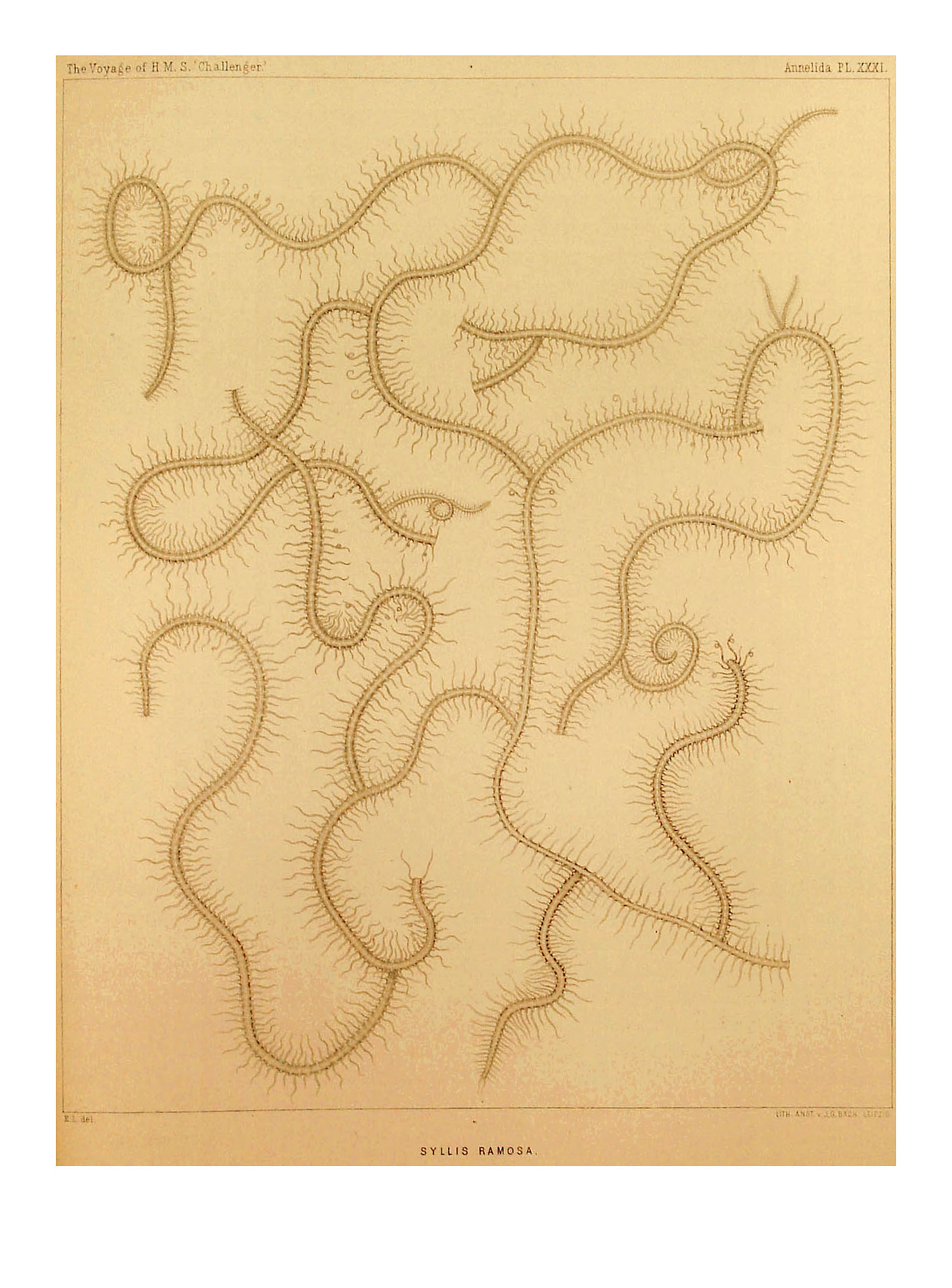
Comparative data for Syllis ramosa came from McIntosh (Reference McIntosh1879, Reference McIntosh1885, pl. XXXIII, figures 11–13, pl. XVA, figures 17 & 19; pl. XXXIVA, figures 9, 10 & 12) and Imajima (Reference Imajima1966) and specimens of the same lodged at the Natural History Museum, London (BMNH) and the Oxford University Museum of Natural History, Oxford (OUM), as follows: Syntypes BMNH 1885: 12: 1: 150–154, Challenger Expedition, M79 – specimen from Challenger Station 209, 95 fathoms, near Cebu, Philippines, M85 – specimen from Challenger Station 192, 140 fathoms, Arafura Sea, SE of Ceram; M85 – male stolon, probably detached, but may include a bit of the stalk (pl. 34A, figure 13) from a sponge in the Oxford Museum of unstated provenance, provided by Professor Mosely. In addition, we examined one specimen of Syllis cf. ramosa from Sagami Bay, Japan, 100 fathoms, which is lodged in the National Science Museum, Tokyo (NSM unreg.).
RESULTS
Segmental asymmetry
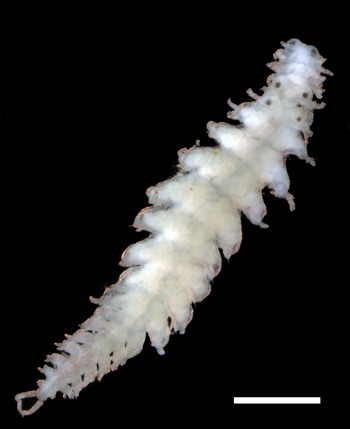
Ramisyllis multicaudata exhibits regional areas of asymmetry revealed by the dorsal parapodial cirri, which as in the post-proventricle segments of many syllids, alternate between long and short forms. The long dorsal cirri in the posterior (distal) region of the present specimens contain vivid white pigment (other worms living with white Petrosia have purple coloured dorsal cirri), as seen in photos of living animals (Figure 1). In this figure, most of the white cirri on each segment are opposite one another. However, in one section of the worm the white dorsal cirri are opposite short, unpigmented dorsal cirri indicating a symmetry shift. This type of asymmetry, referred to as ‘segmental asymmetry’, has also been observed in S. ramosa and is unique to the branching syllid morphotype.

Fig. 1. Ramisyllis multicaudata and host sponge Petrosia sp. in vitro showing tails emerging from excurrent canals. Inset shows dorsal cirri out of phase indicating segmental asymmetry. Scale bar: 5.0 mm.
Description of stolon stalks
Stolon stalk segments resemble the ‘intermediate’ segments (sensu Glasby et al., Reference Glasby, Schroeder and Aguado2012) of the stock in terms of segmental length, dorsal cirri morphology and chaetal form; in particular, the dorsal cirri do not show the typical alternation pattern of long/thick – short/thin that is present in anterior and posterior segments, including those emerging from the sponge (Figure 2). Stolon stalks branch from the stock in the midbody (a zone proximal to the production of terminus-producing branches); they include from 6–25 segments (Figure 3A, B). Each stalk produces a stolon, and we have seen no sign of oogenesis in the stalk.

Fig. 2. Two early stolons of Ramisyllis multicaudata attached to a main branch via a stolon stalk (ss), and below leading to a normal tail. Note alternating enlarged dorsal cirri on the main branch which are not present on the stolon or stalks. Scale bar: 0.5 mm.

Fig. 3. Near mature stolons of Ramisyllis multicaudata and attached stolon stalks (ss), (A) male; (B) female. Scale bars: 0.5 mm.
Female stolon development is initiated when posterior (distal) segments (the future stolon) begin to swell with the onset of vitellogenesis (Figure 2). The proximal segments fail to develop gametes and are soon differentiated from the developing stolon, remaining as the stalk. There does not appear to be a clear relationship between the number of stalk segments and the maturity of the stolon; stalks with about 8 segments can carry immature (Figure 2) or near fully mature stolons (Figure 3B).
Stolons sometimes occur in yoked pairs with one stalk branching from another (Figure 4), although unbranched stalks with well-developed stolons and no branches are usually present as well. At least some of these yoked pairs arise as an initial branch from the axial segments, which proceed to generate a stolon at its growing end; but another branch also forms from this stalk, which also produces a stolon. This newer stolon starts to develop after the first stolon has already developed somewhat, and hence is at an earlier developmental stage (Figure 4). Once this sequence has been recognized, it is possible to detect stalk pairs from which a mature stolon has already departed. The abandoned stalks show a characteristic terminus distinct from the growing tip of a new stolon or of a developing axial branch; it is smaller in diameter and appears as a short stubby appendage on the end of the stalk (Figure 4). This appendage develops a pair of anal cirri (Figure 5); the beginnings of an anal opening can be seen more clearly on better developed stubs.

Fig. 4. Ramisyllis multicaudata, female stolons in yoked pairs. Note terminus at stolon detachment point. Scale bar: 0.5 mm.

Fig. 5. Ramisyllis multicaudata, Scanning electron micrograph of developing female stolon attached to stalk segments of parent-stock, anterior end, ventral view. Note minute developing anal cirri (ac), dorsal cirrus (dc), neuropodia (n) and ventral cirrus (vc) of first chaetiger. Scale bar: 0.1 mm. Modified after Glasby et al. (Reference Glasby, Schroeder and Aguado2012).
Description of stolons
Male stolons ranged between 2.0–2.8 mm long, 0.50–0.65 mm wide (max.) for 18–22 chaetigers. Female stolons ranged between 3.15–3.50 mm long, 0.60–0.75 mm wide (max.) for 17–19 chaetigers. All specimens of R. multicaudata examined were observed to have developing stolons attached to stolon stalks in the midbody region within the sponge. The stolons of any adult specimen were consistently either male or female.
One specimen (QD4) from a sponge which measured ~5.3 × 4.2 cm, was dissected out as completely as possible. It had about 375 branches and over 500 termini, including 36 stolons, all female, most of which contained a full complement of oocytes. Three other sponge specimens that were systematically searched for the presence of worms also showed that, within any host sponge, attached stolons were all of one sex or all of the other. Thus the available evidence indicates that the worms have fixed separate sexes (see Discussion).
The head of the stolons is deeply bifid, has two pairs of eyes and lacks antennae and palps. Male and female stolons differed significantly in appearance (Table 1). The first chaetiger of both male and female stolons is slightly modified, almost fused to the head, and carries dorsal cirri, a neuropodium bearing chaetae and ventral cirri (Figures 5 & 6A, B). The first pair of dorsal cirri of the male is 2–3 times as long as those on the subsequent stolonal segments (in the female it is about the same length as the later dorsal cirri). Female stolons are laden with oocytes, usually well-developed and readily seen by the time the stolon and its branch are easily distinguished from somatic branches. One female stolon (Figure 6B) contained between 250 and 300 oocytes; oocytes develop in the coelom between chaetigers 1–4 and 13–14 (Figures 6A & 7B). In one specimen (CG 16–03 sponge 2) containing about 10 isolated mature female stolons, very few oocytes were remaining, suggesting that spawning had occurred inside the sponge prior to collection (see Discussion).

Fig. 6. Ramisyllis multicaudata Anterior end of male stolon (A) and anterior end of female stolon (B) photographed using differential interference optics (Nomarski). vg, vestigial gut bulb. Scale bars: 0.1 mm.

Fig. 7. Ramisyllis multicaudata, (A) male stolon, dorsal view; (B) female stolon, ventral view. Scale bars: 0.5 mm.
Table 1. Morphological differences between male and female stolons of Ramisyllis multicaudata.

The body of males is slimmer in most cases than that of the females and the parapodia are relatively better developed, including the presence of a lanceolate neuropodial lobe, which is absent in the female (Figure 7; Table 2). Further, the dorsal cirri of the male are unarticulated whereas those of the female are articulated. The ventral cirri of both sexes are unarticulated and lingulate. Fully mature males had their anterior three segments filled with pale material (Figure 3A) – interpreted here as sperm masses – which often caused these segments to appear slightly swollen. Histological sections of less mature, presumed, male stolons (without such apparent sperm localization) showed only gonia at immature stages in the coelom. Since no gametes have been observed in segments of the stalk (above), we believe that gametogenesis occurs within the stolon (spermatogenesis could be easily missed).
Table 2. Comparison of key variable developmental features of Ramisyllis multicaudata with other ribbon clade members.

Data for Ramisyllis from this study; those for other taxa from Potts (Reference Potts1913), Gravier & Dantan (Reference Gravier and Dantan1928), Okada (Reference Okada1933), Aguado & San Martín (Reference Aguado and San Martín2009), Aguado et al. (2012, 2015a, c) and Álvarez-Campos et al. (Reference Álvarez-Campos, San Martín and Aguado2013). Note that Trypanosyllis is very probably not monophyletic, and the gemmiparous species might well be different from those that are scissiparous.
Mature stolons of both sexes have, in addition to typical neurochaetae of the stock (Figure 8A, B), paddle-like notochaetae which differ from the capillary types of other sylline stolons (Figure 8A, B). The chaetae are arranged in two distinct chaetal bundles (each with about 10 notochaetae) adjacent to a thin notoacicula. The paddle-like chaetae are simple, obliquely truncate and distally tapered to a thin, drawn-out tip, very transparent, and difficult to see until one is aware that the notochaetal bundle diffracts light; thus a small rainbow in the parapodium of a preserved specimen under a dissecting microscope indicates the presence of this notochaetal bundle, even though the individual chaetae cannot be easily distinguished (Figure 6B, segment 4). Paddle chaetae appear to develop late in the formation of the stolon, since they are not detectable in those still attached to their stalks or in recently detached male stolons. Their shape and late occurrence suggests a swimming function, in which case they are similar to the natatory chaetae of other Syllidae.

Fig. 8. Ramisyllis multicaudata midbody parapodium of stolons (dorsal side up), showing presumptive natatory chaetae (nc). (A) male; (B) female. Scale bars: 0.1 mm.
One female stolon of 18 chaetigers had three heads anteriorly (Figure 9); we suggest that this represents aberrant growth rather than regeneration of lost anterior segments, because the specimen otherwise conforms to the 17–19 fixed chaetiger number of females, and the general pattern of segmental oocyte development (anterior and posterior region free of oocytes). The posterior-most head and following segments (including the bulb-like vestigial gut) had the appearance of a normally developed female.

Fig. 9. Aberrant polycephalic female stolon of R. multicaudata photographed under a stereo microscope using top and bottom oblique-angle lighting. Scale bar: 0.5 mm.
Comparison of stalks and stolons between R. multicaudata and S. ramosa (pooled data from Japanese and Philippine specimens)
There appear to be few, if any, differences in the form of the stolon stalk between the two species. In both species, chaetiger width is less than adjoining branches of the stock, and the parapodia and chaetae are the same form as those of the adjoining stock segments. Ramisyllis multicaudata has 6–25 stolon stalk chaetigers compared with the slightly greater maximum range of 4–33 for S. ramosa (Table 3).
Table 3. Comparison of key morphological differences in the stolons and stolon stalks of Ramisyllis multicaudata and Syllis ramosa.

Dc, dorsal cirri. (S. ramosa data from observations of syntype material (BMNH, OUM) and Japanese specimens (NSM), and from the literature – McIntosh Reference McIntosh1879, Reference McIntosh1885; Imajima, Reference Imajima1966; all specimens of S. ramosa assumed to be conspecific).
The stolons of the two species have very similar head morphologies with two pairs of similar-sized eyes and lack of palps and antennae. However, there are several differences between the two species not associated with the head. Compared with S. ramosa, the female stolons of R. multicaudata are smaller (3.15–3.5 cf. 9 mm), have fewer chaetigers, 18–22 (male), 17–19 (female) compared with 29–33 (male), 29 (female), and have fewer articulates in the dorsal cirri, 0 (male), 3–4 (female) compared with 4–5 (male), 13 (female) (Table 3).
DISCUSSION
Gametogenesis and stolon aberrance in Ramisyllis multicaudata
The site of gametogenesis in stolon-forming Syllinae is variable. Compared with R. multicaudata where gametogenesis takes place within those segments that form the stolon, the gametes in Haplosyllis spongicola (Grube, Reference Grube1855) originate in the stock and are later transferred to the stolon (Wissocq, Reference Wissocq1966); in Typosyllis antoni Aguado, Helm, Weidhase & Bleidorn, Reference Aguado, Helm, Weidhase and Bleidorn2015b they are developed within a large number of posterior segments including those that are later transformed into the stolon; when the stolon is completely developed, it detaches, but some posterior segments full of gametes still remain for later stolonizations (Aguado et al., Reference Aguado, Helm, Weidhase and Bleidorn2015b; MTA, personal observation).
The finding of a multi-headed stolon in R. multicaudata, while noteworthy, is not unique. The development of aberrant polycephalic stolons in other Syllinae has been explained by the absence of the proventricle (artificially removed in lab conditions) (Weidhase et al., Reference Weidhase, Beckers, Bleidorn and Aguado2016 and references therein). However, we cannot confirm that the specimen of R. multicaudata that developed one aberrant stolon had lost the proventricle. In contrast, this specimen possessed several stolons with all except one showing normal development. This might suggest that other factors, apart from the proventricle and its role in controlling reproduction, are involved in stolon development in R. multicaudata. Such polycepahlic stolons have been produced experimentally in the genera Trypanosyllis (Junqua, Reference Junqua1957) and Typosyllis (Weidhase et al., Reference Weidhase, Beckers, Bleidorn and Aguado2016).
Stolon morphology in Syllinae
The morphology of stolons across the Syllinae is poorly known and based mostly on those attached to the parent stock rather than free swimming forms. Thus, comparison of late-developing stolon features such as the appearance of presumptive natatory chaetae, maximum number of chaetigers, eye development, and pigmentation patterns, all of which may occur or be completed after stolon detachment, is currently not possible. Despite this limitation, several authors including Malaquin (Reference Malaquin1893) and Potts (Reference Potts1911) have attempted a classification of sylline stolons based on head morphology. Five stolon types are currently recognized, in order of increasing complexity: acephalous (no head), acerous (a head but no head appendages), dicerous (palps only), tetracerous (palps and lateral antennae), and pentacerous (palps, lateral antennae and a median antenna). Note that the appendages of dicerous stolons, referred to here as palps because of their position on the head and because they are not apparently articulated, is not universally agreed upon. Potts (Reference Potts1911) originally referred to them as lateral tentacles, and subsequent authors have called them antennae. However, we prefer to confine the concept of stolon antennae to the articulated appendages of tetracerous and pentacerous stolons, which is in keeping with the homologies of prostomial antennae established by Rouse & Fauchald (Reference Rouse and Fauchald1997) for Phyllodocida in general. The nature of the paired appendages on the head of dicerous stolons needs further study. Notwithstanding these somewhat confused concepts, it seems clear that the stolons of R. multicaudata are of the acerous type. This is indicated by the deeply bilobed anterior prostomium, two pairs of eyes, absence of antennae and palps (Potts, Reference Potts1911; San Martín, Reference San Martín and Ramos2003); additionally they show sexual dimorphism.
Comparisons with Syllis ramosa
As in R. multicaudata, the stolons of S. ramosa are most probably acerous. Most of McIntosh's illustrations of heads show the absence of antennae and palps, characteristic of acerous stolons. His figure 10 in pl. XXXIVA shows a stolon with a small pair of ‘antennae’, characteristic of a dicerous stolon; however, McIntosh (Reference McIntosh1885: 201) seems to express some doubt about the species identity of the stolon, as its supposedly young stage (small oocytes) did not match its high number of segments (31).
The presence of a branch bearing a developing stolon at its tip is obviously a feature of branching syllids, since to our knowledge such a structure has only been found in R. multicaudata and S. ramosa. If these taxa are not closely related as the current taxonomy suggests, the branching body pattern might have evolved independently. Both species are schizogamous; however, because multiple stolon producing stalks may appear at the same time, in this sense the species are gemmiparous (see Garwood, Reference Garwood1991 for descriptions of typical examples of these two systems of stolonization). Aguado et al. (Reference Aguado, Glasby, Schroeder, Weigert and Bleidorn2015a) used the term ‘branching gemmiparity’ to more accurately describe this type of reproductive mode.
McIntosh (Reference McIntosh1885) suggested that S. ramosa from the type locality in the Philippines was viviparous. We have found no sign of viviparity in either his material (from the BMNH) or that from Japan. Therefore, S. ramosa was erroneously included in the Schroeder & Herman's (Reference Schroeder, Hermans, Giese and Pearse1975) list of viviparous species. Since the available Japanese specimens do not include stolons, the following observations come from Okada (Reference Okada1937). In the Japanese S. ramosa, the stolon stalk is composed of a variable number of segments, all of which are typical in their structure. They thus differ from McIntosh's (Reference McIntosh1885, pl. XXXIII, figure 11) description of the Challenger material, in which the stolon stalks are formed of four segments, each of which has a pair of small ‘tentacles’, which might represent the dorsal cirri of rudimentary parapodia. Okada (Reference Okada1937) doubted that the condition described by McIntosh (Reference McIntosh1885) actually occurs, suggesting that the two might be different species; however, not enough information is available to answer this question. McIntosh (Reference McIntosh1879, Reference McIntosh1885) did not report how many such stalk-stolon combinations he observed. He notes that some male stolons have gametes in eight segments. McIntosh (Reference McIntosh1885, pl. 33, figure 11) illustrates the attachment of a developing female stolon of his S. ramosa to the main axis of one of the branches. It is attached by a segmental stalk four segments long, on which the parapodia are either incompletely developed or (in the two segments closest to the body) absent.
Comparisons with other members of the ribbon clade and other Syllinae
Like other members of the ribbon clade, R. multicaudata appears to have acerous stolons (see Aguado et al., Reference Aguado, San Martín and Siddall2012, although the stolons of Parahaplosyllis were not known at that time). Okada (Reference Okada1933, Reference Okada1937), Álvarez-Campos et al. (Reference Álvarez-Campos, San Martín and Aguado2013) and Aguado et al. (Reference Aguado, Murray and Hutchings2015c) take a different view on the nature of the head appendages thus coming to the conclusion that both acerous and dicerous stolons are present in members of the ribbon clade. Clearly further comparative studies are required to elucidate the nature of these stolon head appendages.
At present, we can find no exclusive common reproductive characteristics among the ribbon clade taxa. Features such as stolons lacking regionalization, absence of brooding, and embryonic development not connected with the parental body, are simply plesiomorphic absences found in most Syllinae. The reproductive features that are variable within the clade and currently unique to Ramisyllis, such as branching gemmiparity, presence of presumptive natatory chaetae in late stolons and dimorphic males/females (Table 1) may also be present in other Syllinae; however, whether they represent convergences or homologies is uncertain. For example, natatory chaetae somewhat resembling those in R. multicaudata have been found in the only known specimen of Nuchalosyllis lamellicornis Rullier & Amoureux, Reference Rullier and Amoureux1979, although these are pointed rather than truncate (Aguado & San Martín, Reference Aguado and San Martín2009). Nothing is known of reproduction in this species, nor were such chaetae observed in a second species of Nuchalosyllis, N. maitieae Fukuda & Nogueira (Reference Fukuda and Nogueira2013). This taxon also resembles other ribbon clade members in terms of general body form (flattened body), and therefore would be a valuable addition to any future phylogenetic analysis of the group. Ramisyllis appears to have separate sexes producing only male or only female stolons; sequential hermaphroditism is also a possibility and will be the subject of further investigation (below). We do not know whether stolons of both sexes leave the sponge to mate in open water, or if one is visited by the other inside a sponge. We have never observed developing larvae or embryos inside a sponge. Males appear to be less swollen and have fewer sperm-filled segments compared with typical sylline stolons such as Typosyllis pigmentata (PCS, personal observation), which spawn in open water and are usually as stuffed with gametes as are the females. Such a strategy would suggest that spawning occurs within the sponge. An intra-sponge spawning hypothesis is also supported by the finding of mature female stolons inside the sponge having very few eggs remaining inside the coelom. A population study using DNA microsatellites (or the whole genome) to compare adults, detached stolons and juveniles from different sponges would contribute to elucidating parentage as well as to assessing the probable spawning site.
ACKNOWLEDGEMENTS
The authors would like to thank Maureen Metcalf (Francheschi Imaging and Microscopy Center, Washington State University) and the staff of the Histology Laboratory (School of Veterinary Medicine, Washington State University). This study is a contribution of the project ‘Macroevolutionary transitions in Syllidae’ CGL2015-63593-P MINECO/FEDER. This research received no specific grant from any funding agency, commercial or not-for-profit sectors. The comments and suggestions of the referees and IPC editors are gratefully acknowledged.
FINANCIAL SUPPORT
This research received no specific grant from any funding agency, commercial or not-for-profit sectors.


