


1. Introduction
Echinoderms have diverse body plans, living exclusively in marine environments since the Cambrian Period (Sprinkle, Reference Sprinkle1976). Many Cambrian echinoderms lack pentaradial symmetry and mobility, which are characteristic of modern echinoderms; their primitive body plans, many of which became extinct before the end of Cambrian Period, are not well understood due to a lack of modern analogues. Taphonomy can provide insight into better understanding of the palaeobiology of Cambrian stalked echinoderms, commonly dominated by gogiids. Furthermore, gogiid taphonomy can be better constrained when compared to analogues of post-Cambrian echinoderms (e.g. Gahn & Baumiller, Reference Gahn and Baumiller2004; and references therein).
A profound change of physical and chemical sedimentary conditions in substrates occurred during the late Neoproterozoic to post-Cambrian transition, and the Cambrian interval represents a critical transitional period for substrate change (Bottjer, Hagadorn & Dornbos, Reference Bottjer, Hagadorn and Dornbos2000). Thus, the constructional morphology of Cambrian sessile echinoderms reflects the confluence of niche competition (Wilbur, Reference Wilbur2005a, Reference Wilburb) and substrate conditions, but the specific controls responsible for the Cambrian echinoderm diversity are subject to considerable debate. Some studies (Sprinkle & Guensburg, Reference Sprinkle and Guensburg1995; Wilbur, Reference Wilbur2006) focused on the availability of skeletal substrates, whereas other studies emphasized the substrate conditions, such as the coverage of microbial mats and development of a mixed layer (Bottjer, Hagadorn & Dornbos, Reference Bottjer, Hagadorn and Dornbos2000; Dornbos & Bottjer, Reference Dornbos and Bottjer2000). The present study of early and middle Cambrian stalked gogiid echinoderms is a multidisciplinary approach that provides additional information to understand better the nature of Cambrian substrates in the fine-grained siliciclastic settings.
Reports on Cambrian echinoderm faunas in China are uncommon. Putative echinoderm skeletal elements (Xue, Tang & Yu, Reference Xue, Tang and Yu1992) had been reported from the Doushantuo Formation (Ediacaran), but these phosphatized microelements do not contain stereomic microstructure. Zhang & Jiang (Reference Zhang and Jiang1983) reported isolated echinoderm plates, but they misidentified them as sponge spicules. Huang, Zhao & Gong (Reference Huang, Zhao and Gong1985) reported the first discovery of articulated gogiids in Guizhou Province, South China. However, specimens were not formally described until much later (Zhao, Huang & Gong, 1994; Zhao et al. Reference Zhao, Yuan, Zhu, Yang, Guo, Qian, Huang and Pan1999; Parsley, Zhao & Peng, Reference Parsley, Zhao and Peng2005; Parsley & Zhao, Reference Parsley and Zhao2006). Han et al. (Reference Han, Tang, Wei and Wang2000) reported the first articulated stylophorans from the late Cambrian strata of Guangxi Province, South China. Recently, new early Cambrian gogiid faunas were discovered in Guizhou (Peng et al. Reference Peng, Zhao, Wu, Yuan and Tai2005a,b) and Yunnan Province (Hu et al. Reference Hu, Luo, Hou and Erdtmann2006). The abundance of articulated Guizhou gogiids (n > 1000) from the Balang and Kaili formations collectively provides substantial information to test hypotheses concerning the taphonomy of Cambrian erect echinoderms (e.g. Sprinkle, Reference Sprinkle1973, Reference Sprinkle1976; Dornbos & Bottjer, Reference Dornbos and Bottjer2001). The substrate conditions and settlement strategy for gogiid echinoderms from the Kaili Formation are addressed in Lin, Ausich & Zhao (in press). This paper focuses on taphonomy and preservation of gogiids exemplified by Cambrian material from two faunas in Guizhou Province, South China. The objectives here are to address the following questions:
(1) What kind of palaeocurrent information can we discern based on gogiid entombment orientation (e.g. Sprinkle, Reference Sprinkle1976)?
(2) Dornbos & Bottjer (Reference Dornbos and Bottjer2001) hypothesized that primitive echinoderms exemplified by helicoplacoids are most commonly preserved with partial disarticulation due to the combination of pre-burial and post-burial decay. Can Guizhou gogiid material provide more insight or alternative interpretations about the decay and disarticulation of Cambrian stalked echinoderms in general?
(3) What are the potential causes of any of the unusual disarticulation patterns of Balang echinoderms?
(4) What are the main chemical elements causing the colour differences among Guizhou gogiids?
(5) This study reports for the first time two articulated gogiid species with stereom preservation. What types of skeletal microstructure (e.g. Smith, Reference Smith and Carter1990; Clausen & Smith, Reference Clausen and Smith2005) are preserved in the Guizhou gogiids?
(6) Finally, does our study of stereom preservation support the ‘crystal caskets’ hypothesis proposed by Dickson (Reference Dickson2001)?
2. Stratigraphy and localities
Two major Cambrian gogiid faunas with at least five known localities have been reported from eastern Guizhou Province, China, and both faunas were deposited in fine-grained siliciclastic facies in the Jangnan Slope Belt of the South China plate during the Cambrian Period (Fig. 1). The first reports of articulated Cambrian gogiids from South China were from the Kaili Formation, which also contains a wealth of Burgess Shale-type organisms (see Zhao et al. Reference Zhao, Yang, Zhu, Yuan, Peng and Zhao2002; Lin, Reference Lin2006; Lin et al. Reference Lin, Gon, Gehling, Zhao, Zhang, Hu, Yuan, Yu and Peng2006). Temporal correlation of the Kaili Formation is well constrained by trilobite biostratigraphy (Yuan et al. Reference Yuan, Zhao, Li and Huang2002). Stratigraphy and chemostratigraphy of the Kaili Formation were thoroughly studied in Guo et al. (Reference Guo, Strauss, Liu, Zhao, Pi, Fu, Zhu and Yang2005). Two gogiid species, Sinoeocrinus lui Zhao, Huang & Gong, 1994 and ‘Sinoeocrinus globus’ Zhao et al. Reference Zhao, Yuan, Zhu, Yang, Guo, Qian, Huang and Pan1999 (see Parsley, Zhao & Peng, Reference Parsley, Zhao and Peng2005), are from the Orytocephalus indicus Zone, which is in the middle Cambrian portion of the Kaili Formation (Sundberg et al. Reference Sundberg, Yuan, McCollum and Zhao1999; Zhao et al. Reference Zhao, Yuan, Mccollum, Sundberg, Yang, Guo, Zhu and Yang2001). Recently, new localities in the lower Cambrian Balang Formation, which is known for its trilobite fauna (see McNamara, Feng & Zhou, Reference McNamara, Feng and Zhou2006), has yielded an abundant gogiid fauna in association with the index trilobite Redlichia (Pteroredlichia) murakamii in eastern Guizhou (Peng et al. Reference Peng, Zhao, Wu, Yuan and Tai2005a,b). Potentially, two new gogiid species occur in the Balang fauna, but systematic descriptions have not been completed. Balang gogiids are designated as gogiid sp. 1 and sp. 2 in this study and undoubtedly represent the richest early Cambrian gogiid fauna with the most abundant articulated specimens (Peng et al. Reference Peng, Zhao, Wu, Yuan and Tai2005a). Both faunas contain abundant trace fossils (Yang, Reference Yang1994; Yang & Zhao, Reference Yang and Zhao1999; Wang et al. Reference Wang, Zhao, Lin and Wang2004; Peng et al. Reference Peng, Zhao, Wu, Yuan and Tai2005b; Wang et al. Reference Wang, Zhou, Zhao and Yu2006; this study) and lack evidence for microbial mats that are characteristic of Precambrian and some Cambrian substrates (Bottjer, Hagadorn & Dornbos, Reference Bottjer, Hagadorn and Dornbos2000). In general, the benthic palaeoenvironments of the Balang and Kaili faunas consist of soft substrates composed of silt- and clay-size sediments and parautochonous skeletal debris with relatively shallow mixed layers.

Figure 1. Map of China indicating position of Guizhou Province (upper left), and map of Guizhou Province with five important localities for Cambrian echinoderm faunas and the depositional settings during the Cambrian Period (modified from Lin, Reference Lin2006). Illustrated Kaili gogiids are from Wuliu-Zengjiayan (site 1) and Miaobanpo (site 2) sections. Site 3 is the location of the stratotype of the Kaili Formation, and there is one articulated gogiid reported from this site (Lin et al. Reference Lin, Yuan, Wang, Zhao, Peng, Babcock and Zhu2005). Sites 4 and 5 are lower Cambrian gogiid localities from the Balang Formation (Peng et al. Reference Peng, Zhao, Wu, Yuan and Tai2005a,b).
3. Material, terminology and methods
Illustrated specimens are deposited at the following institutes: Orton Geological Museum of the Ohio State University (OSU), Columbus, Ohio, USA and the Guizhou Museum of the Guizhou University, Guiyang, Guizhou Province, China. Kaili Formation specimens from the Wuliu-Zengjiayan section (site 1 in Fig. 1) are abbreviated as GTBJ, and specimens from the Miaobanpo section (site 2 in Fig. 1) are abbreviated as GM. Lower Cambrian gogiids from the Balang Formation at the Wenglingtang section (site 4 in Fig. 1) are abbreviated as KW, and those from Jianggu section (site 5 in Fig. 1) are abbreviated as ZJ. In addition, some specimens of Balang gogiids are on permanent display at the Kaili No. 4 Middle School (KMS4), Kaili, Guizhou Province, China, curated by Yishan Wu. These specimens contain unparalleled information about the taphonomy of Balang gogiids; thus, they are illustrated here and assigned depository numbers.
Terminology for the major gogiid body elements is illustrated in Figure 2. Other terminology follows Sprinkle (Reference Sprinkle1973). Gogiid echinoderms do not have a true ‘stem’; thus, the usage of ‘stem’ is not recommended in Ubaghs (Reference Ubaghs and Moore1967a). Instead, ‘holdfast’ (see Sprinkle, Reference Sprinkle1973) is used to describe the plates below the theca. The holdfast is further divided into the stalk and attachment structure (Lin, Ausich & Zhao, in press). For ‘Sinoeocrinus globus’ (Fig. 2d, f), the stalk consists of small polygonal plates, and the attachment structure consists of tiny plates (Sprinkle, Reference Sprinkle1973, text-fig. 26) or platelets (Parsley & Prokop, Reference Parsley and Prokop2004). The Sinoeocrinus lui (Fig. 2c, e) stalk has highly ornamented plates surrounded by tiny plates and gradually becomes an attachment structure with only platelets. The attachment structure in ‘S. globus’ is robust and disc-shaped but much less distinctive than in other species. For lower Cambrian Balang gogiids (Fig. 2a, b), holdfasts consist mainly of relatively small plates compared to thecal plates, although the attachment structure (Fig. 2a) may be similar to S. lui. The main difference between the dominant species, gogiid sp. 1 (Figs 2b, 5a–e), and the possible new species, gogiid sp. 2 (Figs 2a, 5f–j), is that gogiid sp. 2 contains a very distinct break at the junction between the theca and holdfast, whereas gogiid sp. 1 has a gradual transition from holdfast to theca. This distinction is present in both juvenile and adult specimens of the same thecal height.

Figure 2. Illustration of morphological terminology used for Cambrian gogiids from Guizhou Province, China, based on latex casts. See text for details. All scale bars = 5mm. (a) New early Cambrian species (gogiid sp. 2) with distinctive break in the junction between the theca and holdfast from the Balang Formation, KW-8–229. (b) New early Cambrian species (gogiid sp. 1) with gradational transition between the theca and holdfast from the Balang Formation, ZJ 001. (c) Sinoeocrinus lui from the Kaili Formation, GM-9–5-3701. (d) ‘Sinoeocrinus globus’ from the Kaili Formation, GM-9–4-445. (e) Enlarged view of the holdfast of a S. lui, GTBJ-16–2-26. (f) Enlarged view of the holdfast of a ‘S. globus’, which is a different specimen on the same slab as (d) GM-9–4-445.
Specimens were imaged with optical digital photography and scanning electron microscopy (SEM) at the School of Earth Sciences, Ohio State University. Due to the rarity of exceptionally preserved specimens, specimens were uncoated but wrapped with aluminum foil for SEM imaging (see Figs 9, 10). Images were taken at medium resolution to avoid charging effects due to long exposure of specimens without coatings. Both secondary electron (SE) and backscatter electron (BSE) images were taken for comparison. Seven specimens (e.g. Figs 11, 12) were analysed further with an energy dispersive X-ray (EDX) analyser attached to the SEM to determine the chemical composition. Stereom pore size and trabecular thickness were measured on enlarged SEM printouts with a digital caliber. Source data for inferring palaeocurrent information came from the examination of 381 specimens, including museum specimens, new material collected in the field, literature (Zhao, Huang & Gong, 1994; Zhu, Erdtmann & Zhao, Reference Zhu, Erdtmann and Zhao1999; Parsley & Zhao, Reference Parsley, Zhao, Heinzeller and Nebelsick2004), an unpublished work (J. Peng, unpub. M.S. thesis, Guizhou Univ., 2005), and a scanned booklet deposited at a permanent online address (http://hdl.handle.net/1811/24227). Data obtained for taphonomic classification are based only on new material collected near Kaili City (site 4 in Fig. 1), Guizhou Province, China.
Due to the nature of preservation, most studied specimens photographed digitally in colour (e.g. Figs 3, 7, 8, 13) have little contrast after conversion to greyscale. Thus, a non-standard colour filtering technique is adapted to enhance the morphology in greyscale. Each digital colour image was first opened with the GIMP 2.2.10 imaging software (available at: http://www.gimp.org) and the following procedure was applied. On the Filters menu, Colors option was chosen, then the Decompose function. After applying this filter effect, each colour image was converted into a stack of three greyscale layers according to the three principal colour elements RGB (red, green and blue). By scrolling down the Layer menu, individual layers could be deleted manually in order to see the greyscale layers individually. Layers could be restored by clicking the Undo function under the Edit menu. The layer with the best contrast was selected, and the new layer was saved as a JPEG or TIFF file after applying the function Flatten Image under Image menu. Each image was then imported into the drawing software CorelDraw 12 for further assembly of figures. The same procedures were applied to most of the greyscale images in this study (Figs 3, 5, 13).

Figure 3. Specimens of ‘S. globus’ showing different current orientations. Scale bars = 10mm. (a) Specimen GM-17–298 preserved in non-feeding posture; both brachioles and theca aligned with current direction. (b) Specimen GM-9–3-180 contains two individuals preserved in non-feeding postures; body axes perpendicular to each other. (c) Specimen GM-9–5-1709 contains three individuals preserved in non-feeding posture; body axes oriented in clockwise rotation. (d) Specimen GM-9–2-3530b contains two individuals preserved in a possible feeding posture; body axes pointing opposite to each other.
4. Taphonomy of gogiid echinoderms
4.a. Entombment patterns
Gogiid and other eocrinoids have an elongated body that if transported could provide palaeocurrent indication. Gogiids lived on the seafloor by attaching to skeletal debris (Sprinkle, Reference Sprinkle1976; Guensburg & Sprinkle, Reference Guensburg and Sprinkle1992; Wilbur, Reference Wilbur2005b; Lin, Ausich & Zhao, in press). Articulated specimens could be toppled over and buried immediately so that the body axis aligned in parallel with the dominant current direction. However, in the material studied, there are multiple orientations preserved on the same slabs of Guizhou material (Fig. 3). For example, on a single slab, the body axes can be oriented parallel to one another in the same direction (see Fig. 13), perpendicular to one another (Fig. 3b), parallel to one another but pointing in opposite directions (Fig. 3d), or in apparent clockwise rotation among neighbouring individuals (Fig. 3c). Furthermore, unlike the Laurentian gogiid faunas (Robison, Reference Robison1965; Sprinkle, Reference Sprinkle1973, Reference Sprinkle1976; Durham, Reference Durham1978), which contained a few specimens attaching to skeletal substrates and a general lack of attached specimens, preservation of articulated gogiids still attached to skeletal debris is common in the Guizhou material, especially for ‘S. globus’ (Lin, Ausich & Zhao, in press). This indicates a short transport history for most of the Guizhou gogiids.
Articulated specimens were buried in five basic orientations or entombment patterns (Fig. 4; see Table 1). These include: (1) gogiids laid in parallel to one another with enclosed brachioles; (2) gogiids laid in parallel to one another with brachioles splayed out in a fan-shaped pattern; (3) gogiids with body axes perpendicular to one another; (4) gogiids with body axes parallel to one another but pointing to the opposite directions; (5) gogiids with body axes oriented in a clockwise or anticlockwise rotation (which is a combination of Type 3 and Type 4 entombment patterns). This method only applies to articulated remains in this study, and the results are given in Table 2. Our study shows that the majority (92 out of 139 slabs) of Guizhou gogiid-bearing hand samples contain the Type 2 entombment pattern (Figs 2a, 4b). Slabs and hand samples containing gogiids with body axes oriented in clockwise or anticlockwise rotation among neighbouring individuals (Type 5 entombment pattern) (Figs 3c, 4e) are sometimes present in both Balang and Kaili faunas although not in every gogiid species. The presence of three or more burial orientations on the same bedding surface (or the Type 5 entombment pattern) suggests that gogiids were probably buried with high turbulent flows generated or induced by storm events.
Table 1. Types of entombment patterns exemplified by Chinese gogiid echinoderms

Table 2. Summary of gogiid-bearing slabs (n = 139) based on entombment patterns

Subtotals of each type for each fauna are indicated in bold.

Figure 4. Schematic drawings of entombment patterns exemplified by Guizhou gogiid echinoderms. Types of entombment patterns are explained in Table 1.
4.b. Decay/disarticulation
4.b.1. Taphonomic classification
Dornbos & Bottjer (Reference Dornbos and Bottjer2001) restudied helicoplacoids and described the quality of helicoplacoid preservation with reference to three taphonomic groups. Although gogiids and helicoplacoids are the earliest known erect echinoderms, gogiids differ considerably from helicoplacoids in having brachioles and subdivisions of the main body (theca and holdfast). We modified Dornbos & Bottjer's (2001) concept of taphonomic groups to derive meaningful and practical taphonomic information for gogiid echinoderms. Group 1 contains gogiids with fully articulated theca and holdfast and relatively complete brachioles. Group 2 includes gogiids with partial disarticulation of the theca and/or holdfast but missing a considerable portion of the brachioles. Group 3 contains specimens with the theca nearly completely disarticulated. Although many museum specimens could be used for this taphonomic comparison, Lin (2006, p. 17) cautioned about biases in collecting. Thus, only new material collected from the Balang fauna for this study are included (see Table 3). Unlike helicoplacoids (Dornbos & Bottjer, Reference Dornbos and Bottjer2001), which are mostly preserved with partial disarticulation, the majority of Balang gogiids (79%) are preserved with only a slight amount of disarticulation in both the theca and holdfast. Combined with results from entombment patterns (see Section 4.a), Balang gogiids are interpreted to have been buried rapidly in episodic obrution deposits with turbulent currents more frequently than were the helicoplacoid faunas.
Table 3. Comparison of three lower Cambrian echinoderm faunas based on taphonomic groups

Helicoplacoid specimens are deposited at University of Southern California (USC) and the Los Angeles County Museum of Natural History (LACMNH) and gogiid specimens from the Balang Formation (site 4 in Fig. 1) are deposited at the Orton Museum, The Ohio State University (OSU).
4.b.2. Disarticulation sequence
Although the taphonomic groupings are informative, it is also useful to study the disarticulation sequence of the major gogiid body parts. We predict that the relative timing of decay follows this sequence: (1) detachment of brachioles, (2) disarticulation of brachioles, (3) disarticulation of theca and (4) disarticulation of holdfast. Comparisons are made to taphonomic studies of post-Cambrian crinoids because the tripartite body design of gogiids is similar to that of crinoids, and fossil and modern crinoid disarticulation patterns are well studied (see Ausich, Reference Ausich, Jangoux and Lawrence2001; Gahn & Baumiller, Reference Gahn and Baumiller2004, and references therein). In general, crinoids are rarely preserved in a feeding posture because the disturbance that results in the burial typically causes a retraction of the arms into a trauma posture. Once a crinoid is dead, the disarticulation sequence varies depending on whether or not muscles existed in the arms (Ausich & Baumiller, Reference Ausich and Baumiller1993). If muscles were present, the arms disarticulate first, followed by the column disarticulating into pluricolumnal segments, the calyx plates if sutured by connective tissue, and finally pluricolumnal segments into individual columnals. If the only connective tissues in the arms were ligaments, arm disarticulation and column disarticulation into pluricolumnal segments began at approximately the same time (Ausich & Baumiller, Reference Ausich and Baumiller1998). Pluricolumnal segments may remain articulated for months in modern experiments (Oji & Amemiya, Reference Oji and Amemiya1998) and are common in the fossil record (Baumiller & Ausich, Reference Baumiller and Ausich1992).
Based on a similar body plan and no evidence for muscular articulations in gogiids, gogiids should disarticulate in a pattern similar to crinoids that lack muscles in arms, but the gogiid decay differs in several important ways. First, the brachioles were very narrow, and mesodermal connective tissue (Smith, Reference Smith and Carter1990) must have played a major role in binding adjacent brachiolar plates. At least one specimen (Fig. 5i) contains articulated segments of brachioles and a disarticulated theca; thus, segments of brachioles may remain articulated on the seafloor for some period of time. Furthermore, approximately 70% of surveyed Chinese gogiids are preserved with brachioles splayed in a fan-shaped pattern (Type 2 entombment pattern) (Fig. 5a, b, f; Table 2). Even the coiling axes of helical brachioles are preserved typically straight in transported specimens (Fig. 5c, d, i). These observations lead us to suggest that Chinese gogiid brachioles were relatively stiff in life with limited maneuverability compared to crinoid arms.

Figure 5. Schematic drawings of inferred disarticulation of Cambrian stalked echinoderms exemplified by ‘S. globus’, in chronological order. (a) Living position of ‘S. globus’ attached to a skeletal bioclast (indicated by a hyolith). (b) ‘S. globus’ with a few broken brachioles lying on the substrate at death. (c) Relatively simultaneous disarticulation of brachioles, theca and holdfast. (d) Isolated thecal plates, which are the most common gogiid and other eocrinoid elements, preserved in the fossil record, during a late stage of decay.
Second, the priority of theca versus holdfast decay is unclear. Specimen OSU 52615 (Fig. 5g) clearly has a mostly disarticulated theca and a mostly articulated holdfast. A single isolated attachment structure of the ‘G. globus’ type from the Kaili Formation was noted in the field. Sprinkle & Collins (2006, pl. 7, fig. 2) illustrated one specimen of Gogia stephenensis with a similar disarticulation pattern. However, two Balang specimens (KW-5–76, ZJ-15–3; unillustrated) are preserved with an articulated theca, partially disarticulated stalk plates, and missing attachment structures. Furthermore, Ubaghs & Vizcaïno (Reference Ubaghs and Vizcaïno1990) illustrated a specimen of Gogia (Alanisicystis) andalusiae with attached brachioles, a partially disarticulated theca, but a fully articulated anal pyramid and holdfast. The rarity of partially disarticulated specimens, and no apparent trend of decay priority of particular body parts, lead us to reject our initial decay hypothesis. Alternatively, there is no strong decay preference among gogiid body parts. This interpretation agrees with Dornbos & Bottjer's (2001) conclusion for helicoplacoid taphonomy.
Third, upon death of living stalked crinoids, the first decay step is the detachment of the crown (calyx plus arms) from the columnal (Oji & Amemiya, Reference Oji and Amemiya1998), and detached crowns are relatively common in the fossil record (e.g. Gahn & Baumiller, Reference Gahn and Baumiller2004). However, this is not the case in gogiids, due to the general lack of specimens preserved as only the articulated theca and brachioles, which would be morphologically equivalent to a crinoid crown. Similarly, other closely related Cambrian eocrinoids, including Akadocrinus (Sprinkle, Reference Sprinkle1973) and Lyracystis (Sprinkle & Collins, Reference Sprinkle and Collins2006), bear a long stalk but do not disarticulate like crinoids. Therefore, the taphonomy of Cambrian stalked echinoderms collectively has an intermediate disarticulation pattern between helicoplacoids and crinoids.
Based on material for this study, gogiid literature and the literature on post-Cambrian stalked echinoderms, a general decay model for gogiids is proposed (Fig. 6). Once a gogiid was dead and laid down on the seafloor, decay of volatile tissues leading to disarticulation of body plates proceeded rapidly (Fig. 6b, c). However, ratios of isolated plates derived from brachioles, theca and holdfast that survived in a transported assemblage of gogiid remains are disproportionate because they have different masses and shapes among body plates leading to differential susceptibilities to turbulence. Therefore, unlike crinoids, from which the most commonly recognized plates are columnals, we hypothesize that for gogiids the isolated thecal plates (Fig. 6d) are the most common elements recognized in the Cambrian sediments (e.g. Sprinkle, Reference Sprinkle1973; Álvaro, Vennin & Vennin, Reference Álvaro, Vennin and Vennin1997).

Figure 6. Unusual burial postures and disarticulation patterns of gogiids from the Balang Formation. (a–e) Gogiid indet. sp. 1. (f–j) Gogiid indet. sp. 2. (a) Well-preserved specimen in normal burial position, ZJ-15–5-10. (b) Gogiid specimen in a sunburst pattern, OSU 52612. (c, d) Specimen with mostly articulated plates, selective disarticulation between the basal brachioles and the upper theca, and some disoriented segments of brachioles, OSU 52613. (e) Specimen with mostly articulated plates, partially disarticulated brachioles, and missing the upper middle portion of the theca, OSU 52614. (f) Well-preserved specimen in normal burial position, KW-8–229. (g) Specimen with disarticulated brachioles, partially disarticulated thecal plates scattered around nearby matrix, and a dislocated portion of the holdfast stalk, OSU 52615. (h) Specimen in S-shape posture, OSU 52616. (i) Specimen with partially articulated segments of brachioles and completely disarticulated theca and holdfast (only a few thecal plates remaining), OSU 52617. (j) Two individuals with post-mortem elongation, KMS4–30. Scale bars = 20mm (a, e); 10mm (b, f, g); 15mm (c, d); 5mm (h, i); 25mm (j).
4.b.3. Summary
The proposed entombment patterns and disarticulation sequence is based on well-preserved material from South China and can be applied to study closely related gogiid genera, including Gogia (Walcott, Reference Walcott1917; Robison, Reference Robison1965; Sprinkle, Reference Sprinkle1973; Durham, Reference Durham1978; Sprinkle & Collins, Reference Sprinkle and Collins2006) and Gogia (Alanisicystis) (Ubaghs & Vizcaïno, Reference Ubaghs and Vizcaïno1990; Gil Cid & Domínguez Alonso, Reference Gil Cid and Domínguez Alonso1999), Marjumicystis (Ubaghs & Robison, Reference Ubaghs and Robison1985), Sinoeocrinus (Zhao et al. Reference Zhao, Huang and Gong1994) and undescribed lower Cambrian material from both Guizhou (Peng et al. Reference Peng, Zhao, Yuan, Wang and Wu2005a; this study) and Yunnan Province (Hu et al. Reference Hu, Luo, Hou and Erdtmann2006). In addition, our gogiid decay sequence indicates that thecal plates should be the most recognizable body plates among transported gogiid or closely related eocrinoid remains (e.g. Sprinkle, Reference Sprinkle1973, pl. 25, figs 2–7, 9–22; Berg-Madsen, Reference Berg-Madsen1986, fig. 4A; Smith, Reference Smith and Carter1990, fig. 12A; Skovsted, Reference Skovsted2006, fig. 4A). Columnals (Berg-Madsen, Reference Berg-Madsen1986, figs 5–7) and the ‘basal cups’ (e.g. Cymbionites; Whitehouse, Reference Whitehouse1941; Smith, Reference Smith1982; Ubaghs, Reference Smith and Carter1967b) do not exist in gogiids.

Figure 7. Evidence of bioturbation, co-occurring trace fossils, and potential trace makers from the Balang Formation. (a, b) Part and counterpart of a slab containing Planolites burrows associated with an articulated gogiid, KMS4–31. (c) An arthropod resting trace Rusophycus, OSU 52618. (d) A trilobite Redlichia (Pteroredlichia) murakamii, KW-12–701. (e) A large bivalved arthropod Tuzoia sp., KW 582. Scale bars = 20mm (a, b, d, e); 5mm (c).
Study of disarticulation patterns gives us a sense of the articulation of the gogiid when it was alive. By comparing taphonomic studies on Cambrian (helicoplacoids: Dornbos & Bottjer, Reference Dornbos and Bottjer2001) and post-Cambrian (crinoids: Ausich, Reference Ausich, Jangoux and Lawrence2001, and references therein) erect, sessile echinoderms, it is evident that gogiid and helicoplacoid echinoderms share a similar biological grade: no differentiation of connective tissue in binding skeletal plates. In other words, although gogiids bear a similar tripartite body plan (brachioles, theca and holdfast) to those of crinoids (arms, calyx and columnal), the holdfast in gogiids is essentially composed of immature thecal plates and represents the continuation of the main body chamber; unlike crinoid columnals that are constructed and developed distinctively different from the calyx plates.
4.c. Unusual entombment/disarticulation patterns
A dozen specimens exhibit rather unusual preservation patterns and each deserves a detailed description. All specimens except those noted in Section 4.c are from the Balang fauna, near Kaili City (site 4 in Fig. 1), Guizhou Province, South China.
4.c.1. Sunburst pattern
As described in Section 4.b, most gogiids are preserved lying on their sides. However, one Balang gogiid (Fig. 5b) and two specimens of Sinoeocrinus lui have the summit (top surface) preserved with brachioles radiating out in all directions in a sunburst orientation. The simplest explanation for the sunburst orientation in gogiids is that they were buried in situ and compressed vertically. Alternatively, individuals could be transported rapidly via storm-generated currents and positioned upside down at the burial site, analogous to some burial postures of crinoids (see starburst pattern in Gahn & Baumiller, Reference Gahn and Baumiller2004, and references therein). Where bedding information was recorded, the only preserved orientation is a sunburst gogiid compressed right side up showing the oral surface. Sunburst patterns are also known from Laurentian gogiid faunas (Sprinkle, Reference Sprinkle1973, text-figs 15, 30, pl. 21, fig. 5).
Two helicoplacoid specimens illustrated in Dornbos & Bottjer (2000, fig. 3B, C) are similar to the gogiid sunburst pattern described here, although helicoplacoids lack brachioles. Dornbos & Bottjer (Reference Dornbos and Bottjer2000) interpreted helicoplacoids in a mud-sticker habitat based on a few specimens (only two specimens illustrated) preserved in this position. The Balang specimens (gogiid sp. 1) reached the average adult size of helicoplacoids, but the sunburst pattern exhibited by gogiids is not explained as evidence for a mud-sticker habit. Rather, ‘S. globus’ has unambiguous evidence of attaching to skeletal debris (Fig. 3b–d), and its normal entombment patterns (Table 2) are more typical of an attached habit. Instead of being interpreted as a mud-sticker, this single Balang specimen must have been buried vertically.
4.c.2. S-shape posture
Gogiids are commonly preserved with a gently curved holdfast. This is true for those in the Balang Formation; however, a single specimen is preserved with a distinct bend in the upper portion of the holdfast stalk (Fig. 5h). Gogiids from North America have various degrees of similar curvature (Sprinkle, Reference Sprinkle1973), but not one of those specimens has the true S-shape posture illustrated here. Due to the evidence of relatively moderate bioturbation in the Balang Formation, this posture is interpreted here as the result of post-burial bioturbation (see Sections 4.c.3 and 5).
4.c.3. Selective disarticulation of body parts
Evidence of bioturbation is further supported by specimens exhibiting selective disarticulation (Fig. 5c, d, e, g, i). Specimen OSU 52613 (Fig. 5c, d) has the basal portion of brachioles and the summit of theca disarticulated, and brachioles are disoriented, pointing toward the side and posterior. Specimen OSU 52614 (Fig. 5e) exhibits both disarticulating brachioles and an obvious missing portion of the theca. Specimen OSU 52615 (Fig. 5g) contains a partially articulated theca with isolated thecal plates scattered in the matrix and is missing the upper portion of the holdfast. Finally, specimen OSU 52617 (Fig. 5i) contains a few isolated thecal plates and some articulated brachioles. These specimens collectively have evidence of decay, post-mortem bio-disturbance, or a combination of both.
4.c.4. Post-mortem elongation
One slab from the Balang Formation contains two individuals with plates separated slightly one from another with signs of elongation along the longitudinal body axis (Fig. 5j). This is not an artefact of tectonic distortion because Balang gogiids are commonly preserved in different orientations on the same surface (Figs 3, 5), and there are no specimens with elongation along the latitudinal body axis. In addition, these two elongated clusters of plates cannot be mistaken for a longitudinal, cross-sectional profile of a single individual because each cluster and the spacing between clusters are approximately the same width as the maximum width of a typical gogiid holdfast in the Balang fauna. Close examination of the specimens shows that most of the theca and holdfast plates remain in relative position (holdfast plates and platelets are still partially articulated toward the base), however, these individuals are at least twice as long as the average adult theca (e.g. Fig. 5f) in the absence of brachioles. It is improbable that any traction processes at the seafloor could result in elongation of partially disarticulated plates of two individuals in parallel. Due to the absence of brachioles, these individuals probably had undergone some short period of decay prior to final burial. The cause cannot be determined with any certainty, but it must be explained by some biological or taphonomic factors.
5. Potential biological causes of unusual burial postures and disarticulation patterns
All illustrated gogiids with unusual burial postures and disarticulation patterns in Figure 5 are from the same locality (site 4 in Fig. 1) of the Balang Formation. In this particular locality, trace fossils occur commonly in the same horizon as gogiid faunas. The two most common trace fossils are Planolites and Rusophycus. Fodichnia (feeding traces) are dominated by Planolites, and one slab (Fig. 7a, b) contains one Planolites burrow intersecting one articulated and slightly deformed gogiid. The most probable cause for the deformation of this gogiid is the infaunal activity of the Planolites producer. After death and decay of ligaments, a loosely sutured echinoderm would be easily attacked by infaunal scavengers/predators. The gogiid specimens with selective disarticulation are missing various parts of the body, holdfast, theca or brachioles (Fig. 5c, d, e, g, i), and some of the missing sections (e.g. Fig. 5e, g) resemble portions of Planolites burrows. Whether infaunal organisms (indicated by the presence of burrows) were scavengers searching for decaying organisms or deposit feeders devouring nutrient-rich sediments in the Balang Formation is uncertain based on this study, but our field observation shows that most burrows are in close proximity to body fossils, suggesting an ecological association between infaunal organisms and decaying carcasses exemplified by gogiids. Similar post-Cambrian evidence was reported in Maples & Archer (Reference Maples and Archer1989). Our finding suggests that an ecological association between infaunal organisms and decaying echinoderms has occurred since the beginning of Phanerozoic times.
In addition, cubichnia (resting traces) characterized by Rusophycus (Fig. 7c) indicate epifaunal or shallow infaunal activities. Pre-burial or shallow burial bio-disturbance of carcasses could also contribute to the preservation anomalies described in Section 4.c, above. Benthic arthropods, such as redlichiid trilobites (Fig. 7d) and large bivalved arthropods (e.g. Tuzoia) (Fig. 7e), from the same deposit could have produced either enough current turbulence to enhance preferential sorting of certain ossicles or knock down erect living gogiids accidentally while moving through them. Thus, we hypothesize that a combination of in situ decay, pre-burial bio-disturbance, and post-burial bioturbation results in the diverse burial postures and selective disarticulation patterns exemplified by Balang gogiids.
6. Elemental analyses
During the course of studying stereom preservation on Guizhou echinoderms (Figs 8–10) under SEM, a total of 80 spot measurements in seven gogiid specimens were analysed for chemical composition and results are summarized herein (Figs 11–12). In general, the matrix of gogiid-bearing strata contains trace amounts of titanium and manganese (Fig. 11f) in association with clay minerals (e.g. illite) (Fig. 12d). The gogiid-bearing lithofacies of the Balang Formation collectively contain more mica (e.g. biotite; Fig. 12e) than those of the Kaili Formation. Important element concentrations are addressed separately.


Figure 9. ‘Sinoeocrinus globus’ (GM-9–4-1524) with stereomic microstructure from the Kaili Formation. (a) Brachioles. (b) Close-up of a brachiole section. (c) A twisted region of a single brachiole. (d) A portion of thecal plates with stereom in adjacent plates infilled with syntaxial calcite cement. (e) Close-up of thecal plates with stereom. (f) Close-up of brachiolar plates. (g) Close-up of a single thecal plate with stereom. (h, i) Details of stereom in thecal plates. (a, b, g, h) Secondary electron images; (c–f, i) backscatter electron images. Scale bars = 4mm (a); 400μm (b); 600μm (c, f); 1mm (d, e, g); 100μm (h, i). Abbreviations: b.s. – brachiolar stereom; c.c. – carbonate cement.

Figure 10. Sinoeocrinus lui (GM-9–2-3676b) with stereomic microstructure from the Kaili Formation. (a) Region of the theca with exposed stereom. (b) Close-up of (a) showing a thecal plate with stereom. (c, d) Close-ups of stereom. (e, i) Middle portions of the holdfast revealing stereom around the margins of stalk plates. (f) Close-up of (e) showing the boundary (indicated by the arrow) between the edge of a plate with stereom and the cleavage surface of a plate infilled by syntaxial calcite cement. (g, h) Portions of the lower holdfast with almost all stalk plates preserved with stereom. (j) Close-up of a single, ornamented stalk plate with stereom. (k) Basal portion of holdfast preserved as a natural mould showing the boundary between holdfast stalk and attachment structure (e.g. Fig. 2). (l) Close-up of (k) with a small remnant of stereom. (a, d, e, i, h, l) Backscatter electron images; (b, c, f, g, j, k) secondary electron images. Scale bars = 1mm (a, e, g, h, i, k); 600μm (b, f, j); 100μm (c, d); 300μm (l). Abbreviations: c.p. – clay-filled plate; c.s. – cleavage surface; h.p. – holdfast plates; p.m. – plate moulds; s.m. – stereomic microstructure.
6.a. Carbon (C)
Carbon is concentrated as inorganic and organic carbon. Inorganic carbon is stored in the carbonate molecules (calcite in this case), which can be verified if a calcium peak is also present (Figs 11a–d, 12b). Organic carbon is commonly preserved as a black region outlining the position where volatile organs and/or non-biomineralizing cuticles occurred in various taxa from the Kaili Biota (Lin, Reference Lin2006). In the case of gogiid echinoderms, organic carbon (Fig. 12a) is only present in a few fresh samples from the Balang Formation (Fig. 13). Trace amounts of sulphur and iron (possibly in the form of pyrite) are indicative of the microbial decay associated with the preservation of organic carbon
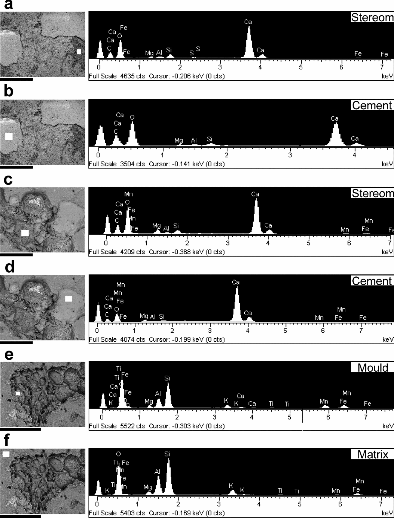
Figure 11. Chemical analyses (indicated by white rectangles) of calcite-bearing gogiid specimens from the Kaili Formation. (a) Thecal plate of ‘S. globus’ (GM-9–4-1524) with stereom. (b) Thecal plate of ‘S. globus’ (GM-9–4-1524) infilled with calcite cement. (c) Stalk plate of S. lui (GM-9–2-3676B) with stereom. (d) Stalk plate of S. lui (GM-9–2-3676B) infilled with calcite cement. (e) Platelets of attachment structure of S. lui (GM-9–2-3676B). (f) Matrix of a S. lui-bearing slab (GM-9–2-3676B). Scale bars = 1mm (a–d); 600μm (e, f). See Gaines, Kennedy & Droser (Reference Gaines, Kennedy and Droser2005, p. 196, fig. 5) or Skinner (Reference Skinner2005, p. 169, fig. 1b) for methods.

Figure 12. Chemical analyses (indicated either by white rectangles or white arrows) of gogiid specimens from the Balang Formation. All scale bars = 1mm. (a) Organic carbon concentration in the dark region of a gogiid (OSU 52611) illustrated in Figure 13d. (b) Manganese concentration within a gogiid cavity (OSU 52619). (c) Iron concentration of a gogiid holdfast (OSU 52611). (d) Matrix of a gogiid-bearing slab (OSU 52611). (e) Biotite grain associated with a gogiid-bearing slab (OSU 52611).
6.b. Calcium (Ca)
The climate in the study region is humid with high annual precipitation (http://english.people.com.cn/data/province/guizhou.html); thus, calcium carbonate is relatively rare in weathered samples. However, the calcium peaks (Fig. 11) in fresh samples are indicative of carbonates, mainly calcite, in these siliciclastic settings. In general, there should be three sources of calcium carbonate for these fossils: (1) original calcareous gogiid plates, (2) syntaxial, magnesian and/or ferroan calcite cements (Dickson, Reference Dickson2001) and (3) calcium carbonates induced by microbial activities on decaying organisms (Briggs & Wilby, Reference Briggs and Wilby1996). Calcium carbonate is concentrated mostly in either the gogiid plates with preserved stereom (Fig. 11a, c) or plates filled with syntaxial or sparry calcite cements (Ausich, Reference Ausich, Jangoux and Lawrence2001) (Fig. 11b, d). Some Chinese gogiid moulds contain residual amounts of calcium (Fig. 11e) compared to the surrounding matrix (Figs 11f, 12b).
6.c. Manganese (Mn)
Manganese oxides (e.g. pyrolusite) form diagnostic dendritic patterns on bedding surfaces and are commonly associated with the cracks in the rock. Spectrum analyses (Fig. 12b) show that manganese concentrations within the cavities of gogiid moulds are probably a mixture of several species of manganese oxides and hydroxides (e.g. birnessite: Post, Reference Post1999). In some cases, manganese oxides or hydroxides form pseudomorphs replacing calcite plates (indicated by the trace amount of calcium) (Fig. 12b). In addition, there are dark margins, which are also manganese-rich, around gogiids, suggesting the foreign origin of manganese concentration within the gogiid cavities.
6.d. Iron (Fe)
Iron is the most common element associated with gogiid plates and skeletal elements of other organisms in the studied region. Due to preservation of soft parts and non-biomineralizing cuticles, iron sulphide should be the dominant species of iron-bearing minerals associated with Burgess Shale-type deposits (Gabbott et al. Reference Gabbott, Hou, Norry and Siveter2004). However, iron sulphide is rarely associated with Guizhou gogiids except when organic carbon is also present. Instead, iron oxides (e.g. limonite) are formed via chemical weathering processes and give echinoderm plates a yellow to yellowish-brown colour, distinctly different from the grey, greenish grey to yellow-green matrix. Due to preservation of numerous articulated gogiids (Table 2) suggesting a rapid burial of those individuals alive or soon after death, microbial-induced pyrite should be the precursor of some of the iron oxides because in situ decay would have been unavoidable (Lin, Reference Lin2006).
7. Stereom preservation and diagenesis of gogiids
7.a. Previous studies on stereom
Studies of the microscopic three-dimensional network of trabeculae or stereomic microstructure began on modern echinoderms (Macurda & Meyer, Reference Macurda and Meyer1975; Roux, Reference Roux1975). Fossil echinoderm stereom has also been studied, but preservation of this microstructure is not common. Typically, stereom must be replaced by early mineralization or secondarily enhanced by laboratory techniques (see Smith, Reference Smith and Carter1990, table 5). Examples include the following: (1) preservation with secondary mineralization (e.g. silicification, phosphatization, pyritization and coatings of iron oxides: Lane & Sevastopulo, Reference Lane and Sevastopulo1982; Berg-Madsen, Reference Berg-Madsen1986; Ausich & Babcock, Reference Ausich and Babcock2000; Clausen & Smith, Reference Clausen and Smith2005), and specimens in such preservation can be obtained from acid residues; (2) slight etching of well-preserved calcite plates (Lapham, Ausich & Lane, Reference Lapham, Ausich and Lane1976; Ausich, Reference Ausich1977) and (3) hydrofluoric acid treatments of plates preserved in an argillaceous matrix, transforming the stereom from calcium carbonate to calcium fluorite (Sohn, Reference Sohn1956; Sprinkle & Gutschick, Reference Sprinkle and Gutschick1967; Sevastopulo & Keegan, Reference Sevastopulo and Keegan1980; Lane & Sevastopulo, Reference Lane and Sevastopulo1981). These techniques are used to develop the three-dimensional aspect of stereom on facet surfaces. Three-dimensional fabrics have also been traced into plates using thin-sections (e.g. Whitehouse, Reference Whitehouse1941; Smith, Reference Smith1982; Ausich, Reference Ausich1983; Riddle, Wulff & Ausich, Reference Riddle, Wulff, Ausich, Burke, Mladenov, Lambert and Parsley1988).
7.b. Stereom preservation in gogiid echinoderms
Sprinkle (1973, p. 7) and Álvaro, Vennin & Vennin (Reference Álvaro, Vennin and Vennin1997) reported eocrinoids preserved with calcite, and Gil Cid & Domínguez Alonso (Reference Gil Cid and Domínguez Alonso2002, fig. 4.3, 4.6, 4.7, 4.9) first interpreted surface stereom preservation on latex casts of Ubaghsicystis segurae preserved as natural moulds, but the present study reports the first occurrence of articulated gogiids with stereomic microstructure preserved in calcite (Fig. 11). From the large collection (n>1000) of Kaili echinoderms, only two specimens representing two species are known with preserved stereom (Figs 8–10). According to Dickson (2001), there are two principal pathways for the Mg calcite transformation that occurs in echinoderm stereom. Our SEM/EDX results show that examples of Type 1 (stereom preservation) (Figs 9b, c, e, f, g–i, 10a–d, g–j), Type 2 (non-stereom preservation) (Figs 9d, 10e, i) and combined transformations (Fig. 10f) are present in articulated samples from the Kaili fauna. Dickson (2001, p. 774) hypothesized that the Mg calcite transformation can be triggered by a temperature rise during burial. Kaili samples also show evidence of thermo-alternation during diagenesis based on the presence of both stereom types containing irregular pores (Figs 9h, 10c) and trabeculae composed of secondary carbonate crystals (fig. 10d). Due to the rarity of this exquisite preservation, only the surface stereom of thecal plates in both ‘S. globus’ and S. lui is measured and given in Table 4. Other types of stereom (see Smith, Reference Smith and Carter1990) may be present on the broken surface of brachiolar and stalk plates (Figs 9b, 10g, h, j), but this needs to be confirmed with additional material. A few specimens of Balang gogiids are preserved with calcite, but preserved stereom is not known.
Table 4. Perforate skeletal microstructure measurements obtained from Kaili gogiids (illustrated in Figs 8, 9h, i, 10c, d)

See Smith (1990, v. 2, p. 69) for methods.
A – mean maximum pore diameter in microns; na – number of measurements; SEa – standard error of mean; t – mean minimum distance between adjacent pores in microns; nt – number of measurements; SEt – standard error of mean.
7.c. Timing of calcite transformation and calcite dissolution
According to Dickson (Reference Dickson2001), early cementation must take place prior to the beginning of Type 1 transformation, and these syntaxial carbonate cements acted as ‘crystal caskets’ to protect the stereom shape. However, in the two Kaili samples with stereom preservation, the cement is largely absent, especially evident in backscatter electron images (Figs 9i, 10d) where the pore space is largely empty (indicated by the black colour) within the three-dimensional network of trabeculae (indicated by the whitish grey colour). Furthermore, traces of labyrinthic stereom beneath the pores on the surface stereom are visible. There must be some protecting medium to preserve the stereom shape. Although syntaxial cementation is virtually unavoidable if stereom is unprotected, we suggest that the protecting medium does not always have to be carbonate cements, or ‘crystal caskets’ proposed by Dickson (Reference Dickson2001), during Type 1 Mg calcite transformation. In order to explain the preservation of stereom shape with open pores in the Kaili deposit, the surface stereom must have been buried and partially infilled with fine mud or clays prior to the formation of syntaxial cements. This was very localized (individual plate-specific) preservation because Type 1, Type 2 and combined Mg calcite transformations occurred adjacent to one another. Alternatively, surface stereom could have been filled with syntaxial cements and then etched out during weathering. The latter case is less probable because adjacent calcite plates that underwent Type 2 transformation were virtually unaffected on the broken and crystal surfaces (Fig. 10e, f).
In siliciclastic settings, echinoderms, particularly Cambrian echinoderm faunas, are preserved primarily as natural moulds (Sprinkle, Reference Sprinkle1973). Weathering by-products preferentially stained echinoderm cavities providing good colour contrast on gogiid moulds compared to the matrix. Based on both the quality of the latex moulds that contain exceptionally fine details of plate ornamentation and rare stereom preservation, the dissolution of calcite must have taken place late during diagenesis, at least after the Cambrian successions were exposed to surface weathering in Guizhou.
8. Soft-part preservation
Preservation of soft parts in echinoderms (e.g. Donovan, Reference Donovan and Donovan1991; Ausich, Reference Ausich, Jangoux and Lawrence2001; Glass, Reference Glass2006, and references therein) is rare and previously unknown in gogiids. During the summer field excursion of 2005, one field assistant split open a slab containing nine gogiid individuals from the Balang Formation (Fig. 13). Close examination of both part and counterpart shows that eight out of nine individuals are well exposed and contain dark stains in the centre of theca. EDX analyses of a small portion of the slab containing two complete gogiids (Fig. 13b, c) indicate that there are high carbon concentrations (fig. 12a) associated with these dark regions. Thus, these organic smudges are interpreted as traces of inner organs housed within the gogiid theca. Carbon films have been reported in fossil crinoids (Springer, Reference Springer1901; Meyer, Milson & Webber, Reference Meyer, Milson and Webber1999; Meyer & Milson, Reference Meyer and Milson2001), but these typically occur in a discrete layer rather than a diffused stain as described in this study.

Figure 13. Gogiids (OSU 52611) with organic preservation of soft parts from the Balang Formation. (a) The entire specimen with nine gogiid individuals (numbered). (b) Close-ups of the group of five individuals, each of which contains a black region in the centre of the theca. A fragment of (b) was analysed under SEM/EDX for chemical composition (see Fig. 12a, c–e). (c) Interpretive drawing of the gogiids (nos 4–8) illustrated in (b) with organic soft-parts (indicated by the dotted polygons). Scale bars = 20mm (a); 10mm (b, c).
9. Conclusions and implications
A wealth of information recorded in both the articulated and disarticulated Guizhou gogiid specimens provides a rare opportunity to document some taphonomic processes of Cambrian stalked echinoderms in more detail. The taphonomic history of Guizhou gogiids was complicated, but some important taphonomic processes, including decay, entombment, burial history, bio-disturbance and diagenesis, can be interpreted.
This study shows that, regardless of the morphological differences between helicoplacoids and gogiids, these two groups belong to a similar biological grade with undifferentiated mesodermal connective tissue binding the endoskeleton. In contrast, although the basic body plans of gogiids are in some ways similar to those of crinoids, gogiid disarticulation is distinctively different from crinoid decay.
Our study confirms that the most common yellow to yellowish-brown colour associated with Cambrian echinoderm moulds is indicative of iron oxides (e.g. limonite), and the relatively less common dark brown colour is due to a concentration of manganese oxides and hydroxides (e.g. pyrolusite and birnessite: Post, Reference Post1999). Black stains in the centre of some Balang gogiids are organic carbon and are interpreted as evidence of volatile organs within gogiids. It is worth noting that although calcium carbonate is rare in both deposits, it is preserved with various colours. One specimen (fig. 8a) contains plates with light yellow stereom in contrast to ossicles with white calcite. The other analysed specimen (Fig. 8b) contains both dark grey stereom and calcite plates. The latter colour may reflect the impurities (trace amounts of manganese and iron) incorporated into the calcite structure during decay of both volatile organs in the body cavity and mesodermal connective tissue within the ossicle.
Although surface stereom preservation in Early Palaeozoic carpoid faunas is common in shale facies (Smith, Reference Smith and Carter1990), stereomic preservation in similar facies is rarely reported from Cambrian strata. The preservation of stereomic microstructure from Guizhou Province, South China is one of the oldest reported examples of stereom on articulated gogiid echinoderms. The timing of dissolution of Cambrian echinoderm stereom is fairly recent. This raises the possibility that more well-preserved gogiids with stereom should be preserved in South China and Laurentia (e.g. Spence Shale: Sprinkle, Reference Sprinkle1973, Reference Sprinkle1976; Gunther & Gunther, Reference Gunther and Gunther1981).
Acknowledgements
This study is a contribution from Lin's dissertation at The Ohio State University, Columbus. Many thanks to R. L. Parsley (Tulane University, New Orleans) for making latex moulds and providing one image presented in this study; S. Bhattiprolu for taking SEM images and conducting EDX analyses; Yishan Wu (Kaili No. 4 Middle School, Kaili City) and Yaoping Zhang (Kaili Science and Technology Committee, Kaili City) for field assistance; and two anonymous reviewers for constructive reviews on earlier drafts of this manuscript. This project was funded by 2006 Ohio State University International Dissertation/M.A. Thesis Research Travel Grant and Alumni Grants For Graduate Research and Scholarship; 2006 Geological Society of America Graduate Student Research Grant; 2003 National Science Foundation East Asia Summer Research Fellowship (to Lin); National Natural Sciences Foundation of China (40672018) (to Zhao); the 2005 Key Project of International Cooperation of Guizhou Science and Technology Department (Gui Co. No. 2005–400106) (to Peng).


