


Introduction
The tympanic membrane or eardrum has been for centuries the only window through which to inspect the hearing organ. From early antiquity and the polymaths’ first descriptions, little progress was made until the great anatomists of the Renaissance. Major breakthroughs in the understanding of the tympanic membrane anatomy later took place in the nineteenth century.
This paper aimed to trace the progress of the description of the tympanic membrane from antiquity to the early twentieth century. We chose to focus on the key steps, to identify how underlying historical context (beliefs, misconceptions or technology) influenced the understanding of the tympanic membrane anatomy.
Materials and methods
A search for relevant scientific medical or historical literature was conducted using PubMed and Google Scholar search engines, and the Bibliothèque Inter-Universitaire Santé catalogue of the medical faculty of the Université de Paris. Screening was performed using the following key words: eardrum, tympanic membrane, tympanum, tympanoplasty, hearing theory, audition, otolaryngology history, Hippocrates, Aristotle, Galen, Avicenna, Vesalius, Fabricius, Fallopio, Shrapnell, Toynbee and Politzer. For primary sources, when a text was identified as pertinent, we used the Medica® digital library of the Bibliothèque Inter-Universitaire Santé of the medical faculty of the Université de Paris to access and read the full text. If the text was not available, the internet was searched to find the manuscript. For texts written in Latin, Greek, Arabic, French, Italian or German, official translations to English were used. Selected texts from primary sources and data from secondary articles were gathered and organised in a chronological order, and then organised by a connected theme.
Results and discussion
From ancient polymaths to Renaissance anatomists
In antiquity, the tympanum was thought to be an interface between two worlds: the human being (outer) and the soul (inner) worlds. This concept remained entrenched for centuries.Reference Gamatsi, Nikolopoulos and Lioumi1
It is uncertain whether the Egyptians described the tympanic membrane, but they discussed ear diseases in medical treaties thousands of years before Hippocrates,Reference Ebbell2 although it is he who is generally credited with the first description of the tympanic membrane, depicting a dry thin-spun web.Reference Weir3 Ancient Greeks discussed the anatomy and function of the ear as philosophers, in their quest to understand the mystery of the senses. Hearing was considered a singular sense, likely thought to perceive the visible (human) as well as the invisible (the gods’) worlds. Harmony was an audible gift given to humanity by the gods to soothe disturbances of the human soul.Reference Baskevitch4
Ancient Greeks understood that sounds were vibrations which could spread in the air and be perceived by the hearing organ. Empedocles (fifth century BC) described the ear as a bell consisting of a ‘fleshy twig’Reference Macauley5 able to receive sounds and, by its proper vibration, transmit them to an aerial cavity (the middle ear). Tympanic membrane perforation was thought to induce deafness as the inner air could leak out: the tympanic membrane had an incidental role, sealing off the inner ear like a lid on a pot. In their treaties, Aristotle and Galen described the tympanic membrane as an interface (it was named meninx or meninga for membrane) and mentioned its vulnerability. However, neither individual gave the tympanic membrane (or the ossicles) a distinct anatomical terminology, and failed to identify its crucial role in transmitting vibrations to the inner ear. The concept of vibration was understood, but the organ was yet to be identified.
Throughout the Middle Ages, the legacy of the meninga lived on. The great Persian physician Avicenna (tenth to eleventh century AD) bestowed on the tympanic membrane a key role in his theory of audition, but considered it a perceptive organ proceeding from the nervous system like the cranial nerves: ‘And the ear hole leads to a pocket of stagnant air (the middle ear) and covered with a bundle of nerves (the ear drum)’.Reference Sina6 By analogy, he compared the eardrum to the iris.Reference Hamidi, Sajjadi, Boroujerdi, Golshahi and Djalilian7
The first breakthrough occurred in sixteenth century Renaissance Italy with the development of human cadaveric dissections.
Gabriel Fallopius is often credited with the birth of the term ‘tympanum’ in De observationes anatomicae (1562): ‘tympanum … ego appelavi’ from the Greek tumpanon (drum) as an analogy to its function.Reference Storrs8–Reference Falloppio10 However, Andreas Vesalius (1514–1546) coined it in his 1543 edition of De humani corporis fabrica. ‘Quemadmodum in tympanis fidem’, said Vesalius, describing the first ossicle (the malleus) anchored in the tympanic membrane's width.Reference Vesalius11 In the 1543 edition of De humani corporis fabrica, Vesalius drew ‘Figure 8’, from ‘Book I’, illustrating the hearing organ, with the tympanic membrane captioned as ‘B: Membrane drawn across the foramen that leads from the ear into the cavity carved in the temporal bone for the organ of hearing’ (Figure 1).Reference Garrison and Hast12

Fig. 1. In the 1543 edition of De humani corporis fabrica,Reference Vesalius11 Vesalius drew ‘Figure 8’, from ‘Book I’, featuring the temporal bone and tympanic membrane, with the following caption ‘B: Membrane drawn across the foramen that leads from the ear into the cavity carved in the temporal bone for the organ of hearing’.
The exploration of the tympanic membrane enabled by these newly lawful human dissections, all taking place in a close geographical area, allowed rapid progress. Within a few decades, this progress had surpassed centuries’ worth of ancient knowledge (mostly obtained from animal dissection). The understanding of the importance of percussion in the transmission of sound, from scant tympanic membrane vibrations, transmitted to the ossicles and then to the oval window, arose simultaneously.Reference O'Malley and Clarke13,Reference Massa14
Duverney's Traité des maladies de l'ouïe (1683)Reference Duverney15 was the first otological treatise in a vernacular language; it combined prior anatomical discoveries and his own observations. Duverney thoroughly detailed the tympanic membrane (Figure 2): ‘This septum is an almost round membrane, dry, thin, firm, transparent and engaged in a groove carved in the circumference of the end of the bony duct…. Although this membrane is stretched, it does not make a straight plane, but it is curved inside being pulled by the handle of the hammer’ (translation from French by the authors). Duverney called the tympanic membrane ‘tambour’ (drum) and correctly conveyed the concept of vibration and resonance. He did not perceive the tympanic membrane to be an extension of nervous tissue and correctly identified its epidermic nature. The tympanic membrane still had a protective role, but also a proper mechanical role, amplifying and transmitting vibrations: the tympanic membrane was the first component of the hearing machine. However, he was wrong when he wrote that the tympanic membrane could be bent by the tensor tympani muscle to alter vibrations (its protective effect against acoustic trauma was unknown).

Fig. 2. Duverney's Traité des maladies de l'ouïe (1683)Reference Duverney15 was the first otological treaty in a vernacular language. Duverney thoroughly detailed the tympanic membrane he called a ‘tambour’ (drum), and correctly conveyed the concept of vibration and resonance. Note that in ‘Fig. V’, the bone marked as ‘B’ is mistakenly considered a fourth ossicle (but which is in fact the lenticular process of the incus). Courtesy of Bibliothèque Inter-Universitaire Santé, Université de Paris.
Indeed, as detailed tympanic membrane observation in vivo was not possible in those times, cadaveric specimen studies – subject to desiccation or to poorly controlled preservation procedures – could lead to erroneous observations. A famous example is the so-called Rivinus’ foramen.Reference Mudry16 Johannes Augustus Rivinus the Younger (1692–1723) reported in his book De Auditus Vitiis (1717) an observation his father Augustus Quirinus Rivinus the Older (1652–1723) made in a letter: ‘I studied the organs of hearing by dissection. At length, in the year 1689, in the month of September, I found the opening that I had long sought, it was in the tympanic membrane itself (…) near the head of the malleus…’.Reference Rivinus17 The controversy lasted almost three centuries and this foramen existence was widely accepted as plausible. However, the contemporary Antonio Maria Valsalva (1666–1723) – who had been informed of Rivinus’ theory before his son published the letter in 1717 – thought it was an artefact brought about by the specimen conservation before post-mortem examination, and did not draw it in his book De Aure Humana Tractatus (1704) (Figure 3).Reference Valsalva18 In 1869, Adam Politzer (1835–1920) wrote: ‘Above the short process, according to Bochdolek, is a short canal, the foramen of Rivini, which was formerly considered an artificial opening in the membrane. This foramen, however, does not appear to be constant’.Reference Politzer19 Use of the first otoscopes, and more so the use of microscopic otoscopy, provided, in the second half of the twentieth century,Reference Griffith20 confirmation that Valsalva was right about the Rivinuses, father and son. Nowadays, any tympanic perforation is considered pathological: the first otologists, even the greatest, would not have unanimously thought so.

Fig. 3. Antonio Maria Valsalva's De Aure Humana Tractatus (1704),Reference Valsalva18 from ‘Table III’. Valsalva was aware of the contemporary theory of Rivinus’ foramen, but thought it was an artefact brought about by the specimen conservation before post-mortem examination and did not draw it in his book. A = glandulae meatus auditoria cum corpore suo reticulari; B = incus; C = malleus; D = processus brevior incudes (short process of the incus); E = chorda tympani; F = membrana tympani
Nineteenth century and birth of otology
A second surge in knowledge took place during the nineteenth century, following technological progress. Otologists such as Floriano Caldani, Joseph Toynbee, Henry Jones Shrapnell, Alexander Prussak and finally Adam Politzer were amongst the first to use self-lit endoscopes to examine the tympanic membrane. While aurists previously had to examine ears with cones and lenses reflecting the sun's rays, it was only in 1730 that Archibald Cleland (1700–1771), a Scottish physician in the British army, invented the first candle-lit otoscope to examine soldiers exposed to loud explosions.Reference Rolls21 Later, the French military surgeon Jean-Pierre Bonnafont (1805–1891) invented the first lateral light source with an opening in the reflecting mirror, allowing the direct examination of the tympanic membrane.Reference Segal and Willemot22 This invention was then improved with the Edison electric light filament.
In 1794, Floriano Caldani (1772–1836), anatomist from the famous University of Padua where Andreas Vesalius taught, published his ‘Observations on the eardrum membrane and new research on animal electricity’ (under the title: Osservazioni sulla membrana del timpano e nuove ricerche sulla elettricita animale lette nell' Accademia di Scienze Lettere ed Arti di Padova).Reference Caldani23 These reports, read in academic sessions in 1790 and 1791, were the first to describe the fibrous structure of the tympanic membrane, with two layers (‘due strati di fibre’) of concentric and radial fibres (‘Uno degli strati è di circolari concentriche, 1’altro di radiate’). He rejected the nervous origin of Mondino – still taught in the early nineteenth century (page 5) – and insisted on the novelty of his observation having used a microscope (‘non ponno vedersi senza 1’ajuto del microscopio’ (page 6)).Reference Caldani23 Initiating the shift in progress from Italy to northern Europe, the German anatomist Samuel Thomas Sömmerring (1755–1830) further described the tympanic membrane with accurate illustrations in his atlas, Abbildungen des Menschlichen Hörorgans, published in 1806 (Figure 4).Reference Von Sömmerring24
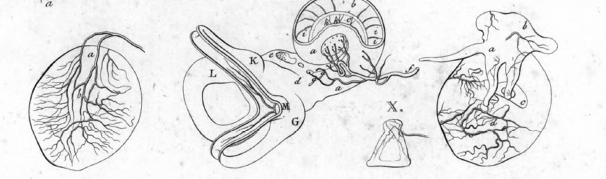
Fig. 4. Samuel Thomas Sömmerring's Abbildungen des Menschlichen Hörorgans (1806),Reference Von Sömmerring24 from ‘Table IV’. He described the tympanic membrane with accurate illustrations in his atlas, particularly its vascularisation. Note this illustration is one century later than Valsalva's De Aure Humana Tractatus (1704),Reference Valsalva18 with considerably more detail.
Shortly afterwards, Henry Jones Shrapnell (1792–1834) published a detailed description of the tympanic membrane in the London Medical Gazette (1832).Reference Shrapnell25 Shrapnell identified the pars flaccida, which ‘occupies the anterior superior angle, and so much of the membrana tympani as is above the tubercle of the malleus, and the ligament in which it is hung’, and which still bears his name (Figure 5).Reference Shrapnell25 He further described the fine three-layered structure of the tympanic membrane, and reported that the pars flaccida microstructure was not composed of muscle fibres as contemporaries said (such as Sir Edward Homes),Reference Shrapnell25 but of a tangle of conjunctive fibres. For Shrapnell, the tympanic membrane had elastic properties, but no contraction capability.

Fig. 5. ‘Mr. Shrapnell on the membrana tympani’, published in the London Medical Gazette (1832).Reference Shrapnell25 Shrapnell identified the pars flaccida, which still bears his name.
Famous during his lifetime and considered as the father of British otology, Joseph Toynbee (1815–1866) was well known for his collection of over 2000 temporal bones, allowing him to identify numerous anatomical variations. In 1838 and 1839, he anonymously published letters in The Lancet challenging those he called quack aurists.Reference Mudry26 His approach was based on methodical and accurate observation of his patients, combined with meticulous specimen dissection, drawing, for the first time, pathology and treatment inferences from tympanic membrane anatomy.Reference Toynbee27
Toynbee described the tympanic membrane as consisting of five superimposed layers (from outside to inside: ‘the epidermis, the dermoid layer, the radiate fibrous layer, the circular fibrous layer and the mucous layer, with its epithelium’), and identified the cutaneous nature of the external layers: ‘Between the epidermoid and radiating fibrous layers of the membrana tympani, there is a distinct and completed lamina of membrane which is continuous with the dermoid layer of the meatus’. He understood and emphasised the impact of this observation: ‘A knowledge of the existence of the membrane here described is of interest to the anatomist, who recognizes in it the secreting organ of the epidermoid layers of the membrana tympani; and to the surgeon, who by its presence is able to understand phenomena occurring in certain diseases of the ear which have been hitherto incomprehensible to him’. He described the retraction pocket phenomenon, but was wrong in assigning it to an ‘ulceration of the fibrous laminae’ portrayed as: ‘the … inflammation of the dermoid layer, which spreads first to the radiate fibrous and thence to the circular lamina. The laminae, being weakened by ulcerative process, fall inwards as far as the promontory, to which they often ultimately adhere, and, when an orifice has been thus produced, its margins are not unfrequently drawn into the shape of a funnel, whose inner part adheres to the tympanic walls’.Reference Toynbee28 These observations led him to propose an ingenious way to restore hearing in tympanic membrane perforation cases, using an Indian rubber disc which would be placed on the eardrum and was removable thanks to an attached silver wire.Reference Mudry26
In the late nineteenth century, progress shifted back to the European continent with Alexander Prussak and the great Adam Politzer (Figure 6). Politzer (1835–1920) synthesised knowledge about the tympanic membrane. He definitively described its normal shape: ‘not stretched as a perfect plane upon the end of the external meatus, but … arched in such a manner that its concavity is presented outward, while its convexity is turned toward the inner wall of the tympanum…’. Contrarily to Toynbee, he reduced the number of layers to three: ‘a middle fibrous – the so-called lamina propria membranae tympani, an external dermoid and an internal mucous layer’. However, he acknowledged that the lamina propria had ‘two separable laminae – an external radiate, and an internal circular’.Reference Politzer19

Fig. 6. Adam Politzer (1835–1920) photographed by Rudolf Krziwanek (1843–1905). He synthesised in detail the knowledge of the normal tympanic membrane anatomy and provided the first pathological tympanic membrane description, within the first otoscopy atlas, published in 1865. He is known to have also been one of the first historians of otolaryngology.
Although Politzer progressed significantly in understanding Eustachian tube permeability and catarrh,Reference Politzer29,Reference Mudry30 he did not understand its link with so-called cholesteatoma disease. This disease was coined by Johannes Müller (1801–1858) in 1838 for what he considered to be a ‘glandular neoplasm of middle ear mucosa’.Reference Soldati and Mudry31
The famous Russian otologist Alexander Federovitch Prussak (1839–1897), who attended Politzer's lessons in Vienna, pursued the anatomical study of the tympanic membrane. He gave his name to the ‘triangular shaped free space’ in the outer part of the attic between Shrapnell's membrane, the neck of the malleus medially and the lateral process of the malleus inferiorly.Reference Prussak32,Reference Proctor33 His work completed the microscopic anatomical understanding of the pars flaccida, and helped to end the debate on the foramen of Rivinus.
Politzer was not only the first modern otologist, but also the first historian of otolaryngology: he recognised the decisive works of his predecessorsReference Politzer34 and helped to trace progress in the understanding of the tympanic membrane. He synthesised in detail knowledge of normal tympanic membrane anatomy, now established. In particular, in 1865, he published the first pathological tympanic membrane description, in the first otoscopy atlas: Die Beleuchtungsbilder des Trommelfells im gesunden und kranken Zustande (Figure 7).Reference Mudry30,Reference Politzer35 Otology as a specialty was born with the tympanic membrane.

Fig. 7. Illustrations from Adam Politzer's English-language book The Membrana Tympani in Health and Disease (1869).Reference Politzer19 This was followed four years later by his first otoscopy atlas: Die Beleuchtungsbilder des Trommelfells im gesunden und kranken Zustande (1865).Reference Politzer35 At that time, otology was born as a specialty. ‘Fig. 2.’ = membrana tympani with crescentic chalky with deposit; ‘Fig. 3.’ = calcareous deposit in the inner side of a 24-year-old female's tympanic membrane; ‘Fig. 4.’ and ‘Fig. 5.’ = calcareous deposits and perforations.
Conclusion
From early antiquity, when polymaths poetically portrayed the tympanic membrane as a gateway to other worlds, to the great anatomists of the Renaissance who identified the ear as parts of a complex mechanical object, the understanding of the eardrum remained limited for centuries. A great technological leap was possible in the nineteenth century with new self-lit otoscopes. This contributed to the emergence of otologists, clinicians who would dedicate their life to the study of the ear. A reproducible scientific approach, combining microscopic anatomical studies with patient care, progressively shed light on the tympanic membrane in all its complexity. This provided better insight in tympanic membrane physiology and pathology, paving the way for modern otology.
Competing interests
None declared


