


Introduction
A pneumocoele is a pathologically expanding, air-containing paranasal sinus.Reference Wolfensberger and Herrmann1 Pneumocoeles are uncommon entities, with only 25 cases reported in the literature since 1969.Reference Bachor, Weber, Kahle and Draf2 Although the aetiology remains unclear, the most plausible theory is that of a one-way valve effect allowing air into the sinus under increased pressure without allowing pressure equilibration. However, the location and nature of this valve has not been well described. Such a valve effect is often created by an obstructing mass, such as an osteoma, but normal variants of anatomy are rarely cited.
We present a patient with chronic sinusitis who developed a pneumocoele of her left frontal sinus with erosion into her orbit. This patient was a habitual, chronic nose-blower with a large type III frontal cell adjacent to an intersinus septal cell. We propose the new theory that these cells formed a one-way valve causing air trapping and pathological expansion of our patient's frontal sinus. To our knowledge, there are no previously published cases that establish an association between large frontal cells and expansion of a pneumocoele.
Case report
A patient evaluated and treated at our institution for a pneumocoele of the left frontal sinus and chronic sinus disease underwent endoscopic sinus surgery.
This 31-year-old woman presented with a history of severe sinus symptoms for many years, with thick nasal discharge and a constant need to blow her nose. Over the previous three months, she had experienced an acute increase in left frontal sinus pain and had noticed changes in the appearance of her left eye. She reported a feeling of pressure in her left eye when she blew her nose. She had no history of sinonasal surgery or head trauma. No altitude changes were reported.
Examination of the patient revealed a noticeable mass effect on her left brow causing asymmetry and depression of her orbit.
Nasal endoscopy revealed slight rightward deviation of the patient's nasal septum, significant mucosal inflammation and bilateral turbinate hypertrophy. A computed tomography scan demonstrated significant thickening in all eight sinuses and a pneumocoele of the left frontal sinus with erosion into the left orbit. There was an intersinus septal cell and a type III frontal cell on the left side (Figure 1).

Fig. 1 Coronal computed tomography scan demonstrating superior orbital roof erosion secondary to a left frontal pneumocoele, a type III frontal cell (long arrow), and an intersinus septal cell (short arrow).
Operative intervention included bilateral endoscopic sinus surgery with a meticulous frontal recess dissection (Figure 2).
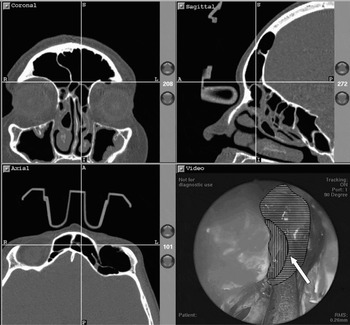
Fig. 2 Triplanar, intra-operative, computed tomography (CT) imaging plus 70° nasal endoscopy view, demonstrating partial dissection of the posterior aspect of the type III frontal cell and entry into the frontal sinus. In the CT views, the vertical line represents the intersinus septal cell and the horizontal line delineates the type III frontal cell. The proposed site of the one-way valve is at the junction of these two cells (arrow).
Post-operatively, the patient reported a dramatic improvement in her orbital pain. Four months post-operatively, her eye had returned to a normal position.
Discussion
Urken et al. Reference Urken, Som, Lawson, Edelstein, Weber and Biller3 differentiated enlarged, aerated sinuses into three categories: hypersinus, pneumosinus dilatans and pneumocoele.
Hypersinus refers to a sinus that is larger than normal but with normal wall thickness. It does not extend beyond the normal boundaries of the surrounding bone. Patients are completely asymptomatic, and there is no encroachment on surrounding structures.
Pneumosinus dilatans refers to an aerated sinus that has expanded beyond the normal boundaries of the surrounding bone. There may be frontal bossing, intracranial extension, or ethmoid, nasal or orbital encroachment. However, the thickness of the sinus wall is normal. Symptoms may result from pressure on surrounding structures and may include headache, diplopia and decreased visual acuity.
Pneumocoele refers to a sinus that is aerated beyond the normal margins with either focal or generalised thinning of the bony wall. Symptoms and signs are similar to pneumosinus dilatans. Although pneumocoeles and pneumosinus dilatans may be variations of the same disease, no cases monitoring the progression of pneumosinus dilatans into a pneumocoele have been reported in the literature.Reference Walker and Jones4
The aetiology of pneumocoeles remains unclear, although a few mechanisms have been proposed.
One theory that has fallen out of favour is infection with a gas-forming organism, as no such organism has ever been isolated.
A mechanism that could account for some cases is a mucocele that spontaneously drains. However, most patients do not report diffuse nasal discharge prior to diagnosis.Reference Walker and Jones4
Another theory is that pneumocoeles have a developmental aetiology, but resolution with surgery argues strongly against this mechanism.Reference Flanary and Flanary5
Currently, the most credible theory is that of a one-way valve effect at the sinus ostium. Air enters the sinus when outside pressure is increased (i.e. during nose-blowing) but is unable to escape and thus allow pressure equilibration. There is much evidence supporting this mechanism. Wolfensberger and HerrmannReference Wolfensberger and Herrmann1 measured increased pressure in a maxillary sinus pneumocoele and discovered a lack of normal fluctuation with the respiratory cycle. After performing a Valsalva manoeuvre against a closed nose, a persistent increase in pressure was noted, indicating partial obstruction and supporting the trap-valve theory. Further evidence is provided by the fact that pain is usually exacerbated by changes in atmospheric pressure, such as during flying or diving, presumably because the pressure does not equilibrate. Perhaps the best evidence supporting the valve theory is that surgery to open the sinus results in arrest of sinus expansion. Unfortunately, this mechanism does not explain the absence of mucocele formation; one would expect that a one-way valve would prevent mucus drainage.Reference Walker and Jones4 Nevertheless, the one-way valve theory has thus far garnered the most support. However, the location and nature of this valve have not been well described.
• A pneumocoele is a pathologically expanding, air-containing paranasal sinus
• This paper describes a case of frontal sinus pneumocoele secondary to a frontal recess cell
• The case was successfully managed via an endoscopic approach
The present case supports the one-way valve mechanism of pneumocoele development. This patient suffered from severe, chronic sinus disease and characterised herself as a ‘habitual’ nose-blower. Several studies have mentioned nose-blowing as a possible factor contributing to pneumocoele development.Reference Wolfensberger and Herrmann1, Reference Walker and Jones4, Reference Flanary and Flanary5 In our patient's case, we believe that air was forced between the type III frontal cell and intersinus septal cell when she blew her nose. The cells created a valve such that the air was not able to escape, and the chronicity of this event caused expansion of the pneumocoele.
Frontal sinus cells are thought to develop from anterior ethmoid cells after the development of the frontal sinus itself. Their incidence has ranged from 20 to 41 per cent in different studies.Reference DelGaudio, Hudgins, Venkatraman and Beningfield6 These cells complicate frontal recess anatomy and can cause sinus ostia obstruction. In our patient, removal of these cells and opening of the frontal sinus relieved her symptoms and returned her eye to a normal position, providing evidence that the cells were the cause of the sinus expansion.
The present case supports the one-way valve theory of pneumocoele development and provides a plausible location for a valve created by two cells within the frontal recess. We believe that large frontal cells should be considered a contributing factor in the development of pneumocoeles in some individuals.


