


Introduction
Alkaptonuria is a rare autosomal recessive metabolic disorder caused by deficiency of homogentisic acid oxidase within the tyrosine and phenylalanine degradation pathway.Reference Fernandez-Canon, Granadino, Beltran-Valero de Bernabe, Renedo, Fernandez-Ruiz and Penalva1 Homogentisic acid is a metabolic product of phenylalanine and tyrosine. In patients with alkaptonuria it accumulates, polymerising to form a dark pigment that is selectively deposited in connective tissues.Reference Keller, Macaulay, Nercessian and Jaffe2 This pigment has a high affinity for fibrillary collagens surrounded by abundant mucopolysaccharide substances, in various tissues including hyaline cartilages, intervertebral discs, skin, sclera, and the concha and helix of the ear.Reference Hamdi, Cooke and Hassan3–Reference Maathuis and Driessen6 However, tympanic membrane involvement has previously been described in only one case report.Reference Pau7
We report otoscopic and audiological findings for a patient with alkaptonuria, and we discuss the probable association between this condition and hearing loss.
Case report
A 58-year-old man was admitted to our department complaining of tinnitus and hearing loss in both ears for one year. The patient had no history of otological surgery, infection, trauma, otorrhoea, tympanic membrane perforation or drug use. Upon questioning, he also complained of a 10-year history of chronic, nonspecific lower back pain and stiffness. In 2005, he had been admitted to the department of physical medicine and rehabilitation for lower back pain, and had been diagnosed with alkaptonuria, with typical dark urine.
Physical examination showed bluish-black pigmentation on both sclerae and on the helixes of both ears. There was no other skin discolouration.
Otoscopic examination revealed dark discolouration of both tympanic membranes (Figures 1 and 2).
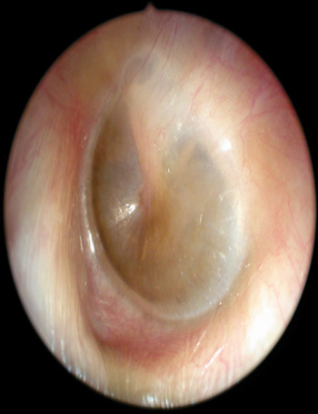
Fig. 1 Dark pigmentation on the anterior-inferior and anterior-superior part of the left tympanic membrane.

Fig. 2 Dark pigmentation on the anterior-inferior and anterior-superior part of the right tympanic membrane.
Audiological evaluation revealed mixed high frequency hearing loss in both ears.
Tympanometric examination revealed type A tympanograms bilaterally, and the absence of acoustic reflexes both ipsilaterally and contralaterally.
Computed tomography of the temporal bones showed no abnormality.
Discussion
Clinical manifestations of alkaptonuria include ochronosis and arthritis. Patients with alkaptonuria are generally asymptomatic until 30 years of age, after which ochronosis develops. The major clinical features of ochronosis relate to deposition of homogentisic acid within connective tissues: ochronotic pigment deposits are seen in connective tissue in hyaline cartilage, tendons, ligaments and muscles. Consequently, these tissues become weak, brittle, and prone to chipping, cracking and rupture, leading to rapid degeneration of the joints.Reference Keller, Macaulay, Nercessian and Jaffe2 The condition involves large joints such as the spine, hip and knee; rarely, small joints can also be involved.Reference Zhao, Chen, Shao de and Zhang8
The present report reveals peculiar ochronotic pigment deposition on the tympanic membrane, in addition to previously diagnosed lower back involvement. Our patient also had mixed high frequency hearing loss. The conductive component of this hearing loss would have developed as a consequence of progressive ochronotic pigment deposition and consequent altered tympanic membrane elasticity, or as a consequence of ochronotic impairment of the incudomalleolar and incudostapedial joints.
This pattern of abnormal tympanic membrane discolouration can result from a variety of causes, including haemotympanum, cholesterol granuloma, jugular bulb dehiscence, glomus tumours and prolonged drug use (e.g. minocycline).Reference Balatsouras, Dimitropoulos, Fassolis, Kloutsos, Economou and Korres9–Reference Suwannarat, Phornphutkul, Bernardini, Turner and Gahl13
• Alkaptonuria is a rare autosomal recessive metabolic disorder; homogentisic acid oxidase deficiency results in dark pigmentation of connective tissues
• The presented, alkaptonuric patient had dark tympanic membrane pigmentation and mixed high frequency hearing loss
• The conductive hearing loss component can be explained by ochronotic alteration of tympanic membrane elasticity or ossicular joint mobility
• Alkaptonuria should be considered in the differential diagnosis of abnormal tympanic membrane pigmentation with hearing loss
The diagnosis of alkaptonuria is verified by detection of homogentisic acid in the urine.Reference Peker, Yonden and Sogut14 Pigmentation of the ear auricle and sclera, and/or altered urine colour, are typical clinical manifestations and can aid diagnosis. Our patient had pigmentation of the sclerae and ear auricles, together with altered urine colour.
There is no effective treatment for alkaptonuria, although various therapeutic modalities have been employed, such as ascorbic acid, nitisonine and a low protein diet.Reference Ibrahim, El-din Selim and Al-Ansari5 However, there is no evidence that these drugs can prevent the development of ochronosis, treat existing pigmentation, or affect bone metabolism. Currently, symptomatic treatment of alkaptonuria appears to be the sole therapeutic option.Reference Keller, Macaulay, Nercessian and Jaffe2
This case emphasises the fact that clinicians should consider alkaptonuria within the differential diagnosis of dark tympanic membrane pigmentation coexisting with hearing loss.


