


Introduction
An aberrant internal carotid artery (ICA) is a rare vascular anomaly in the middle ear. In this anomaly, the ICA takes an aberrant lateral course in the temporal bone and passes through the middle-ear cavity.
The clinical symptoms and signs of aberrant ICA are often non-specific, and include hearing loss, pulsatile tinnitus and a retro-tympanic mass behind the anteroinferior part of the tympanic membrane.Reference Windfuhr1, Reference Eryilmaz, Dagli, Cayonu, Dursun and Gocer2 The condition needs to be distinguished from glomus tumour, high jugular bulb and other vascular malformations.
Every otologist should be aware of this rare entity, as a misdiagnosis can result in fatal bleeding complications or catastrophic consequences.Reference Botma, Kell, Bhattacharya and Crowther3, Reference Sauvaget, Paris, Kici, Kania, Guichard and Chapot4 When aberrant ICA and dehiscent high jugular bulb are simultaneously encountered in the operated ear, the risk of serious bleeding is elevated.
We report a very rare case in which an aberrant ICA was present in combination with a dehiscent high jugular bulb; this combination has not previously been reported.
Case report
A 24-year-old man presented with a five-year history of intermittent right-sided aural fullness, pulsatile tinnitus and mild hearing impairment.
Otoscopy revealed a protruding vegetation on the right tympanic membrane and a curved, reddish mass behind the tympanic membrane (Figure 1), with normal findings in the left ear.
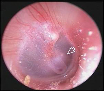
Fig. 1 Otoscopic view showing protruding vegetation on the tympanic membrane and a curved, reddish mass (arrowhead) behind the anteroinferior part of the tympanic membrane in the right ear.
Pure tone audiometry showed mild conductive hearing loss in the right ear.
High resolution computed tomography (CT) of the temporal bone revealed protrusion of the internal carotid artery (ICA) into the middle ear together with a dehiscent high jugular bulb (Figure 2). The left temporal bone was normal.

Fig. 2 Axial high resolution computed tomography scan of the temporal bone, showing protrusion of the right internal carotid artery (arrow) into the middle-ear cavity, together with an ipsilateral dehiscent high jugular bulb (asterisk).
Magnetic resonance angiography (MRA) showed a reduced diameter and lateralisation of the right ICA compared with the left ICA (Figure 3). The left ICA was normal, and there was no intracranial aneurysm, arteriovenous malformation or other persistent embryological vessel.

Fig. 3 Magnetic resonance angiography showing reduced diameter and lateralisation of the right internal carotid artery (ICA) (arrowhead) compared with the left ICA. The left ICA is normal, and there is no intracranial aneurysm, arteriovenous malformation or other persistent embryological vessel.
A diagnosis of an aberrant ICA in the middle ear with a dehiscent high jugular bulb was made. The patient was managed with conservative treatment. He was also informed of the danger of manipulation of the external auditory canal, and was advised that he would be reviewed yearly for audiometric and otoscopic examination.
Discussion
Vascular anomalies of the temporal bone are rare, but some are significant in the event of middle-ear surgery. These vascular variants are aberrant internal carotid artery (ICA), high jugular bulb, persistent stapedial artery, dehiscent carotid artery canal and dehiscent high jugular bulb.Reference Eryilmaz, Dagli, Cayonu, Dursun and Gocer2, Reference Koesling, Kunkel and Schul5 An aberrant ICA has an incidence of less than one per cent.Reference Botma, Kell, Bhattacharya and Crowther3, Reference Koesling, Kunkel and Schul5 Dehiscent high jugular bulb has a 1 per cent occurrence rate,Reference Sauvaget, Paris, Kici, Kania, Guichard and Chapot4, Reference Koesling, Kunkel and Schul5 and the frequency of a high jugular bulb varies between 6 and 22 per cent.Reference Koesling, Kunkel and Schul5, Reference Wang, Shi, Liu, Wang, Huang and Chen6 Thus, aberrant ICA is the rarest of these vascular anomalies.Reference Koesling, Kunkel and Schul5 There has been no previous report of an aberrant ICA occurring together with a dehiscent high jugular bulb.
The clinical symptoms and signs of an aberrant ICA are often non-specific. Hearing loss is the most common presenting symptom: others include pulsatile tinnitus, serous otitis media, otalgia and aural fullness.Reference Windfuhr1, Reference Botma, Kell, Bhattacharya and Crowther3, Reference Sauvaget, Paris, Kici, Kania, Guichard and Chapot4 The usual clinical sign is a reddish, retro-tympanic mass behind the anteroinferior part of the tympanic membrane on otoscopy. The audiometric examination may or may not show conductive hearing loss. The clinician should therefore be aware of the possible significance of a combination of symptoms and signs such as pulsatile tinnitus, conductive hearing loss and a retro-tympanic mass.
Radiological investigations should be performed to evaluate middle-ear vascular anomalies.Reference Windfuhr1, Reference Sauvaget, Paris, Kici, Kania, Guichard and Chapot4 High resolution CT of the temporal bone will identify an aberrant ICA as a retro-tympanic mass in the hypotympanum, with an enlarged inferior tympanic canaliculus, a deficient bony plate along the tympanic portion of the ICA, and absence of the vertical segment of the ICA canal.Reference Windfuhr1–Reference Sauvaget, Paris, Kici, Kania, Guichard and Chapot4 Magnetic resonance angiography is a useful tool providing excellent angiographic visualisation of intracranial and extracranial vascular structures, facilitating the diagnosis of aberrant ICA. In such cases, it shows a reduced diameter of the aberrant part of the ICA, and on the frontal view, the vertical segment of the ICA is lateral to a line drawn vertically through the vestibule. Magnetic resonance angiography has almost superseded the need for conventional angiography. Conventional angiography is an invasive procedure, which should be performed in cases of bleeding or aneurysm.Reference Windfuhr1, Reference Sauvaget, Paris, Kici, Kania, Guichard and Chapot4
Most authors recommend a conservative attitude toward a proven but asymptomatic aberrant ICA, although the necessity for treatment is controversial.Reference Windfuhr1, Reference Sauvaget, Paris, Kici, Kania, Guichard and Chapot4
Reasons for surgical treatment include relief of troublesome symptoms, prevention of possible destruction of middle-ear structures, and in the case of aneurysm formation. Ruggles and Reed recommended separating the ICA from the middle-ear space by covering the vessel with fascia and then compressing it into the promontory defect and covering it with a bone graft. However, this may further compromise the blood flow through these already narrowed vessels, with the risk of consequent neurological disorders.Reference Ruggles and Reed7 The benefit of surgery must be weighed against the risk of possible neurological deficits and serious bleeding complications.
In cases of aberrant ICA with a dehiscent high jugular bulb which present without bleeding complications, we believe that the best treatment is conservative management with regular follow up, in order to avoid manipulation of the external auditory canal.
Life-threatening haemorrhages from an aberrant ICA, and massive bleeding from a dehiscent high jugular bulb injury during middle-ear surgery, have both been described in the literature.Reference Windfuhr1, Reference Sauvaget, Paris, Kici, Kania, Guichard and Chapot4, Reference Huang, Wang and Young8 When aberrant ICA and dehiscent high jugular bulb are simultaneously encountered in the operated ear, the risk of serious bleeding is elevated.
• Aberrant internal carotid artery (ICA) is a rare middle-ear vascular anomaly
• The reported case had this anomaly together with a dehiscent high jugular bulb
• This combination carries a high risk of bleeding
• Symptoms and signs of aberrant ICA are often non-specific, so imaging is required for diagnosis
• A retro-tympanic mass with hearing loss and pulsatile tinnitus should arouse suspicion
• Correct diagnosis is essential as misdiagnosis can have catastrophic consequences
One may hypothesise that the existence of an anomaly in one of these great vessels may be associated with the formation of an anomaly in the other. The presence of carotid canal dehiscence had been found to be significantly correlated with high jugular bulb presentation.Reference Wang, Shi, Liu, Wang, Huang and Chen6 One of the hypotheses about the genesis of an aberrant ICA postulates that carotid canal dehiscence may result in the passing of an aberrant ICA through the middle-ear space.Reference Botma, Kell, Bhattacharya and Crowther3, Reference Sauvaget, Paris, Kici, Kania, Guichard and Chapot4 Therefore, the possibility of a study assessing the relationship between aberrant ICA and dehiscent high jugular bulb requires further investigation.
Conclusion
The described case showed an extremely rare combination of aberrant internal carotid artery together with a dehiscent high jugular bulb; this combination has not previously been reported in the literature. In this condition, the risk of serious bleeding is elevated. Every otologist must be aware of this rare entity in order to avoid catastrophic consequences. Where such cases present without bleeding complications, we recommend a conservative approach.


