


Introduction
A laryngocoele is a cystic dilatation of the laryngeal saccule, which arises from the laryngeal ventricle and extends into the paralaryngeal space.Reference Felix, Felix and Mello1, Reference Dursun, Ozgursoy, Beton and Batikhan2 The aetiology is unknown though it is probably related to both congenital and acquired factors, such as increased airway pressure. A possible association between laryngocoele and laryngeal cancer has also been reported.Reference Dursun, Ozgursoy, Beton and Batikhan2
Based on the anatomical features, laryngocoeles can be divided into internal, external and mixed laryngocoele. The former is confined to the interior of the larynx and extends postero-superiorly into the false cord and the aryepiglottic fold. An internal laryngocoele is observed during laryngoscopy as a smooth swelling of the supraglottis. It is traditionally removed though a transoral approach. External laryngocoele extends superiorly and appears laterally in the neck through the opening for the superior laryngeal nerve and vessels in the thyrohyoid membrane. This laryngocoele type presents as a swelling in the neck at the level of the hyoid bone anterior to the sternocleidomastoid muscle. It is excised though an external cervical approach. A ‘mixed’ (combined) laryngocoele is defined by the simultaneous existence of both internal and external features. This can be excised though a combined cervical-transoral approach.Reference Dursun, Ozgursoy, Beton and Batikhan2, Reference Martinez Devesa, Ghufoor, Lloyd and Howard3
Surgical excision through a cervical approach is the treatment of choice for large (external and mixed) laryngocoeles.Reference Dursun, Ozgursoy, Beton and Batikhan2 In this paper, we report the first case in the literature of a large mixed laryngocoele treated with transoral robotic surgery without cervical incision.
Case report
A 69-year-old female patient presented to the Department of Otolaryngology – Head and Neck Surgery at San Donato Civil Hospital, Arezzo, Italy, with a 3-month history of hoarseness, dysphonia and foreign body sensation. This was associated with left cervical soft and compressible tumefaction, which increased in size during the Valsalva manoeuvre.
Videolaryngoscopy showed a round bulging of the left ventricular fold which changed its size during phonation and Valsalva manoeuvre. Computed tomography (Figure 1) showed a paralaryngeal saccular dilatation of the left laryngeal ventricle, measuring approximately 30 × 25 × 18 mm in size, in communication with the laryngeal air column. A diagnosis of mixed laryngocoele was made on the basis of these findings.
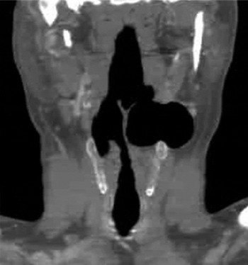
Fig. 1 Coronal computed tomography image showing paralaryngeal saccular dilatation of the left laryngeal ventricle (approximately 30 × 25 × 18 mm in size) in communication with the laryngeal air column, which is suggestive of a large mixed laryngocoele.
The patient was offered transoral robotic surgery for excision of the laryngocoele, which was described to her as being a ‘not currently approved’ alternative to traditional cervical access. Full informed consent was obtained from the patient. The hospital ethics committee approved the transoral robotic surgery approach for laryngocoele excision even though this procedure was not among the currently approved indications for robotic surgery.
The transoral robotic surgery was performed using the da Vinci S surgical robotic system (Intuitive Surgical, Sunnyvale, California, USA). The system consists of a surgeon's console, a surgical cart, a manipulator unit with two laterally placed instrument arms, and a centrally located endoscopic arm holding a three-dimensional (3D) camera.
As previously described by O'Malley et al.,Reference O'Malley, Weinstein, Snyder and Hockstein4 the patient was placed in the supine position on an operating theatre table. After standard endotracheal intubation, the da Vinci robot was positioned at a 308–458 degree angle with respect to the operating table. Oral cavity retraction was achieved using the Feyh-Kastenbauer Retractor (Gyrus, Maple Grove, Minnesota, USA).Reference O'Malley, Weinstein, Snyder and Hockstein4 The retractor was suspended with a retraction arm fastened separately to the operating theatre bed. The 3D camera endoscope was centrally inserted through the oral cavity and two twisted robotic arms were introduced transorally on each side into the laryngeal lumen (Figure 2). Surgical resection was performed using 5-mm robotic instruments, including monopolar cautery and Maryland dissector. Bipolar forceps were used for haemostasis.

Fig. 2 Intra-operative view of the larynx during the transoral robotic procedure (pre-surgery). Note the bulging of the left ventricular fold (arrow). A = twisted robotic arms; T = orotracheal tube
The anterior edge of the left aryepiglottic fold was incised with monopolar cautery (Figure 3). The left ventricular medial wall was then grasped and pulled medially. A Maryland dissector was used to free the laryngocoele sac from the surrounding tissue by progressively pulling the laryngocoele wall medially into the laryngeal lumen. The laryngocoele mucosal wall was dissected from the thyrohyoid membrane, which was kept intact, and the whole laryngocoele sac was removed through the laryngeal lumen (Figure 4). The robot docking time was 15 minutes, while the surgical procedure lasted 20 minutes.

Fig. 3 Intra-operative view during transoral robotic surgery showing the incision of the left aryepiglottic fold anterior edge using monopolar cautery (arrow).

Fig. 4 Intra-operative view during transoral robotic surgery showing dissection of the laryngocoele sac from the surrounding tissue (as described in the Case report section).
Oral feeding was started on the first post-operative day and the patient was discharged on the second day after surgery. The patient's symptoms improved and no post-operative complications were reported. Histological examination confirmed the diagnosis of laryngocoele.
Discussion
A laryngocoele is a common dilatation of the laryngeal ventricle lined by pseudostratified, columnar, ciliated epithelium, with occasional foci of stratified squamous epithelium, and a mixture of submucosal serous and mucous glands. It is usually benign, although an association with local tumour or chronic inflammation may be apparent.Reference Dursun, Ozgursoy, Beton and Batikhan2, Reference Martinez Devesa, Ghufoor, Lloyd and Howard3 Patients usually complain of symptoms such as hoarseness or dysphonia, dyspnoea, foreign body sensation, and cough. In the case of a large external or mixed laryngocoele, there is cervical swelling, which consists of a laterocervical soft compressible mass below the hyoid bone level. An internal laryngocoele is traditionally treated using an endoscopic endolaryngeal approach, whereas external and mixed laryngocoeles have required cervical access for the complete removal of a laryngocoele sac.Reference Dursun, Ozgursoy, Beton and Batikhan2, Reference Martinez Devesa, Ghufoor, Lloyd and Howard3
Since the development of the first surgical robot in 1988 by Kwoh et al.,Reference Kwoh, Hou, Jonckheere and Hayati5 robotic technology has been applied to several surgical fields in an attempt to improve the accuracy and efficiency of surgery.Reference Davies6 In particular, O'Malley et al. and Hockstein et al. showed the value of transoral robotic surgery using the da Vinci Surgical System for the management of upper aerodigestive tract tumours in humans.Reference O'Malley, Weinstein, Snyder and Hockstein4, Reference Hockstein, Nolan, O'Malley and Woo7, Reference Hockstein, O'Malley and Weinstein8
Laryngocoele excision is traditionally performed through a cervical approach (for external and mixed laryngocoeles) or by using a microscopic CO2 laser-assisted technique (for internal laryngocoeles). Some authors have proposed that microscopic laser treatment should also be used for mixed laryngocoeles in an attempt to avoid cervical scars.Reference Dursun, Ozgursoy, Beton and Batikhan2, Reference Martinez Devesa, Ghufoor, Lloyd and Howard3 However, this procedure can at times be surgically awkward because of: long and poorly functional instruments; limited vision associated with the placement of microscopic optics outside the oral cavity; and the straight laser beam, which does not enable one to reach lateral hidden areas.Reference Dursun, Ozgursoy, Beton and Batikhan2, Reference Martinez Devesa, Ghufoor, Lloyd and Howard3
• This paper describes the first reported case of a large mixed laryngocoele treated with transoral robotic surgery
• This technique allows a minimally invasive approach without cervical incisions
• Transoral robotic surgery enabled accurate dissection with complete removal of the large mixed laryngocoele
Transoral robotic surgery offers several advantages and benefits over traditional approaches for the treatment of laryngocoeles. For instance, optics are placed in the oral cavity, thus allowing closer, angulated vision of the surgical field. In addition, rather than using traditional laryngoscopes, instruments are introduced though mouth gags, which offer a wider view and range of motion. Furthermore, miniaturised, angulated, ‘tremor-filtered’ robotic instruments with ‘wristed-tips’ enable one to reach far lateral (hidden) areas.Reference Hockstein, Nolan, O'Malley and Woo7, Reference Hockstein, O'Malley and Weinstein8 In cases of large external and mixed laryngocoeles, transoral robotic surgery could allow accurate dissection with complete removal of the lesion via a minimally invasive, low morbidity approach. In addition, this technique is associated with a reduced operative time, short hospital stay and low cost. Furthermore, in comparison with a traditional cervical approach, the absence of cervical incisions results in a better overall functional outcome, the prevention of cervical scars and improved aesthetic results. Our experience demonstrates the utility of transoral robotic surgery for laryngocoele excision and indicates that this type of procedure should be included in the indications for transoral robotic surgery.


