


Crossref Citations
This article has been cited by the following publications. This list is generated based on data provided by Crossref.
Dinkel, Anke
Njoroge, Ernest M
Zimmermann, Anja
Wälz, Marcus
Zeyhle, Eberhard
Elmahdi, Ibrahim E
Mackenstedt, Ute
and
Romig, Thomas
2004.
A PCR system for detection of species and genotypes of the Echinococcus granulosus-complex, with reference to the epidemiological situation in eastern Africa.
International Journal for Parasitology,
Vol. 34,
Issue. 5,
p.
645.
OBWALLER, A.
SCHNEIDER, R.
WALOCHNIK, J.
GOLLACKNER, B.
DEUTZ, A.
JANITSCHKE, K.
ASPÖCK, H.
and
AUER, H.
2004.
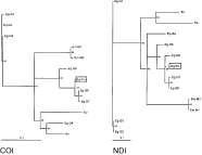
Echinococcus granulosusstrain differentiation based on sequence heterogeneity in mitochondrial genes of cytochromecoxidase-1 and NADH dehydrogenase-1.
Parasitology,
Vol. 128,
Issue. 5,
p.
569.
Daniel Mwambete, K
Ponce-Gordo, F
and
Cuesta-Bandera, C
2004.
Genetic identification and host range of the Spanish strains of Echinococcus granulosus.
Acta Tropica,
Vol. 91,
Issue. 2,
p.
87.
KAMENETZKY, L.
MUZULIN, P. M.
GUTIERREZ, A. M.
ANGEL, S. O.
ZAHA, A.
GUARNERA, E. A.
and
ROSENZVIT, M. C.
2005.
High polymorphism in genes encoding antigen B from human infecting strains of Echinococcus granulosus.
Parasitology,
Vol. 131,
Issue. 06,
p.
805.
Xiao, Ning
Qiu, Jiamin
Nakao, Minoru
Li, Tiaoying
Yang, Wen
Chen, Xingwang
Schantz, Peter M.
Craig, Philip S.
and
Ito, Akira
2005.
Echinococcus shiquicus n. sp., a taeniid cestode from Tibetan fox and plateau pika in China.
International Journal for Parasitology,
Vol. 35,
Issue. 6,
p.
693.
YANG, Y. R.
ROSENZVIT, M. C.
ZHANG, L. H.
ZHANG, J. Z.
and
MCMANUS, D. P.
2005.
Molecular study of Echinococcus in west-central China.
Parasitology,
Vol. 131,
Issue. 04,
p.
547.
Jenkins, D.J.
Romig, T.
and
Thompson, R.C.A.
2005.
Emergence/re-emergence of Echinococcus spp.—a global update.
International Journal for Parasitology,
Vol. 35,
Issue. 11-12,
p.
1205.
Maillard, S.
Benchikh-Elfegoun, M. C.
Knapp, J.
Bart, J. M.
Koskei, P.
Gottstein, B.
and
Piarroux, R.
2006.
Taxonomic position and geographical distribution of the common sheep G1 and camel G6 strains of Echinococcus granulosus in three African countries.
Parasitology Research,
Vol. 100,
Issue. 3,
p.
495.
Capuano, F.
Rinaldi, L.
Maurelli, M.P.
Perugini, A.G.
Veneziano, V.
Garippa, G.
Genchi, C.
Musella, V.
and
Cringoli, G.
2006.
Cystic echinococcosis in water buffaloes: Epidemiological survey and molecular evidence of ovine (G1) and buffalo (G3) strains.
Veterinary Parasitology,
Vol. 137,
Issue. 3-4,
p.
262.
Romig, Thomas
Dinkel, Anke
and
Mackenstedt, Ute
2006.
The present situation of echinococcosis in Europe.
Parasitology International,
Vol. 55,
Issue. ,
p.
S187.
NAKAO, M.
McMANUS, D. P.
SCHANTZ, P. M.
CRAIG, P. S.
and
ITO, A.
2006.
A molecular phylogeny of the genus Echinococcus inferred from complete mitochondrial genomes.
Parasitology,
Vol. 134,
Issue. 5,
p.
713.
Cringoli, G.
Veneziano, V.
Rinaldi, L.
Capuano, F.
and
Garippa, G.
2006.
Cystic Echinococcosis in Water Buffaloes from the Campania Region of Southern Italy.
Veterinary Research Communications,
Vol. 30,
Issue. S1,
p.
31.
Cringoli, G.
Veneziano, V.
Rinaldi, L.
Capuano, F.
and
Garippa, G.
2006.
Cystic Echinococcosis in Water Buffaloes from the Campania Region of Southern Italy.
Veterinary Research Communications,
Vol. 30,
Issue. S1,
p.
245.
THOMPSON, R. C. A.
BOXELL, A. C.
RALSTON, B. J.
CONSTANTINE, C. C.
HOBBS, R. P.
SHURY, T.
and
OLSON, M. E.
2006.
Molecular and morphological characterization ofEchinococcusin cervids from North America.
Parasitology,
Vol. 132,
Issue. 3,
p.
439.
Varcasia, A.
Canu, S.
Lightowlers, M. W.
Scala, A.
and
Garippa, G.
2006.
Molecular characterization of Echinococcus granulosus strains in Sardinia.
Parasitology Research,
Vol. 98,
Issue. 3,
p.
273.
Naidich, Ariel
McManus, Donald P.
Canova, Sergio G.
Gutierrez, Ariana M.
Zhang, Wenbao
Guarnera, Eduardo A.
and
Rosenzvit, Mara C.
2006.
Patent and pre-patent detection of Echinococcus granulosus genotypes in the definitive host.
Molecular and Cellular Probes,
Vol. 20,
Issue. 1,
p.
5.
2007.
Diagnostic Medical Parasitology.
p.
381.
Cruz-Reyes, A.
Constantine, C.C.
Boxell, A.C.
Hobbs, R.P.
and
Thompson, R.C.A.
2007.
Echinococcus granulosus from Mexican pigs is the same strain as that in Polish pigs.
Journal of Helminthology,
Vol. 81,
Issue. 3,
p.
287.
Busi, M.
Šnábel, V.
Varcasia, A.
Garippa, G.
Perrone, V.
De Liberato, C.
and
D’Amelio, S.
2007.
Genetic variation within and between G1 and G3 genotypes of Echinococcus granulosus in Italy revealed by multilocus DNA sequencing.
Veterinary Parasitology,
Vol. 150,
Issue. 1-2,
p.
75.
Varcasia, A.
Canu, S.
Kogkos, A.
Pipia, A. P.
Scala, A.
Garippa, G.
and
Seimenis, A.
2007.
Molecular characterization of Echinococcus granulosus in sheep and goats of Peloponnesus, Greece.
Parasitology Research,
Vol. 101,
Issue. 4,
p.
1135.