


Introduction
Contemporary neonatal intensive care methods have resulted in dramatic improvements in the survival of the low birth weight (LBW) infants (<1500 g).Reference Eichenwald and Stark1, Reference Fanaroff, Hack and Walsh2 Since 2000, however, the overall survival rate has remained stable and no additional improvement in these rates has occurred (Table 1). During the 1990s, the clinical approach for the care of the LBW infants became more aggressive with increased use of antenatal and postnatal steroids, cesarean section delivery, surfactant therapy, assisted ventilation and the use of a number of pharmacological interventions some tested and others untested in this population. These have been associated with an increase in morbidities including bronchopulmonary dysplasia, sepsis and high rates of neurodevelopmental impairment.Reference Hack3, Reference Wilson-Costello, Friedman, Minich, Fanaroff and Hack4 Since these developments in clinical care and therefore improvement in survival rates have been recent in terms of chronology, these babies are only in their 20s or 30s; there are insufficient data to relate the development of non-communicable disease of adults to the nutritional, environmental and other influences early in life. As the oldest preterm survivors enter young adulthood, the vast majority of them live free from neurosensory impairment but may suffer from a variety of other medical problems such as asthma, hypertension, obesity, type 2 diabetes, hyperlipidemia and cancer.Reference Doyle and Anderson5–Reference Hovi, Andersson and Raikkonen7 Furthermore prematurity has been implicated in the origin of adult depression and behavioral problems.Reference Hack3, Reference Wilson-Costello, Friedman, Minich, Fanaroff and Hack4 Whether these non-communicable disorders are the consequence of programming remains to be determined. Data from studies in experimental animals showing a relationship between changes in intrauterine and neonatal nutritional and other environmental perturbations and long-term consequences in the offspring suggest that such may be the case also in the prematurely born human newborn.
Table 1 Rainbow Babies and Children's Hospital neonatal intensive care admission and survival rates by GA for children with birth weight <1000 g
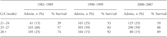
GA, gestational age.
The evaluation of programming effects in prematurely born babies is difficult because of lack of large prospective data specifically in relation to the development of non-communicable diseases. However, identification of potential contributors for whom animal data exists, for example influence of methyl group load, or the effects of pharmacological interventions like vitamin A, glucocorticoids, etc., can help identify programming-related changes. Finally the available outcome data can be correlated retrospectively to the potential events early in life that could have programming effect. However, the changes in demography of the study population in combination with changes in the algorithms of clinical care have made such evaluation difficult. In the present review we will discuss the available data in humans with particular emphasis on the contribution of interventions in the neonatal intensive care unit (NICU). Where applicable, reference is made to the contributing data from studies in animal models. However, it should be underscored that premature birth, neonatal intensive care and survival of extremely immature babies are phenomena that are historically recent and unique to humans. Until recently there were no animal models of prematurity and the data of animal models of premature birth were limited and exclusively of short duration.
The major events that could potentially result in programming effects in a prematurely born infant can be listed as follows:
Intrauterine environment
Premature interruption of pregnancy
Neonatal interventions
Nutritional care during infancy
Contributors to programming: (1) Intrauterine environment
The critical role of intrauterine environment on fetal growth and programming has been documented in a large number of studies in humans and in animal models.Reference Whincup, Kaye and Owen8–Reference Simmons10 Data from studies in animal models show that various nutrient and environmental manipulations during pregnancy result in fetal growth restriction, lower birth weight and programming of the offspring.Reference Whincup, Kaye and Owen8, Reference Simmons10 Studies in humans have confirmed the original observation of Barker et al. that intrauterine growth restriction as evidenced by lower weight at birth is associated with the higher incidence of non-communicable disease in the offspring in adult life.Reference Warner and Ozanne9 In this context, it is interesting to note that a significantly high proportion of LBW babies have been reported to be growth retarded in utero and born small for gestational age in various studies.Reference Ehrenkranz, Younes and Lemons11 However, the long-term programming consequence of the intrauterine growth restriction early in gestation cannot be easily separated from the confounding influence of other contributors to programming, particularly in studies in humans.
Contributors to programming: (2) Interruption of pregnancy
The impact of premature interruption of pregnancy on the immature baby remains undefined. The transition from fetus to neonate is accompanied by changes in respiration, circulation, glucose and energy homeostasis, thermoregulation and a host of other endocrine, metabolic and physical responses that allow for independent extra-uterine existence. Significantly, there is a marked change in the redox state and oxygenation from a relative hypoxic and markedly reduced fetal state to a highly oxygenated extra-uterine life.Reference Philippidis and Ballard12, Reference Filippi, Messeri and Dani13 These changes are accompanied by generation of reactive oxygen species (ROS) and consequently changes in metabolism to compensate for the oxidant insult. ROS can cause oxidative modification of major cellular macromolecules, that is, lipids, proteins and DNA. A number of molecular targets of ROS have been identified.Reference Avery14 In the context of programming, one carbon metabolism or the methyl transfer plays a critical role in the methylation of proteins, lipids and the methylation of DNA in the transcriptional control of gene expression and metabolism.Reference Kalhan and Marczewski15 The key constituent parts of the one carbon metabolism are displayed in Fig. 1. Folate and methionine cycle form the key components of the one carbon metabolism or methyl transfers and are present ubiquitously in every cell in the body. Methyl groups from serine and glycine entering the folate cycle are transferred to homocysteine to form methionine. Methionine, an essential amino acid, is the immediate source of the methyl groups required for the methylation of proteins, nucleic acids, biogenic amines and phospholipids, etc. Methionine is converted to the bioactive compound, s-adenosyl methionine (SAM), the ubiquitous methyl donor, catalyzed by methionine adenosyl transferase. The transfer of methyl groups from SAM is catalyzed by several different methyltransferases and result in the formation of s-adenosyl homocysteine and subsequently homocysteine. Homocysteine can either be methylated back to methionine or can enter the methionine catabolic pathway, the transsulfuration cascade, wherein it condenses with serine to form cystathionine. Cystathionine, through a series of metabolic steps is converted to cysteine and ultimately to taurine. As shown in the figure, the ROS and change in redox can influence the key regulatory steps of methionine metabolism, that is, the synthesis of SAM and the transsulfuration cascade involved in the synthesis of cysteine and ultimately glutathione.Reference Prudova, Bauman and Braun16–Reference Kabil and Banerjee18 Changes in redox and ROS could potentially affect the generation of SAM and glutathione and consequently impact the methylation process. Although not studied, perturbations in the one carbon metabolism at the time of birth could have long-lasting effect on gene expression and cause programming in the immature baby. It is important to underscore that the expression of genes regulating metabolism is an extremely orchestrated phenomenon, so that several of the metabolic processes appear at critical predetermined points during development. For example, hepatic cytosolic PEPCK and gluconeogenesis appears for the first time after birth in rodent and other mammalian species.Reference Kalhan and Parimi19 Similarly, cystathionine gamma lyase is not present in fetal liver and appears after birth.Reference Kalhan and Bier20 Premature interruption of pregnancy and premature birth will result in early expression of these pathways for independent existence. The long-term consequence of these changes or programming is not known. This could be akin to modification of gene expression demonstrated in the newborn rats as a result of changes in the diet in the neonatal period.Reference Srinivasan, Laychock, Hill and Patel21

Fig. 1 The one carbon (methyl group) metabolism in vivo. The interrelationship between folate and methionine metabolism and its regulation are displayed. As shown, endogenous methyl groups originating from serine during its conversion to glycine are incorporated into the folate cycle to be transferred to homocysteine for the formation of methionine and ultimately s-adenosyl methionine (SAM), the universal methyl donor. The transfer of methyl groups is catalyzed by several different methyltransferases and result in the formation of s-adenosyl homocysteine (SAH) and methylated products of proteins, lipids, DNA, creatine, phosphotidyl choline, etc. SAH is then hydrolyzed to form homocysteine. Homocysteine is the branch point of the methionine cycle, is either methylated back to methionine or participates in the transsulfuration pathway whereby the sulfhydryl group is transferred to serine to form cysteine. Cysteine is a component of the tripeptide, glutathione, the key intracellular antioxidant. The one carbon metabolism requires folate, vitamin B12 (B12) and vitamin B6 (B6). Homocysteine can lead to protein modification, induce inflammation, endothelial damage, thrombosis, etc. The formation of SAM from methionine (catalyzed by methionine adenosyl transferase) is sensitive to reactive oxygen injury (ROS).Reference Prudova, Bauman and Braun16–Reference Kabil and Banerjee18 Transsulfuration is regulated by change in redox state, as well as by hormones such as insulin and glucagon. Thus at the time of birth, both transmethylation and transsulfuration can be effected by the rapid change in redox, by the increased generation of ROS and by the birth associated acute surges in several hormones. GNMT, glycine n-methyltransferase. THF, Tetrahydrofolate; GSH, glutathione; MS, methionine synthase; CbS, cystathionine beta synthase; CgL, cystathionine gamma lyase.
Contributors to programming: (3) Clinical care protocols in the NICU
Evaluation of the contribution of the clinical protocols, used in the NICU, to programming is confounded by a number of variables, making it difficult to establish cause and effect relationship. The potential variables include:
(1) Heterogeneity of the infant population. As shown in Table 1, infants admitted to the NICU represent a wide range of gestational age and therefore development. These data also show that, over the last 20 years, the relative proportion of various gestation groups and their survival rate has not changed significantly. Since the potential for vulnerability to environmental and other insults may be different at different stages of development, these data underscore the need to examine these gestation groups separately when examining the possible programming effects.
(2) The clinical intervention for cardio-respiratory support, for treatment of sepsis, fluid therapy and other problems related to prematurity such as hyperbilirubinemia, necrotizing enterocolitis, patent ductus arteriosus, hypoglycemia and anemia, could have added modifying influences. In addition, these interventions can be institution specific depending on the local expertise and bias and are not easily quantifiable in data analysis.
(3) The protocols for clinical care have continuously evolved over time by incorporating new innovations including pharmacological, clinical, nutrition and other technologies. In addition, certain pharmacological interventions, considered to be useful early on, have been discontinued as the potential harmful effects were recognized. This has been specifically true for the postnatal use of glucocorticoids (dexamethasone) for the prevention and treatment of bronchopulmonary dysplasia after reports of neurodevelopmental impairment were published.
(4) The algorithms of clinical care have other variations introduced by individual care providers based upon the clinical condition of the baby, or the result of individual bias and expertise resulting in the achieved goal being quite different from that planned or desired. These changes in the clinical protocols make it difficult to correlate the observations in the physiological phenotype in adult with the neonatal intervention.
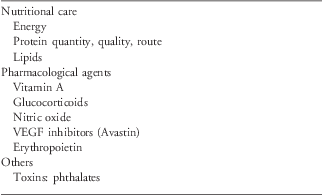
The commonly used clinical interventions in the NICU that could have programming effects on the baby are listed in Table 2. For most of these, the actual cause and effect has not been demonstrated. In the following, we will discuss some of the scientific evidence for potential programming and epigenetic effects of these interventions. Additionally, the LBW babies are exposed to environmental factors such as noise (from the isolette and other equipment), light and other pharmacological interventions like caffeine, oxygen, etc. Their effects, if any, in relation to programming have not been determined.
Table 2 Clinical interventions that may have programming effects
Nutritional care in the NICU
Nutrition has been recognized as the major or critical influence in programming during development. Nutrient imbalance at critical periods during development has been suggested to result in long-term programming by causing epigenetic changes in the DNA and by modifications of the histones.Reference Symonds, Sebert, Hyatt and Budge22 Over the past many years the protocols and algorithms for the nutritional management of the LBW baby have been evolving continuously with the goal of providing enough energy producing substrates and proteins in order to achieve a rate of accretion of lean body mass and body weight that resembles the rate of growth and protein accretion of fetus in utero. Whether such a goal is sufficient for optimal growth and development, or is it desirable or can it be achieved has been a subject of clinical debate and scientific inquiry for many years and continues to be discussed in the contemporary literature.Reference Hay and Thureen23 As discussed below these nutritional protocols have resulted in varying rates of growth of the babies in the neonatal period and possible long-term consequences.
Parenteral and enteral protein intake
The quality and quantity of protein, the route (parenteral v. enteral) of administration and the duration of parenteral nutrition have seen the most dramatic change in the last 20–30 years. With the increasing emphasis on early introduction of enteral feeding, due to its recognized clinical benefits, the average duration of parenteral nutrition has decreased remarkably from an average of ~30 to 40 days to the current duration of ~10 days in most NICUs.Reference Ehrenkranz, Younes and Lemons11 In addition, as the protein requirements of the LBW babies were recognized and the potential impact of low protein intake were documented, there has been a greater emphasis on early provision of larger amounts of parenteral amino acids to the LBW babiesReference Hay and Thureen23, Reference Hay and Thureen26–Reference Ibrahim, Jeroudi, Baier, Dhanireddy and Krouskop30 such that a higher proportion of LBW babies are now given 3–4 g/kg day amino acids starting on day one after birth, with no clinically measurable adverse consequences.Reference te Braake, van den Akker, Wattimena, Huijmans and van Goudoever28–Reference Ibrahim, Jeroudi, Baier, Dhanireddy and Krouskop30 This is in contrast to the clinical practice of 20 and 30 years ago when these infants routinely did not receive amino acids in their parenteral nutrient solutions on the first 2–3 days after birth or received very low quantities (0.5–1.0 g/kg day) often accompanied by low caloric intake in the form of glucose alone.
Low protein intake
The impact of low protein intake on long-term programming in humans has not been reported in part because the follow-up period has been relatively short and because of the confounding effect of other clinical and biological variables on this population. In this context, it is important to underscore the differential effect of low protein with low energy intake as compared with low protein intake in the presence of isocaloric energy intake. Although the combination of low energy intake along with low protein intake results in metabolic responses akin to starvation, that is, increased rate of lipolysis from the adipose tissue and an increased rate of whole body proteolysis in order to meet the energy requirements, the isocaloric protein deficiency results in markedly different responses. In the rodent, dietary protein restriction during pregnancy results in fetal growth retardation and metabolic programming resulting in hypertension, impaired beta cell function and mass, impaired insulin sensitivity and other pathological responses in the adult offspring.Reference Warner and Ozanne9 These have been associated with a change in the hypothalamic–pituitary–adrenal axis, changes in renin–angiotensin system and alterations in the concentrations of catecholamines and adrenoreceptors.Reference Warner and Ozanne9, Reference Augustyniak, Singh, Zeldes, Singh and Rossi31 The perturbations in the maternal metabolism that lead to the observed programming responses in the fetus have not been delineated. In the non-pregnant adult rat, isocaloric protein restriction for 7–10 days resulted in significant differential expression of a number of genes involved in cell cycle, cell differentiation, transport, transcription and metabolic processes in the liver.Reference Kalhan, Uppal and Moorman32 The expression of genes involved in fatty acid oxidation and serine biosynthesis was higher while those involved in urea synthesis and fatty acid synthesis was lower in the protein restricted animals. Tracer isotope studies showed that protein restriction caused a 50% increase in de novo synthesis of serine. Since serine is the primary source of methyl groups for transmethylation in vivo, it was interesting to note that the measured rates of transmethylation of methionine and the methylation potential [SAM/s-adenosyl homocysteine (SAH) ratio] was significantly increased in the protein restricted animals. These data were interpreted to suggest a high methylation demand placed on the organism as a result of changes in cell cycle and differentiation. Chronic protein malnutrition in humans identified by low plasma transthyretrin levels was observed to be associated with hyperhomocysteinemia and possibly changes in one carbon metabolism.Reference Ingenbleek, Hardillier and Jung33 Whether the LBW baby responds to dietary protein restriction in a similar manner has not been evaluated. In addition, the long-term consequences of such a response or its programming effects, if any, have not been determined. It can only be speculated that isocaloric protein restriction at this critical period of development, potentially, can cause programming effect by changes in the one carbon metabolism, transmethylation and methylation potential. However, the clinical data from earlier studies in the LBW babies do not show evidence of isolated low protein intake but show both low protein and energy intake during the first few days after birth.Reference Olsen, Richardson, Schmid, Ausman and Dwyer24, Reference Embleton, Pang and Cooke34 The early protein energy malnutrition in the NICU has been related to neurodevelomental deficits at age of 18 months.Reference Stephens, Walden and Gargus35 Perhaps these developmental deficits also should be considered programming affects.
High protein intake
As discussed above, there has been a recent concerted effort to promote higher protein intake by both enteral and parenteral route and to initiate enteral feedings as early as possible. The higher intake of protein via the enteral route has not been reported to cause any significant problems. In contrast, intravenously administered amino acids mixtures because of their unique composition can cause immediate metabolic changes which may have programming affects. The common amino acid mixture used in the NICU in United States was formulated to result in plasma amino acid concentration resembling that of a term to 1-month-old breastfed infant and does not resemble the amino acid composition of any protein.Reference Heird, Hay and Helms36 The additional criteria for the formulation were maintenance of solubility and stability of the mixture. In general the commonly used amino acid mixtures (e.g. Trophamine) are high in branched chain amino acids and in methionine and low in cysteine. The early administration of larger (3 g/kg day) amounts of amino acids results in higher plasma concentrations of all amino acids in particular essential amino acids for a variable period without causing any toxic effects.Reference Blanco, Gong and Green37 In addition, aggressive early administration of higher amount of amino acids did result in positive nitrogen balance and growth of the neonate.Reference te Braake, van den Akker, Wattimena, Huijmans and van Goudoever28, Reference Ibrahim, Jeroudi, Baier, Dhanireddy and Krouskop30 Studies done by Battista et al.Reference Battista, Price and Kalhan38 showed that the majority of excess branched chain amino acids (BCAA) in Trophamine are oxidized to CO2 and urea and are not retained in body proteins (Fig. 2). Similarly, essential amino acid methionine, also administered in larger quantities than its estimated daily requirement (~28 μmol/kg h as compared with the estimated requirement of 16 μmol/kg h) is also transsulfurated and oxidized to CO2.Reference Thomas, Gruca and Bennett39 However, for it to enter transsulfuration pathway, the excess methionine enters the transmethylation cycle, and is converted to SAM, SAH and homocysteine (Fig. 1). The higher transmethylation will result in an excess methyl load on the organism. Although the consequences of the excess methyl load on the LBW baby are not known, data from animal models show that excess methyl load during pregnancy in mice can lead to the development of allergic asthma in the offspring.Reference Hollingsworth, Maruoka and Boon40 Corresponding data in humans have been inconsistent.Reference Haberg, London, Stigum, Nafstad and Nystad41, Reference Whitrow, Moore, Rumbold and Davies42 However, it is significant to note that bronchial asthma has consistently been identified as a significant morbidity at follow-up in LBW babies in several studies.Reference Hack3, Reference Doyle and Anderson5 The contribution of excess methyl load in the parenteral nutrition on long-term programming requires careful longitudinal follow-up studies.
Fig. 2 Metabolism of leucine and methionine in preterm infants receiving parenteral amino acid nutrition (upper panel). The effect of branched amino acid enriched parenteral nutrition (Trophamine) on rates of leucine oxidation and urea synthesis in preterm babies. The rates of leucine oxidation were measured using [1-13C]leucine tracer and the rate of synthesis of urea using [15N2]urea tracer. As shown, excess BCAA were oxidized to CO2 and urea (lower panel). Rate of appearance of methionine (Ra) in the plasma and estimates of transmethylation and transsulfuration in healthy full-term babies and preterm babies receiving parenteral amino acid nutrition. As shown, the parenteral amino acid containing higher than required methionine (high Ra) resulted in increased transmethylation and transsulfuration. BCAA, branched chain amino acids.
Growth from ‘Birth to Term’
Data reported by a number of investigators from clinical centers across the world have underscored the difficulties in achieving the goal of growth in the neonatal period that will mimic the rate of intrauterine growth. These studies show that in spite of our best efforts in clinical and nutritional care of the LBW baby, these babies at the time of discharge from the hospital do not achieve the median birth weight of the reference fetus at the same postmenstrual age.Reference Ehrenkranz, Younes and Lemons11, Reference Olsen, Richardson, Schmid, Ausman and Dwyer24, Reference Cooke, Ainsworth and Fenton25 Although early and aggressive parenteral and enteral nutritional strategies have been shown to attenuate this postnatal growth failure, the goal of achieving intrauterine rate of growth remains allusive.Reference Cooke, Ainsworth and Fenton25, Reference Embleton, Pang and Cooke34, Reference Wilson, Cairns and Halliday43, Reference Dinerstein, Nieto and Solana44 The postnatal growth retardation or the so called ‘birth to term’ growth failure has been described as an inevitable and universal problem in preterm infants.Reference Embleton, Pang and Cooke34 The potential contributors to the birth to term growth failure, in addition to nutritional support, include neonatal morbidities such as lung disease, necrotizing enterocolitis, intraventricular hemorrhage, treatment with glucocorticoids, etc. Statistical model analysis of the various contributors to the postnatal growth showed that medical morbidities and nutritional factors, in particular protein intake, explained ~50% of the variance in the overall growth velocity.Reference Olsen, Richardson, Schmid, Ausman and Dwyer24 Body composition studies of these infants using dual emission X-ray absorptiometry (DEXA) showed reduced linear growth and reduced fat free mass coupled with increased global and central fat mass.Reference Cooke and Griffin45 Whether such body composition changes at this early stage in life have any relation to the development of insulin resistance and metabolic syndrome later in life remains to be examined. Comparison with the studies from India of ‘fat thin baby’ would suggest a high risk of development of insulin resistance later in life.Reference Yajnik, Fall and Coyaji46 A significant relationship between growth velocity in the NICU and the likelihood of neurodevelopmental impairment has also been reported.Reference Stephens, Walden and Gargus35, Reference Ehrenkranz, Dusick and Vohr47 Whether the impaired neurodevelopment is the consequence of programming or due to an acute irreversible injury to the brain is not clear.
Pharmacological interventions
A number of pharmacological agents, commonly used in the NICU, are listed in Table 2. Data on their long-term consequence are limited. Some of these agents such as caffeine, surfactant are administered to almost all LBW babies and therefore their consequences are difficult to delineate. Others like nitric oxide (NO), erythropoietin, VEGF (vascular endothelial growth factor) inhibitors have only recently been introduced in clinical care and therefore no long-term follow-up data are available as yet. In the following, available data on some of these are discussed in the context of their potential role in programming.
Vitamin A
The plasma concentrations of retinol and retinol binding protein levels in LBW babies have been observed to be low in a number of studies. These low levels do not appear to be influenced by the amount and route of feeding and remain low for several months after discharge from the hospital.Reference Peeples, Carlson, Werkman and Cooke48–Reference Shenai, Chytil and Stahlman51 These data have been interpreted to represent a state of vitamin A deficiency. Data from studies in vivo and in vitro have demonstrated a critical role of vitamin A in the differentiation of pulmonary cells, in surfactant protein metabolism, in airway growth and expression of several genes involved in the development of lungs. In addition, clinical observation studies had suggested that poor vitamin A status in the first months after birth in LBW babies was associated with an increased risk of developing bronchopulmonary dysplasia and long-term respiratory disability.Reference Shenai, Chytil and Stahlman51, Reference Spears, Cheney and Zerzan52 Based upon these data, clinical trials of oral or parenteral vitamin A supplement have been done. These studies show that administration of vitamin A during the first 4 weeks after birth results in significant but small reduction in death or oxygen use at 36 weeks postmenstrual age.Reference Shenai, Kennedy, Chytil and Stahlman53–Reference Tyson, Wright and Oh56 Even though the clinical impact of vitamin A supplement was small, it was recommended that 5000 IU of vitamin A be given intramuscularly to LBW babies who are at risk for developing chronic lung disease or bronchopulmonary dysplasia. However, such a recommendation is not uniformly followed by the clinical neonatologists.Reference Darlow and Graham57, Reference Barros, Bhutta and Batra58
The long-term consequences and potentially programming effects of the administration of vitamin A to the LBW infants have not been delineated. Limited clinical data of relatively short follow-up period suggests that there are no identifiable acute toxicity of high dose vitamin A in the neonate. In addition, no significant impact on mortality and neurodevelopmental impairment at 18–22 months of age was observed.Reference Tyson, Wright and Oh56, Reference Ambalavanan, Tyson and Kennedy59 However, high dose vitamin A could potentially have programming effect by modulating SAM dependent transmethylation reactions (Fig. 1). Studies in rat show that high dose vitamin A and its derivatives (all-trans retinoic acid) caused an increase in abundance and activity of glycine n-methyltransferase (GNMT) and hypomethylation of DNA in the liver.Reference Rowling, McMullen and Schalinske60–Reference Rowling and Schalinske63 Interestingly, the observed responses were both tissue and gender specific, so that there was no effect of retinoids on pancreatic and renal GNMT activity.Reference McMullen, Rowling, Ozias and Schalinske62 In addition, the increase in hepatic GNMT activity was less in female rats when compared with the males. The long-term phenotypic effect of these changes in methylation has not been reported. However, exposure of rats to very high (80,000 IU/kg day) doses of vitamin A during 1–5 days after birth has been shown to result in long-lasting (at 36 weeks of age) defect in learning in water-maze task and depression in heat–pain responses.Reference Kihara, Matsuo, Sakamoto, Yasuda and Tanimura64 Other data suggestive of programming effect on immune system have been reported in response to neonatal exposure to vitamin A in rats.Reference Csaba, Kovacs and Pallinger65 A pattern of motor deficit following late embryonic exposure to all-trans retinoic acid has also been reported.Reference Coluccia, Borracci, Belfiore, Renna and Carratu66 Future studies will examine whether these observations are due to a programming effect related to changes in methylation status caused by perturbation in methionine cycle.
Glucocorticoids
Glucocorticoids have been and remain the most extensively used pharmacological intervention used in the perinatal period. Data from animal models have clearly demonstrated the programing effect of glucocorticoids administration during development. These studies show that exposure to excess glucocorticoids in the prenatal period either due to increased endogenous production as a result of maternal stress, or by exogenous administration or as a result of inhibition of 11-β hydroxysteroid dehydrogenase type 2 (the placental barrier to maternal glucocorticoids) results in lower birth weight and causes hyperglycemia, hypertension, increased HPA axis activity and behavior abnormalities in the offspring.Reference Harris and Seckl67, Reference Drake, Tang and Nyirenda68 These effects have been observed to be transmitted across generations suggesting an epigenetic mechanism. It should be underscored that while animal models are critical for our understanding of the biological and molecular mechanisms of programming, the doses of pharmacological agents used and the duration of administration in these studies have been quite different than those used in clinical practice. In clinical practice, glucocorticoids are either used antenatally to induce pulmonary surfactant synthesis in case of impending premature delivery or in the neonate with respiratory distress syndrome or bronchopulmonary dysplasia in order to wean them from respiratory/ventilator support and minimize the risk of lung injury.
Antenatal glucocorticoids
A single course of glucocorticoids, either betamethasone or dexamethasone, given to the mother has been shown to be effective in reducing morbidity and mortality after premature birth with few immediate side effects. Both of these glucocorticoids are transferred to the fetus in significant amounts without being metabolized in the placenta. Clinical observation studies have not demonstrated any significant biological impact (programming) on the offspring of this single course of steroid administration, although certain subtle and inconsistent effects have been reported. For example, Doyle et al.Reference Doyle, Ford, Davis and Callanan69 observed a significantly higher systolic and diastolic blood pressure at 14 years of age in subjects exposed to antenatal corticosteroids. In contrast, Dessens et al.Reference Dessens, Haas and Koppe70 observed significantly lower systolic blood pressure at 20–22 years of age in subjects exposed to antenatal betamethasone. Long-term follow-up studies from different parts of the world have not shown any impact of a single course of antepartum glucocorticoids on body composition, insulin resistance, lipid profile or other medical or psychological variables.Reference Finken, Keijzer-Veen and Dekker71–Reference Dalziel, Lim and Lambert74 Finken et al.Reference Finken, Keijzer-Veen and Dekker71 had observed a lower glomerular filtration rate at 19 years of age, whereas Dalziel et al.Reference Dalziel, Walker and Parag72 observed higher plasma insulin levels at 30 min following a 75 g oral glucose tolerance test (GTT) at 30 years of age. These later observations require confirmation, by additional follow-up studies. Exposure to a repeat dose of betamethasone during the antepartum period was also not associated with anthropometric or neurocognitive changes; however, the follow-up period of these studies was only 2–3 years.Reference Wapner, Sorokin and Mele75, Reference Peltoniemi, Kari and Lano76
Neonatal glucocorticoids
The data regarding neonatal use of steroids and their programming effects are confounded by the inconsistencies of dosage regimen and follow-up. These data show that postnatal dexamethasone leads to improved ventilation and facilitates weaning from the ventilator support irrespective of dose and timing of administration. As anticipated the use of glucocorticoids in the neonate is associated with variable increase in blood glucose and insulin concentrations, impaired weight gain, clinical complications such as necrotizing enterocolitis, etc.Reference Stark, Carlo and Tyson79 Follow-up data suggest adverse neurological side effects with their use.Reference Yates and Newell77–Reference Stark, Carlo and Tyson79 Neonatal dexamethasone treatment in rats has been shown to cause sustained lung hypoplasia and increased pulmonary arterial pressure in adult rats.Reference le Cras, Markhan and Morris80
Nitric Oxide (NO)
NO, a major endogenous regulator of vascular tone (endothelial vascular relaxing factor) and intracellular signaling molecule, is being used extensively for the treatment of pulmonary hypertension in the neonates born at term or at near-term gestation.Reference Flier and Barrington81 Systematic reviews and meta-analysis of the published literature have confirmed the effectiveness of inhaled NO in reducing death and the need for ECMO (extracorporeal membrane oxygenation) without any measurable systemic toxicity in the doses employed.Reference Flier and Barrington81 Although not approved for use in the prematurely born babies, it is often used outside the licensed indication (off-label) in preterm babies for the prevention and treatment of bronchopulmonary dysplasia. The rational for the use of inhaled NO in the prematurely born infants, and its clinical effectiveness (or lack of it) have been discussed.Reference Subhedar and Dewhurst82–Reference Cole, Alleyene and Barks84 The potential acute toxic effects of inhaled NO include acute lung injury by peroxynitrite formed by the reaction of NO with oxygen, impaired platelet aggregation, methemoglobinemia and possibly extrapulmonary vasodilatation.Reference Weinberger, Laskin, Heck and Laskin85 NO is rapidly inactivated by hemoglobin to form methemoglobin. The extrapulmonary effects of inhaled NO have been attributed to its binding to circulating albumin or hemoglobin and thus delivery of NO in its active form to systemic circulation.Reference Weinberger, Laskin, Heck and Laskin85 In addition, NO is also considered to be an epigenetic molecule.Reference Illi, Colussi and Grasselli86 NO exerts its effect on gene expression and regulation via nitrosylation and tyrosine (tyr)-nitration of a number of nuclear and non-nuclear proteins.Reference Illi, Colussi and Grasselli86 The epigenetic effects of NO have particularly been studied in the developing neurons.Reference Nott and Riccio87 Although not examined in relation to therapeutic doses of NO used in the premature infants, the transfer of NO as protein bound adductReference Keanney, Simon and Stamler88 could potentially allow for its transport to distant organs and consequently result in epigenetic modification of gene expression. Carefully conducted studies in vivo and in vitro would delineate such effects in the future.
Anti-VEGF therapy
Bevacizumab (Avastin), an anti-VEGF therapy, previously used to treat diabetic retinopathy and macular degeneration in adults is now being implemented in preterm NICU infants between 30 and 40 weeks post menstrual age for the treatment of retinopathy of prematurity. It is a recombinant humanized vascular endothelial VEGF antibody that prevents VEGF from binding to its receptors.Reference Lin, Nguyen and Mendoza89 Bevacizumab binds to all isoforms of VEGF,Reference Ferrara90 blocks VEGF-induced angiogenesis and is approved by the US Food and Drug Administration for intravenous treatment of metastatic colon cancer. A recent randomized controlled trial of intravitreal injection of bevacizumab for stage3+ retinopathy of prematurity in the United States has shown lower rates of recurrence of retinopathy (anti-VEGF: 6% v. laser: 42%), rates of macular dragging (3% v. 48%) and retinal detachment (0% v. 6%) in the bevacizumab group compared with the laser therapy group.Reference Mintz-Hittner, Kennedy and Chuang91 Although seemingly an exciting new therapy with great potential to minimize serious sequelae of prematurity, information on the long-term implications of this therapy remains sparse. Intravitreal bevacizumab enters the general circulation, results in prolonged VEGF inhibition and has a half-life of up to 2 weeks in primates. In the fetus, VEGF is expressed in most tissues and is critical for growth and development of vital organs such as kidneys, lungs and brain during the third trimester. In-vitro studies on the effect of bevacizumab-induced inhibition of VEGF have demonstrated dose-dependent inhibition of human umbilical vein endothelial cell proliferation,Reference Wang, Fei, Venderlaan and Song92 increased mortality and impaired liver and cartilage growth in newborn miceReference Gerber, Hillan and Ryan93, Reference Gerber, Vu and Ryan94 and measurable levels of anti-VEGF drug in kidneys, spleen, lung, opposite eye and brain of newborn rabbits receiving intravitreal injection in a single eye.Reference Wu, Lai and Chen95 Preterm infants with retinopathy of prematurity (ROP) have a compromised blood–retinal barrier that may allow even more bevacizumab to enter the blood stream. Given the fact that preterm infants receive treatment for ROP at a time when growth and differentiation is maximal, the potential of such a therapy on programming requires careful evaluation.
Others
Bisphenol-A (BPA), an estrogen-mimic endocrine disrupter, is extensively used in manufacturing polycarbonate plastic and epoxy resins from which food and beverage containers and dental materials are made.Reference Maffini, Rubin, Sonnenschein and Sotoi96, Reference Calafat, Weuve and Ye97 BPA may also be used in polyvinyl chloride (PVC) industry. PVC is used in the manufacture of several medical products used in the NICU such as feeding tubes, umbilical vessel catheters, bags and tubings for parenteral fluids and for their administration, endotracheal tubes, respiratory masks, etc. Di(2-ethylhexyl) phthalate is a plasticizer added to PVC in a number of these medical products to make them soft and pliable. Calafat et al. Reference Calafat, Weuve and Ye97 in a small study examined the exposure to BPA and other phenols in premature infants in two NICUs in Massachusetts, USA. Their data showed that the geometric mean BPA concentration among premature infants undergoing intensive medical interventions was one order of magnitude higher than that seen in the general population. In addition, they found that the conjugated species of phenols were the primary urinary metabolites suggesting that the premature LBW babies had some capacity to metabolize BPA. Whether such an exposure to BPA in the LBW babies has any programming effects continues to be an important area of investigation. Data from studies in animal models show that exposure to BPA during early development can change the phenotype of the offspring by stably altering the epigenome.Reference Dolibnoy, Huang and Jirtle98 Interestingly, administration of methyl donors such as folic acid negated the DNA hypomethylating effect of BPA in this study. Other studies have shown disruptive effects of BPA on the expression of sexually selected traits in adults as a consequence of developmental exposure in the mouse and alterations in the development of mammary gland in the rhesus monkey.Reference Jasarevic, Sieli and Twellman99
Contributors to programming: (4) Catch-up and catch-down growth during infancy
With the improvement in nutritional care of the preterm babies after discharge from the hospital, the frequency of growth failure or stunting has markedly decreased and most prematurely born LBW babies show evidence of catch-up growth.Reference Hack, Merkatz, McGrath, Jones and Fanaroff100–Reference Itabashi, Mishina and Tada102 However, weight gain alone should not be considered evidence of catch-up growth. Uthaya et al. studied the body composition of preterm LBW babies (<32 weeks gestation) using whole body MRI technique.Reference Uthaya, Thomas and Hamilton103 Their data show that at the postconceptional age of term gestation, the preterm babies had significant decrease in subcutaneous adipose tissue and significant increase in visceral adipose tissue while accelerated weight gain in these babies was accompanied by increased total and subcutaneous adiposity. They suggested that preterm baby may be at risk in later life of metabolic complications through increased and aberrant adiposity. However, a study by Cooke et al.Reference Cooke, Griffin and McCormick104 of preterm infants fed a nutrient enriched formula did not find evidence of altered adiposity measured longitudinally during first 6 months using DEXA method. DEXA does not differentiate subcutaneous from visceral fat. A number of other studies, reviews and systematic analysis have examined the impact of after hospital discharge nutrient management of LBW babies and catch-up and catch-down growth.Reference Yeung105–Reference Monteiro and Victora107 In the majority of the studies LBW does not represent the LBW graduate of the NICU, but rather small for gestational age full-term babies.Reference Yeung105–Reference Monteiro and Victora107 Outcome measurements in these infants are confounded by their response to the adverse intrauterine environment. Additionally, catch-up growth in the LBW preterm babies has been observed to continue until 6 years of age.Reference Wehkalampi, Hovi and Dunkel108 Accelerated growth or rapid weight gain has some distinct clinical advantages related to decrease in infections and improved rate of survival.Reference Yeung105 However, it also has been consistently related to the development of obesity in later life in both appropriate for gestational age and small for gestational age full term and preterm infants.Reference Bazaes, Alegría and Pittaluga109, Reference Stettler and Iotova110 A detailed analysis of these data are beyond the scope of this review and the reader is referred to some excellent publications on the subject. Obesity, in particular visceral adiposity, is associated with the insulin resistance and metabolic syndrome.
Clinical outcomes and their correlates
Follow-up data show that the LBW babies have a higher incidence of hypertension, visceral obesity, asthma and neurodevelopmental and behavior problems and perturbations in glucose insulin homeostasis. Whether these non-communicable disorders are the consequence of programming as a result of events during development has been examined only in the case of insulin resistance. These analyses are by necessity statistical correlations of observational data and do not represent causality. The reported data on insulin resistance are discussed below.
Insulin sensitivity and glucose metabolism
The association between insulin sensitivity, altered glucose metabolism at different ages and premature birth (LBW) has been reported in a number of studies from different parts of the world. Hofman et al.Reference Hofman, Regan and Jackson111 examined the parameters of glucose metabolism in 38 prepubertal children aged 4 to 10 years, born prematurely at <32 weeks gestation and compared them with 22 control subjects (appropriate for gestation age and born after 37 weeks gestation). Insulin sensitivity was measured by the use of paired glucose insulin data obtained by frequent sampling intravenous GTT. Their data show that children born prematurely had a lower insulin sensitivity compared with the controls. In addition, the prematurely born subjects had elevated acute insulin release compared with the controls. An altered glucose metabolism (negative correlation between 30 min glucose concentration post standardized glucose load and birth weight) was also observed by Fewtrell et al.Reference Fewtrell, Doherty and Cole112 in children born prematurely and studies between 9 and 12 years of age. Perturbations in glucose and insulin levels and measures of insulin sensitivity have been demonstrated at ages 18–27 years using 75 g oral GTT and HOMA-IR (homeostasis model assesment-insulin resistance),Reference Hovi, Andersson and Eriksson113 and at age 19 years using HOMA,Reference Finken, Keijzer-Veen and Dekker114 at age 22 years using hyperinsulinemic clamp technique.Reference Rotteveel, van Weissenbruch, Twisk and Delemarre-Van de Waal115 All these studies show lower insulin sensitivity in adults born prematurely in different parts of the world.Reference Yeung105–Reference Rotteveel, van Weissenbruch, Twisk and Delemarre-Van de Waal115 In addition, an association between LBW and type 2 diabetes has also been observed in a random sample of middle aged (30–60 years old) Danes.Reference Maffini, Rubin, Sonnenschein and Sotoi96
Whether intrauterine growth restriction is related to insulin sensitivity was also examined by Hofman et al.Reference Lin, Nguyen and Mendoza89 Interestingly, intrauterine growth restriction or small for gestational age did not have any additional effect on the change in insulin sensitivity in the premature group.Reference Hofman, Regan and Jackson111 Similarly, no effect of intrauterine growth restriction was observed on parameters of glucose and insulin metabolism during a standard 75 g GTT in young adults at 18–27 years of ageReference Hovi, Andersson and Eriksson113 and by Rotteveel et al.Reference Rotteveel, van Weissenbruch, Twisk and Delemarre-Van de Waal115 in non-obese young adults aged 22 years who were born prematurely at <32 weeks gestation and weighing <1500 g. Finally, data from a random sample of middle aged Danes showed a similar prevalence of type 2 diabetes in subjects born prematurely and those born small for gestational age.Reference Pilgaard, Færch and Carstensen116 Similar data have been reported by others.Reference Kaijser, Bonamy and Akre117
The effect of growth failure from birth to term (40 weeks) on insulin sensitivity was reported in two studies. Fewtrell et al.Reference Fewtrell, Doherty and Cole112 did not observe any relation between growth during the immediate postnatal period (but before term) and glucose insulin responses at the age of 9–12 years. Similarly, neither a decrease in sd scores for weight at 40 weeks of postmenstrual age (−1.4 ± 1.32) nor the actual sd score at term (−2.6 ⩽ −1.15) were related to glucose or insulin concentration, nor was either related to HOMA-IR index at the age of 18–27 years.Reference Hovi, Andersson and Eriksson113 However, in a group of prematurely born small for gestational age infants, the change in sd scores for weight from birth to term was −0.2 ⩽ −0.69; in this group an increase of 1 sd unit in the score corresponded to a 30.8% increase in fasting insulin concentration and 22.8% increase in the 2 h insulin concentration following a standardized 75 g glucose load. These data suggest that the growth failure from birth to term postmenstrual or postconceptional age does not appear to influence development of insulin resistance in adult life in the prematurely born appropriate for gestational age babies. In contrast, there is some suggestion that weight gain in small for gestational age baby is related to greater insulin responses later. These data require confirmation in specifically designed studies. Childhood weight gain after 18 months of age was the primary determinant of fasting split proinsulin and poststandardized glucose load insulin concentration.
These data suggest that interruption of pregnancy, that is, premature delivery and related events independent of IUGR, neonatal clinical support and growth restriction, is the primary determinant of lower insulin sensitivity later in life. It is compounded by accelerated weight gain (catch-up) in infancy. Although the mechanism or mechanisms responsible for the pancreatic insult are not known, we hypothesize that they may be related to the change in redox or to the exposure of the developing beta cell to parenteral and enteral nutrients administered to the LBW infant to support extra-uterine survival.
Acknowledgments
The authors thank Susan Marczewski R.N. and Manoa Hui for their help in the preparation of this manuscript. The cited work was supported in part by the NIH grant HD042152 and by the Clinical and Translational Science grant award (RR024989) to Case Western Reserve University. We apologize to those authors whose important work we may not have cited or omitted inadvertently due to space constraints.


