


Introduction
Parasitic nematodes are of great agricultural, medical, veterinary and economic importance (Hotez et al., Reference Hotez, Brindley, Bethony, King, Pearce and Jacobson2008), yet despite this relatively little is known about how parasitic nematodes identify and infect their hosts. Growing evidence suggests that chemosensation and other senses such as thermosensation and mechanosensation play important roles in nematode parasite transmission and intra host migration (Dillman et al., Reference Dillman, Guillermin, Lee, Kim, Sternberg and Hallem2012; Tsubokawa et al., Reference Tsubokawa, Hatta, Kikuchi, Maeda, Mikami, Alim, Maruyama and Tsuji2017; Bryant et al., Reference Bryant, Ruiz, Gang, Castelletto, Lopez and Hallem2018). Critical to this process is olfactory sensation, which is also important for and best studied in non-parasitic nematodes, whereby nematodes sense carbon dioxide or other volatile chemicals which attract them towards the correct host, invasion area or food source, or repel them from undesired targets (Bargmann, Reference Bargmann2006; Hallem et al., Reference Hallem, Rengarajan, Ciche and Sternberg2007; Hallem and Sternberg, Reference Hallem and Sternberg2008; Hart and Chao, Reference Hart, Chao and Menini2010; Castelletto et al., Reference Castelletto, Gang, Okubo, Tselikova, Nolan, Platzer, Lok and Hallem2014; Hong, Reference Hong and Sommer2015).
The Strongyloidoidea superfamily (Hunt et al., Reference Hunt, Tsai, Coghlan, Reid, Holroyd, Foth, Tracey, Cotton, Stanley, Beasley, Bennett, Brooks, Harsha, Kajitani, Kulkarni, Harbecke, Nagayasu, Nichol, Ogura, Quail, Randle, Xia, Brattig, Soblik, Ribeiro, Sanchez-Flores, Hayashi, Itoh, Denver, Grant, Stoltzfus, Lok, Murayama, Wastling, Streit, Kikuchi, Viney and Berriman2016) of nematodes contains the important human and veterinary parasitic Strongyloides nematodes (Schär et al., Reference Schär, Trostdorf, Giardina, Khieu, Muth, Marti, Vounatsou and Odermatt2013; Beknazarova et al., Reference Beknazarova, Whiley and Ross2016), the facultative parasitic Parastrongyloides nematodes (Kulkarni et al., Reference Kulkarni, Dyka, Nemetschke, Grant and Streit2013) and the free-living Rhabditophanes nematodes (Dulovic et al., Reference Dulovic, Renahan, Röseler, Rödelsperger, Rose and Streit2020). The phylogeny of this superfamily can be seen in Fig. 1A. Members of the Strongyloides genus are of great research interest due to their life cycle and other biological features, like the availability of closely related culturable facultative and non-parasitic species (Viney and Lok, Reference Viney and Lok2007; Streit, Reference Streit2014, Reference Streit2016; Streit et al., Reference Streit, Wang, Kang and Davis2016; Viney, Reference Viney2017). Within Strongyloides, species often have a narrow range of possible host species which they are capable of infecting, yet the susceptible species do not need to be phylogenetically closely related (Eberhardt et al., Reference Eberhardt, Mayer, Bonfoh and Streit2008; Jaleta et al., Reference Jaleta, Zhou, Bemm, Schär, Khieu, Muth, Odermatt, Lok and Streit2017; Nutman, Reference Nutman2017). A general overview of the life cycles of all species used in this paper can be found at (Eberhardt et al., Reference Eberhardt, Mayer and Streit2007; Viney and Lok, Reference Viney and Lok2007; Kulkarni et al., Reference Kulkarni, Dyka, Nemetschke, Grant and Streit2013; Dulovic et al., Reference Dulovic, Renahan, Röseler, Rödelsperger, Rose and Streit2020) and Fig. 1B. Of note, is the fact that the parasitic generation of Parastrongyloides trichosuri (P. trichosuri) is dioecious (unlike in Strongyloides spp.) and it is therefore possible to generate a high, if not unlimited, number of consecutive free-living generations on agar plates (Grant et al., Reference Grant, Stasiuk, Newton-Howes, Ralston, Bisset, Heath and Shoemaker2006; Kulkarni et al., Reference Kulkarni, Dyka, Nemetschke, Grant and Streit2013), whereas Rhabditophanes diutinus (R. diutinus) is free-living, reproduces asexually and can form dauer larvae as opposed to infective larvae (Dulovic et al., Reference Dulovic, Renahan, Röseler, Rödelsperger, Rose and Streit2020).

Fig. 1. Phylogeny and life cycles of the Strongyloidoidea. (A) Proposed phylogenetic relationship of the Strongyloidoidea based on whole genome sequence, according to Hunt et al. (Reference Hunt, Tsai, Coghlan, Reid, Holroyd, Foth, Tracey, Cotton, Stanley, Beasley, Bennett, Brooks, Harsha, Kajitani, Kulkarni, Harbecke, Nagayasu, Nichol, Ogura, Quail, Randle, Xia, Brattig, Soblik, Ribeiro, Sanchez-Flores, Hayashi, Itoh, Denver, Grant, Stoltzfus, Lok, Murayama, Wastling, Streit, Kikuchi, Viney and Berriman2016). Only the topology is given, the branch lengths in the figure are arbitrary. (B) Life cycles of Strongyloidoidea. Life cycles of S. ratti, S. papillosus, P. trichosuri and R. diutinus. i indicates infective larvae.
In a landmark paper (Castelletto et al., Reference Castelletto, Gang, Okubo, Tselikova, Nolan, Platzer, Lok and Hallem2014), Castelleto et al. performed a comparative analysis of the thermotaxic and chemotactic responses of various parasitic, entomopathogenic and free-living nematodes. This study compared closely related parasitic and entomopathogenic species with similar life cycles but different hosts and phylogenetically remote species either with similar life cycles and ecology or differing life cycles and ecology, finding that parasites with similar hosts respond similarly to odorants. Here, we decided to complement this extensive study with an analysis of phylogenetically close nematodes of the Strongyloidoidea superfamily, which have different life cycles and ecology. Previous research within this superfamily has found that urocanic acid is a strong chemoattractant in Strongyloides stercoralis (Safer et al., Reference Safer, Brenes, Dunipace and Schad2007), while CO2 was shown to be a repellent in contrast to many other parasitic nematodes (Castelletto et al., Reference Castelletto, Gang, Okubo, Tselikova, Nolan, Platzer, Lok and Hallem2014). Differences in olfactory behaviour between S. stercoralis and S. ratti have been reported (Safer et al., Reference Safer, Brenes, Dunipace and Schad2007; Castelletto et al., Reference Castelletto, Gang, Okubo, Tselikova, Nolan, Platzer, Lok and Hallem2014), suggesting that host-seeking behaviour even between these two closely related species is rather different. Using the four species (S. ratti, S.papillosus, P. trichosuri and R. diutinus) of the Strongyloidoidea family that are in permanent culture in our lab, we set out to characterize temperature-dependent and chemotactic behaviours within this family, orienting our experimental approaches after (Castelletto et al., Reference Castelletto, Gang, Okubo, Tselikova, Nolan, Platzer, Lok and Hallem2014).
Materials and methods
Species and strains
Strongyloides ratti (strain ED321) was originally a gift from Mark Viney, University of Bristol, now University of Liverpool, in 2010 and has since been maintained in the laboratory in continuous culture. Strongyloides papillosus (strain LIN) was isolated from sheep at the University of Hohenheim Field Station on the Swabian Alp in 2006 (Eberhardt et al., Reference Eberhardt, Mayer and Streit2007) and has since been maintained in the laboratory in continuous culture. Parastrongyloides trichosuri was originally a gift from Robin Hirst, Ag Research, New Zealand and Warwick Grant, Ag Research, New Zealand, now LaTrobe University, Australia in 2007 and has since been maintained in the laboratory and subjected to 12 rounds of inbreeding by selecting a single gravid female in each round, resulting in strain QA414. Rhabditophanes diutinus (strain KR3021, previously also called Rhabditophanes sp. KR3021) was originally isolated by Ann Rose in 1994 in British Columbia (Dulovic et al., Reference Dulovic, Renahan, Röseler, Rödelsperger, Rose and Streit2020) and was sent to us by Dee Denver, Oregon State University, in 2014 and has since been maintained within the laboratory in continuous culture. Pristionchus pacificus (strain PS312) was a gift from Ralf Sommer, MPI for Developmental Biology and has been maintained in the laboratory in continuous culture. Caenorhabditis elegans (strain N2) was originally obtained from the Caenorhabditis Genetic Center (CGC) and has since been maintained in the laboratory in continuous culture. All Strongyloides, Parastrongyloides and Rhabditophanes species and strains used in this paper are available upon request. Pristionchus pacificus (PS312) and C. elegans (N2) should be requested from Ralf Sommer and the CGC, respectively.
Maintenance of Strongyloides nematodes
Strongyloides ratti are maintained within rats at our in-house animal facility, as described in Dulovic et al. (Reference Dulovic, Puller and Streit2016). Strongyloides papillosus are maintained within rabbits at our in-house animal facility, as described in Eberhardt et al. (Reference Eberhardt, Mayer and Streit2007). Infective larvae for both species were collected by Baermann funnel from water surrounding minimum 7-day-old fecal cultures.
Maintenance of P. trichosuri, R. diutinus, P. pacificus and C. elegans
Parastrongyloides trichosuri, R. diutinus, P. pacificus and C. elegans were all maintained on NGM plates supplemented with a lawn of OP50 bacteria (Stiernagle, Reference Stiernagle2006). Parastrongyloides trichosuri were maintained at 20 °C, and the plates were further supplemented with a piece of autoclaved rabbit feces (Grant et al., Reference Grant, Stasiuk, Newton-Howes, Ralston, Bisset, Heath and Shoemaker2006). Rhabditophanes diutinus was maintained at 15 °C and P. pacificus and C. elegans at 20 °C. To obtain infective P. trichosuri larvae, plates were incubated for 2 weeks post-worms being added, then washed off and the worms concentrated by Baermann funnel for 1 h, following which the infective larvae obtained were used directly for experimentation. To obtain R. diutinus dauer larvae, R. diutinus was grown by liquid culture. Briefly, growing plates of R. diutinus were washed off into S-Medium (Stiernagle, Reference Stiernagle2006) without cholesterol, supplemented with 50 μg mL Streptomycin, 50 μg mL Nystatin and 1% w/v OP50 (from fresh overnight culture that had been pelleted by centrifugation). Cultures were then incubated at 17 °C and shaken at 130 rpm for 12 days. Following this, worms were isolated from the culture by centrifugation, washed once with ddH2O, and then used directly for experimentation. To obtain P. pacificus and C. elegans dauer larvae, plates were incubated for 3 weeks post-worms being added and then washed off with ddH2O and concentrated by Baermann funnel for 1 h.
Dispersal assay
Dispersal assays were performed similarly to those described in Bargmann (Reference Bargmann2006) and Castelletto et al. (Reference Castelletto, Gang, Okubo, Tselikova, Nolan, Platzer, Lok and Hallem2014) with minor modifications. Following a brief pilot study, HEPES agar was determined to allow the largest range of locomotion for all species of Strongyloidoidea examined. In total, 6 cm HEPES agar plates [per litre: 20 g (2%) agar and 3 g (0.05 M) NaCl autoclaved and then supplemented with 10 mL 1 M HEPES, 1 mL 1 M MgSO4 and 1 mL 1 M CaCl2] were marked with a 3.5 cm inner zone, with worms then spotted to the centre of this zone. Following 1 h incubation, the total amount in the inner zone and outer zone (everything outside the inner zone including the rim of the plate) was counted. Dispersal as a measurement was then calculated as follows:

To determine the dispersal response in response to heat stress, the experiment was repeated using a range of temperatures from 4 to 37 °C. Experiments followed the same protocol as above, except the plates were incubated at the required temperature for 1 h. Plates were tested in batches of 10 for each temperature per species, with at least two different species being tested at once. The experiment was repeated to generate three biological replicates.
Chemotactic assay
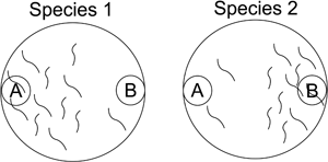
Chemotactic assays were performed similarly to those described in Castelletto et al. (Reference Castelletto, Gang, Okubo, Tselikova, Nolan, Platzer, Lok and Hallem2014) with a few changes. Firstly, 6 cm HEPES Agar (as above) plates were used, following a pilot study to determine the best agar for the experiments. Plates were marked with 1.5 cm circles which were extended to the boundary of the plate in a rectangle (as seen in S1 Fig.). The centre of these scoring zones was then spotted with 5 μL 10 mg mL−1 sodium azide to act as an anaesthetic, followed by 10 μL of either the chemical being tested or a control. Worms (a minimum of 100) were then spotted in the centre of the plate and incubated for 3 h at room temperature after which the number of worms in both scoring zones was counted. A selection of odorants used within (Castelletto et al., Reference Castelletto, Gang, Okubo, Tselikova, Nolan, Platzer, Lok and Hallem2014) were used to allow direct comparison of results, in addition to urea. Odorants were chosen from this paper to comprise a variety of chemical classes and contain a mixture of strong attraction/repulsion or neutral profiles. All chemicals used were undiluted with the exceptions of urea (1 M in water), methyl palmitate (0.05 g in 2.5 mL paraffin oil) and palmitic acid (0.5 g in 10 mL ethanol). Water was used as a control, with the exceptions of ammonia (ethanol), methyl palmitate (paraffin oil) and palmitic acid (ethanol). When odorants were spotted on the lid, small squares (8 mm × 8 mm) of filter paper were stuck to the lid above the scoring zones using double-sided tape, onto which the odorants were spotted. The Chemotactic Index (CI) was calculated as in Castelletto et al. (Reference Castelletto, Gang, Okubo, Tselikova, Nolan, Platzer, Lok and Hallem2014) as follows:

As a result, a positive CI score indicates attraction while a negative CI score indicates repulsion. No minimum value was used for each scoring zone, however at least 10 worms in total had to move into the scoring zone(s) for the plate to be considered valid.
Fur assay
For fur assays, rat fur and mice fur from our in-house animal facilities were collected. Assays were performed as above with the chemotactic assay with either rat fur and mice fur, or rat fur and no fur placed in opposite scoring zones. Following this, S. ratti infective larvae were spotted in the centre and incubated for 1 h at room temperature. The amount of fur (if necessary) used was sufficient to cover the entire scoring zone. The number in both scoring zones and the CI calculated as above.
Fecal assay
HEPES agar plates were again marked as in the chemotaxis assay. Rat and rabbit feces were collected using the same procedure described earlier for the maintenance of S. ratti and S. papillosus, except uninfected rats and rabbits were used. Rat feces was either used freshly (i.e. on the day it was collected) or was stored for a maximum of 2 days at 19 °C prior to being used. Rabbit feces was autoclaved and stored long term at 4 °C. Both feces were warmed to RT for a minimum of 1 h prior to use. A piece of rat or rabbit feces (depending on species tested) was placed in one scoring zone and the other scoring zone left empty. Either adults or infective larvae were spotted in the middle of the plates and then incubated for 1 h at room temperature, after which the worms in both scoring zones were counted. To search for worms in the fecal scoring zone, small amounts of OP50 bacteria in solution were added and the feces broken up. Fecal attraction was determined using the same calculation as for CI.
Statistical analysis and figure generation
Appropriate statistical analysis was performed using Excel or R, with figures generated using Excel and Inkscape. The exact statistical test used is stated in the figure legends where necessary. Significance was determined by P value of <0.05.
Results
Following the example of Castelletto et al. (Reference Castelletto, Gang, Okubo, Tselikova, Nolan, Platzer, Lok and Hallem2014), we first compared the locomotion activity of infective larvae/dauer larvae of the different species on agar plates in the absence of any experimental olfactory stimuli (see Materials and methods ‘Dispersal assay’). We observed substantial differences in the dispersal at room temperature among the four species of Strongyloidoidea tested (Fig. 2). While P. trichosuri [mean Dispersal Index (DI) = 0.47] moved slightly more than S. ratti, S. papillosus (mean DI = 0.13) and R. diutinus (mean DI = 0.17) moved only minimally. Our results for S. ratti (mean DI = 0.40), the only species included in this study and in Castelletto et al. (Reference Castelletto, Gang, Okubo, Tselikova, Nolan, Platzer, Lok and Hallem2014), are very similar to the S. ratti results of Castelletto et al. (Reference Castelletto, Gang, Okubo, Tselikova, Nolan, Platzer, Lok and Hallem2014), confirming that our results are comparable to the results of this publication.

Fig. 2. Dispersal of Strongyloidoidea at room temperature – Strongyloidoidea have different dispersal profiles at room temperature (22 °C). Boxplots of dispersal on plates of S. papillosus, S. ratti, P. trichosuri and R. diutinus. For each species, plates were tested in batches of 10, with three biological replicates. Box and whisker plot shown here is produced from all 30 data points (per species), with the maximum and minimum values shown by the whiskers, and the box representing the interquartile range with the median indicated. Kruskal–Wallis H was performed to determine statistical significance between the samples. P values *<0.05, **<0.01, ***<0.001.
To assay for the effect of temperature on locomotion, and to exclude that the results presented above just reflected different temperature preferences, we examined motility at a range of temperatures from 4 to 37 °C (Fig. 3). For comparison, we also included dauer larvae of the two well-studied, phylogenetically distant, free-living nematodes C. elegans and P. pacificus in the analysis. Consistent with the results in Fig. 2, S. ratti and P. trichosuri showed higher locomotory activity than S. papillosus and R. diutinus over the intermediate temperature range (10–22 °C). Interestingly, iL3s of the two Strongyloides species responded differently to the more extreme temperatures than did the other species tested. All three types of dauer larvae showed peak dispersal at intermediate temperatures and reduced activity at lower temperatures and at high temperature (37 °C). In contrast, S. ratti and S. papillosus showed a significant (P values <0.001) increase in dispersal at 4 °C and at 37 °C

Fig. 3. Strongyloidoidea exhibit differing responses to thermal stress –temperature-dependent motility response of S. papillosus, S. ratti, P. trichosuri and R. diutinus infective/dauer larvae at a range of different temperatures between 4 and 37 °C. As a comparison, the temperature-dependent motility response of P. pacificus and C. elegans are included. For each temperature and species, 10 plates were tested, with all temperatures being tested at the same time. This experiment was repeated to generate three biological replicates. Data shown here in the box and whisker plots comprise the interquartile range with the median indicated, and the maximum and minimum shown from all 30 data points. To determine statistical significance, Mann–Whitney U (two-way) was performed. P values *<0.05, **<0.01, ***<0.001. Significance between neighbouring temperatures is shown only when significant.
To profile the olfactory response of the Strongyloidoidea, infective/dauer larvae were subjected to a variety of odorants known to be emitted by skin, sweat and microbiota on the skin (Fig. 4A). We tested a subset of the odorants used in Castelletto et al. (Reference Castelletto, Gang, Okubo, Tselikova, Nolan, Platzer, Lok and Hallem2014) plus urea. As seen by other authors (Dillman et al., Reference Dillman, Guillermin, Lee, Kim, Sternberg and Hallem2012; Castelletto et al., Reference Castelletto, Gang, Okubo, Tselikova, Nolan, Platzer, Lok and Hallem2014), we found that all species had a unique odorant response profile. Even the two species of the same genus, S. ratti and S. papillosus showed clearly different olfactory response profiles (Fig. 4B), similar to what had been observed in previous research for S. ratti and S. stercoralis (Castelletto et al., Reference Castelletto, Gang, Okubo, Tselikova, Nolan, Platzer, Lok and Hallem2014). Rhabditophanes diutinus had a significant olfactory response (attraction or repulsion) to more and was indifferent to fewer odorants, compared to all of the parasitic species [10 of 13 odorant cues triggered a statistically significant response (Fig. 4C) as opposed to 3–5 of 13 in the parasitic species (Fig. 4B, D)]. Of those odorants tested, only one odorant (acetic acid) triggered a statistically significant response in all four species, acting as an attractant in S. ratti (CI = 0.31) and P. trichosuri (CI = 0.19) and as a repellent to S. papillosus (CI = −0.64) and R. diutinus (CI = −1.00). To confirm that this olfactory preference was olfactory and not hygroscopic or gustatory, some odorants were also tested by using spotted filter paper on the plate lid, such that they could not soak into and spread through the agar. We found the response was near identical for all odorants tested (S2 Fig), confirming that the responses we saw are olfactory and not hygroscopic or gustatory, in agreement with what was found by Castelletto et al. (Reference Castelletto, Gang, Okubo, Tselikova, Nolan, Platzer, Lok and Hallem2014). To test whether Strongyloides spp. would actively seek out fur of its host species, we examined S. ratti's response to rat fur (its natural host) and mouse fur (mice are barely susceptible hosts for S. ratti), such that infections are very short lived and of very low productivity if they are established at all (Gemmill et al., Reference Gemmill, Viney and Read2000). We found that S. ratti has no significant preference for host fur over non-host fur (CI = 0.36 ± 0.40, P value 0.329, S3 Fig). As we noticed the infective larvae were barely crawling towards the fur samples, we then determined whether they were attracted to fur at all. We found that S. ratti is in fact repulsed from fur (CI = −0.63 ± 0.17, P value <0.0001, S3 Fig).

Fig. 4. Olfactory response profiles of the Strongyloidoidea – olfactory response of all four species in response to various volatile odorants is shown as a heatmap (A), with white indicating a strong repulsion and black strong attraction. Olfactory response of R. diutinus (B), P. trichosuri (C), S. ratti (D) and S. papillosus (E) to odorant cues. *P value <0.05, **P value <0.01, ***P value <0.001, if not significant than no star is shown. Two-way ANOVAs between odorant and control for all odorants and species tested. Each odorant was tested in batches of five, with at least three biological replicates. Boxplots show interquartile range of response, with mean response indicated by the middle bar.
Finally, as it is known that Strongyloides infective larvae actively migrate out of feces in search of a new host (Viney and Lok, Reference Viney and Lok2015) while free-living adults tend to stay in the feces. To determine if this is reflected in the chemotactic behaviour of these different stages, we performed chemotaxis assays with adults and infective larvae of S. ratti and S. papillosus using feces of the host they had been raised in (rat for S. ratti, rabbit for S. papillosus) as attractants. As Castelletto et al. (Reference Castelletto, Gang, Okubo, Tselikova, Nolan, Platzer, Lok and Hallem2014) had found for S. stercoralis and S. ratti, we found that adults were attracted towards feces (S. papillosus CI = 0.61, S. ratti CI = 0.59) (Fig. 5) while infective larvae were indifferent (S. papillosus CI = −0.04, S. ratti CI = −0.13).

Fig. 5. Response of Strongyloides larvae to feces – only Strongyloides adults (black) are attracted towards their host feces, infective larvae (white) are indifferent to it. Two-way ANOVA with Bonferroni Post Test performed to determine significance between life stage and species. P value ***<0.001, **<0.01. No significant difference found between the two species. Bars indicate mean response towards feces with errors bars indicating the standard deviation. Each response variable (i.e. host feces for S. ratti) was tested in batches of five, with two biological replicates.
Discussion
Similarly to other authors (Castelletto et al., Reference Castelletto, Gang, Okubo, Tselikova, Nolan, Platzer, Lok and Hallem2014), we identified differences in chemotactic behaviours among closely-related species. These differences within a single genus appear to confirm what other authors previously proposed, in that host-seeking behaviour may have evolved independently even in closely related species in order to accommodate host behaviour and ecology (Dillman et al., Reference Dillman, Guillermin, Lee, Kim, Sternberg and Hallem2012; Castelletto et al., Reference Castelletto, Gang, Okubo, Tselikova, Nolan, Platzer, Lok and Hallem2014).
When examining the effect of temperature on motility, we observed small differences in temperature-dependent motility profiles between the two Strongyloides species, with S. ratti showing higher motility at all examined temperatures. Interestingly both species showed increased motility at 4 °C compared with more moderate temperatures (10–15 °C). This could be due to 4 °C being close to the lower limit of the tolerable temperatures for these two species, leading to them to try to escape and find a new host. Indeed, for S. stercoralis, several authors have noticed that refrigeration is detrimental for the development and the survival of infective larvae (Sultana et al., Reference Sultana, Jeoffreys, Watts, Gilbert and Lee2013; Zhou et al., Reference Zhou, Harbecke and Streit2019). In contrast to the two Strongyloides species, P. trichosuri showed neither a clear increase nor a decrease towards the ends of the temperature range tested. This may mean that the infective larvae of this facultative parasite are less temperature responsive than the two Strongyloides spp. tested. However, it is also possible that for P. trichosuri, the range of tolerable temperatures is wider than for the others and the temperatures tested are all within the comfort zone of this species. Further, we cannot exclude that the low responsiveness to temperature of the P. trichosuri iL3s has arisen in the laboratory. Our culture of P. trichosuri has been maintained exclusively through the free-living cycle for years such that features of developmental stages specific for the parasitic life cycle might have been lost. Further investigation into the temperature response of P. trichosuri is required. The minimal (by comparison) locomotory activity that we saw in R. diutinus is unlikely to be due to its dauer lifestyle (both C. elegans and P. pacificus moved more during the experiment), but instead may be a species trait.
Similar to other authors (Castelletto et al., Reference Castelletto, Gang, Okubo, Tselikova, Nolan, Platzer, Lok and Hallem2014), the species-specific olfactory response we see is consistent with the proposal that olfactory preference is more closely associated with host species than with taxonomic position, with different chemotactic profiles among members of even the same nematode genus. Strongyloides ratti actually appeared to differ more from S. papillosus than from the more distantly related P. trichosuri since all four odorants leading to a significant response in S. ratti also triggered a significant response in the same direction in P. trichosuri. Of the odorants used in this assay, only a single one, acetic acid, produced a significant response for all species tested and this response did not go in the same direction in all four species. Acetic acid is known to be emitted from the skin (Bernier et al., Reference Bernier, Kline, Barnard, Schreck and Yost2000; Gallagher et al., Reference Gallagher, Wysocki, Leyden, Spielman, Sun and Preti2008) and feet (Ara et al., Reference Ara, Hama, Akiba, Koike, Okisaka, Hagura, Kamiya and Tomita2006) which may explain why S. ratti and P. trichosuri are attracted to it as it is their normal entry site for infecting a new host (Grant et al., Reference Grant, Stasiuk, Newton-Howes, Ralston, Bisset, Heath and Shoemaker2006; Viney and Lok, Reference Viney and Lok2007). It is unclear however why it would have an opposite response in S. papillosus. Interestingly R. diutinus, the only non-parasitic species included in our study, responded to more odorants than any of the parasitic species. If this profile is typical for being a dauer rather than an infective larva or is a R. diutinus-specific feature remains to be determined. The lack of response to fur in S. ratti while interesting, is not surprising. It suggests that fur is not a dominant carrier of attractive cues and that a combination of multiple cues (i.e. temperature, odorants) is required to trigger host-seeking behaviour within S. ratti. The lack of attraction towards rat fur in S. ratti could also be partially explained by an inability to penetrate the skin at areas with heavy fur and therefore an adaptive response to increase the efficiency of host-seeking behaviours. These olfactory results, combined with the thermosensory response, indicate that fur alone is not sufficient to generate a response, and that multiple cues from the skin and body temperature are necessary to engage host-seeking behaviour.
It should also be kept in mind that a particular reaction is not necessarily triggered by a single cue but by the combination of all stimuli present. In their natural environments, which are rather different from the laboratory conditions, the species tested might respond differently to the same cues, because they are present in different combinations. Nevertheless, taken together, our results support the notion that the sensing of and responding patterns to cues that are important for host-seeking are predominantly the result of independent evolution within the different species in response to the different lifestyles and hosts and only to a minor extent they are the result of the phylogenetic history of the species in question.
Supplementary material
The supplementary material for this article can be found at https://doi.org/10.1017/S003118202100161X
Data
Data presented within this manuscript are available from the authors upon request.
Acknowledgements
We thank W. Grant, D. Denver, M. Viney and Ralf Sommer for providing nematode cultures. N2 was provided by the CGC, which is funded by the NIH Office of Research Infrastructure Programs (P40 OD010440).
Author contribution
A.D. and A.S. conceived the study. A.D. and M.N. designed the study. M.N., A.D. and D.H. performed experiments. A.D. and M.N. analysed the data. A.D. generated the figures. A.D., M.N. and A.S. wrote the manuscript. All authors approved the manuscript before submission.
Financial support
This work was funded by the Max Planck Society through departmental funding to A.S. M.N. received funding from the DAAD RISE Germany Fellowship Program. The funders had no role in study design, data collection, analysis or interpretation, writing the manuscript or in the decision to/where to submit the article for publication.
Conflict of interest
None.
Ethical standards
Animal care and use adhered to the German Animal Protection Law (Tierschutzgesetz), the German Animal Protection Laboratory Animal Ordinance (Tierschutz-Versuchstierverordnung) and EU Directive 2010/63/EU on the protection of animals used for scientific purposes. The procedures for animal maintenance and experiments were administratively and ethically approved by the local governmental authorities in charge (Regierungspräsidium Tübingen), who also issued the necessary permits (Anzeige vom 15. Juli 2015, AZ35/9185.82-5). The animals were kept in an in-house animal facility, which is regularly inspected by the local veterinary authorities (Veterinäramt Tübingen). This study contains no experiments on human subjects. Care was taken to ensure only the minimum number of animals were used in order to achieve statistically significant results.


