


Case report
Girl P was born at 27.6 weeks’ gestation via spontaneous vaginal delivery to a 31-year-old G1P1 Caucasian mother. The pregnancy was complicated by oligohydramnios, preterm labour, and premature rupture of membranes at 27.5 weeks. She did not receive antenatal steroids and the delivery was complicated by chorioamnionitis. The infant was transferred to a tertiary neonatal intensive care unit for stabilisation and further management. On day 4 of life, she was noted to have free air under her diaphragm on an x-ray, which was confirmed by a repeat left lateral decubitus film. The free air was thought to be secondary to a spontaneous intestinal perforation. She was then started on broad-spectrum antibiotic coverage and underwent placement of a Primrose drain to evacuate the intraperitoneal free air. She became hypotensive with mean arterial pressure in the low 30 mmHg range and was therefore given a total of two 20 ml boluses of normal saline and one unit of fresh frozen plasma. After the fluid resuscitation, her vital signs normalised. She was not provided inotropic support medications for the hypotension at that time.
On day 9 of life, she became tachycardic and tachypneic. Her mean arterial pressure was in the range of 30–40 mmHg, her heart rate varied from 170 to 200 beats per minute, and respiratory rate varied from 60 to 90 breaths per minute. After additional fluid resuscitation, an electrocardiogram was obtained, which showed sinus rhythm with premature atrial contractions, normal ST segments, and possible biventricular hypertrophy. Serum lactate was 4.0 mmol/L. However, no cardiac enzymes were obtained. An echocardiogram demonstrated a globular shape and dyskinesia of the left ventricular apex with a compensatory hyperkinesia of the left ventricular base creating a very distinct hinge point (Fig 1a). In response to this finding, the infant was started on milrinone and dobutamine, which improved the mean arterial pressure. Over the next 3 days, she was weaned off inotropic support. A repeat echocardiogram was obtained on day 13 of life, which showed complete recovery of the apical dyskinesis with normal global left ventricular contractility and no identifiable hinge point (Fig 1b).

Figure 1 Echocardiograms. ( a ) Diagnostic ECHO – Abnormal. ( b ) Recovery ECHO – Normal.
The infant continued to feed, grow, and thrive during the rest of her newborn hospitalisation period. 3 days before discharge, on day 72 of life, a repeat echocardiogram was performed. This demonstrated normal left ventricular chamber dimensions, wall thicknesses, and systolic function. The shape of the interventricular septum had completely normalised. She was discharged on day 75 of life at a corrected gestational age of 38.3 weeks, on room air, and on no medications.
A diagnosis of takotsubo cardiomyopathy was proposed, retrospectively, as the most likely diagnosis based on the clinical history and the echocardiographic findings. There is probably not enough laboratory evidence to confirm a diagnosis of takotsubo cardiomyopathy; however, the presence of cardiogenic shock, which was temporally related to a devastating systemic illness, the transience of the echocardiographic features, and the speedy resolution suggest a diagnosis of takotsubo cardiomyopathy.
Discussion
Takotsubo cardiomyopathy is a transient cardiomyopathy with characteristic findings on the echocardiogram, in addition to specific clinical exclusion criteria. In addition to the echocardiographic findings, there is minimal release of cardiac enzymes and no sign of obstructive coronary disease in spite of electrocardiographic findings suggestive of an acute myocardial infarction.Reference Pernicova, Garg and Bourantas 1 When takotsubo cardiomyopathy syndrome was first described in 1990, it was noted to be more common in postmenopausal women after episodes of intense emotional or physical stress. Only within the last few years has it been recognised, albeit infrequently, in children and even less frequently in infants.
The reported prevalence of takotsubo cardiomyopathy is unknown in children. In adults, it is reported as being accountable for 2% of patients with suspected acute coronary syndrome.Reference Hernandez 2 In adults, the initial presentation may mimic acute coronary syndrome, and therefore the cardiomyopathy may be overlooked. In children, the constellation of findings is often misdiagnosed as myocarditis, dilated cardiomyopathy, or unknown acute ventricular dysfunction. Regardless, takotsubo cardiomyopathy has a very good prognosis.
Clinical features
Diagnostic criteria have been put forth by the Mayo Clinic, although not universally accepted; (Table 1).Reference Madhaven and Prasad 8 When an infant/child presents with heart failure, a typical workup consists of an electrocardiogram, an echocardiogram, and evaluation of serum biomarkers. The electrocardiogram commonly demonstrates diffuse ST segment and T wave abnormalities.Reference Hernandez 2 QTc prolongation has also been reported.Reference Hernandez 2 , Reference Yamaguchi, Nagumo and Nakashima 3 The classic echocardiographic features include “transient hypokinesis, akinesis or dyskinesis of the left ventricle mid segments with or without apical involvement; the regional wall motion abnormalities extend beyond a single epicardial vascular distribution”.Reference Pernicova, Garg and Bourantas 1 , Reference Madhaven and Prasad 8 In adults, it has been shown that left ventricular ejection fraction may decrease as much as 20–49% with complete resolution of the abnormal wall movements over a mean of 18 days.Reference Pernicova, Garg and Bourantas 1 In the acute phase of the syndrome, it is typical to see elevated cardiac enzymes such as troponin and creatinine kinase.Reference Pernicova, Garg and Bourantas 1 , Reference Hernandez 2 There are some reports of elevated Pro-brain natriuretic peptide.Reference Pernicova, Garg and Bourantas 1 However, the elevation in the enzymes is not nearly as high as seen in myocarditis. These elevations in enzymes are not seen in every case.
Table 1 Mayo Clinic diagnostic criteria for Takotsubo cardiomyopathy.Reference Madhaven and Prasad 8
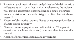
* There are rare exceptions to these criteria, such as those patients in whom the regional wall motion abnormality is limited to a single coronary territory
** It is possible that a patient with obstructive coronary atherosclerosis may also develop takotsubo cardiomyopathy. However, this is very rare in our experience as well as in the published literature, perhaps because such cases are misdiagnosed as an acute coronary syndrome
The pathophysiology of takotsubo cardiomyopathy remains unclear. One theory is that there is a sustained surge of catecholamines with a superimposed exaggerated response by the sympathetic nervous system.Reference Hernandez 2 This is supported by histological studies that have shown that the myocardium looks similar to that seen in catecholaminergic heart toxicity in animals and human subjects.Reference Mohaveh, Reeves and Metha 5 , Reference Frustaci, Loperfido and Gebtiloni 6 In a published case report of an 11-year-old boy with attention-deficit/hyperactivity disorder treated with atomoxetine, he appeared to develop takotsubo cardiomyopathy. Atomoxetine is a norepinephrine reuptake inhibitor and therefore has central and peripheral catecholaminergic effects. This might lend credence to the theory of an overwhelming catecholamine release as a cause of takotsubo cardiomyopathy.Reference Yamaguchi, Nagumo and Nakashima 3 There are also reports of patients with pheochromocytoma qhaving similar left ventricular dysfunction, which again points to the plausibility of excess catecholamine as the cause.Reference Sanchez-Recalde, Costero and Oliver 7
The treatment of takotsubo cardiomyopathy is supportive and should be based on the clinical presentation of each patient.Reference Hernandez 2 , Reference Berton, Vitali-Serdoz and Vallon 4 The management for adult patients typically includes beta blockers and angiotensin-converting enzyme inhibitors. The management of children with takotsubo cardiomyopathy is different from that of adults. In children, congestive heart failure due to low cardiac output is a very common presentation and typically requires inotropic support. Milrinone is commonly used in the paediatric intensive care setting.Reference Hernandez 2 Pulmonary oedema may also be present and furosemide is the drug of choice to optimise diuresis. Mechanical ventilation may also be necessary, depending on the clinical presentation.Reference Hernandez 2
There have been reports of intraventricular thrombosis as a complication of takotsubo cardiomyopathy but this does not appear to be a common complication.Reference Hernandez 2 An association between the development of a thrombus and the presence of elevated C-reactive protein was reported in two case reports. Anticoagulation remains the treatment of choice until resolution of the thrombus.Reference Hernandez 2
Essential to the diagnosis of takotsubo cardiomyopathy is complete recovery of the left ventricular systolic function. The recovery times vary from days to months, with the longest reported recovery period being 4 months.Reference Hernandez 2 In adults, there are reports of recurrence but this has not been reported in children. Our patient has maintained her normal systolic function and remains a healthy growing infant.
Conclusion
In summary, we report a case of a very low birth weight premature infant who developed takotsubo cardiomyopathy after a spontaneous intestinal perforation, with complete recovery of her ventricular systolic function after 4 days. Takotsubo cardiomyopathy is an uncommon cardiomyopathy that has been described more commonly in adults but is starting to be recognised in children.
Acknowledgements
We would like to thank Dr. Kevin Hill for his opinion and his critical review of this brief report.
Financial Support
This research received no specific grant from any funding agency, commercial, or not-for-profit sectors.
Conflicts of Interest
None.


