1. INTRODUCTION
Femtosecond lasers provide unique possibilities for high-precision material processing: thanks to its very fast interaction process, the heat affected zones in the irradiated targets are strongly localized, with minimal residual damage (Gottmann et al., Reference Gottmann, Wortmann and Wagner2008; Boulmer-Leborgne et al., Reference Boulmer-Leborgne, Benzerga, Perriere, Miotello and Ossi2010). In this framework, the generation of laser induced periodic surface structures (LIPSS) from femtosecond pulse direct irradiation has been intensively studied on various materials, including metals, insulators, and semiconductors (Tomita et al., Reference Tomita, Fukumori, Kinoshita, Matsuo and Hashimoto2008; Reif et al., Reference Reif, Varlamova and Costache2008; Okamuro et al., Reference Okamuro, Hashida, Miyasaka, Ikuta, Tokita and Sakabe2010). Among the latter, silicon carbide (SiC) has proved to be a very promising material with high performances in different application fields, as for instance MEMS fabrication and solar cell development (Yu et al., Reference Yu, Leem, Ko and Lee2012; Winkler et al., Reference Winkler, Sher, Lin, Smith, Zhang, Gradečak and Mazur2012; Pecholt et al., Reference Pecholt, Gupta and Molian2011; Reference Pecholt, Vendan, Dong and Molian2008). Although different models have been proposed to explain the generation of periodic self-forming structures in the femtosecond regime of irradiation, the complete process of their generation is still debated (Reif et al., Reference Reif, Varlamova, Varlamov and Bestehorn2011; Bonse et al., Reference Bonse, Rosenfeld and Kruger2011; Derrien et al., Reference Derrien, Torres, Sarnet, Sentis and Itina2012; Sakabe et al., Reference Sakabe, Hashida, Tokita, Namba and Okamuro2009). Recently, using a complex experimental setup, femtosecond near-infrared (NIR) laser pulses were applied to amorphous Carbon (a-C) films together with an extreme ultraviolet (XUV) beam. Under such conditions, although the fluence was kept below the ablation threshold of a-C in all irradiation geometries, a significant increase of both the spatial periodicity and the peak-to-valley depth of LIPSS were observed over that from NIR irradiation alone. LIPSS enhancement was attributed to the combined XUV-NIR action (Mocek et al., Reference Mocek, Jakubczak, Chalupsky, Park, Lee, Kim, Nam, Hajkova, Toufarova, Gemini, Margarone, Juha and Rus2012).
In this paper, we are not reporting on LIPSS formation as usually meant. Instead, we demonstrate that extended periodic structures can be generated on a SiC surface by irradiation from plasma produced by 40 fs laser pulses.
2. EXPERIMENTS
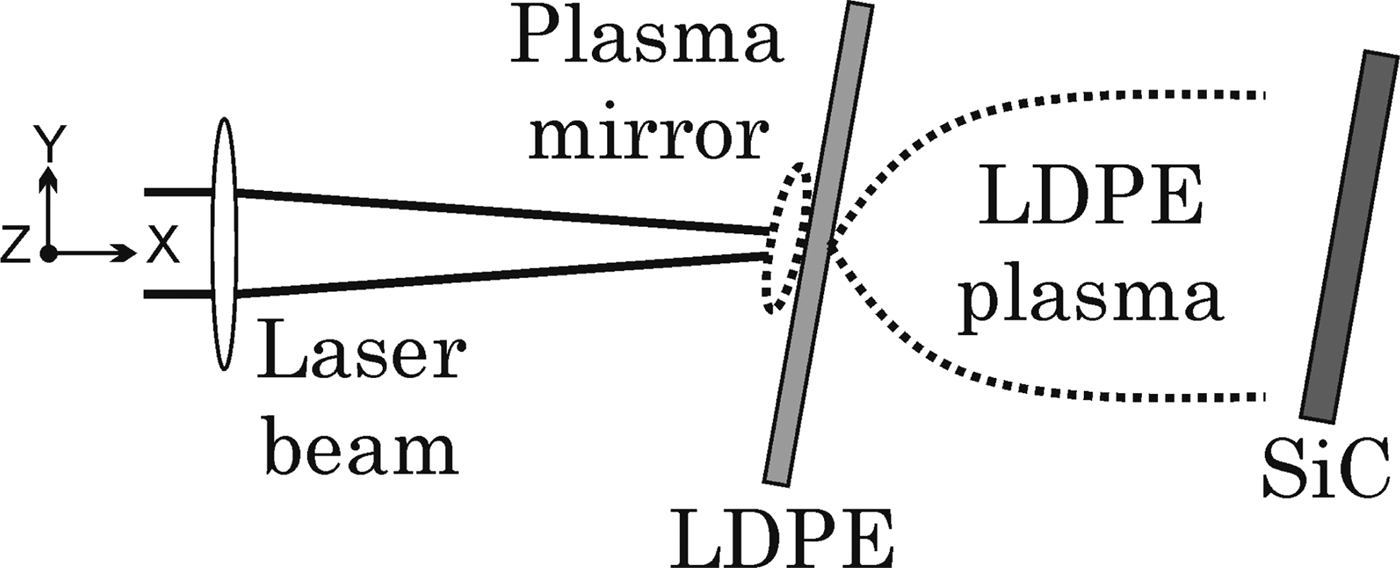
A SiC film about 130 nm thick was deposited by a pulsed laser onto a Si (100) substrate kept at 1173 K using a KrF excimer laser (wavelength 248 nm, pulse duration 25 ns, fluence 6 J cm−2). A homogeneous, compact film, free from surface particulate was obtained. A Ti:Sapphire laser (wavelength 800 nm, pulse width 40 fs, electric field polarization inclined at 6° with respect to the normal incidence direction) was focused on a 15 µm thick low density polyethylene (LDPE) film, directly facing the SiC film at a distance of about 1 cm (Fig. 1). Each laser pulse, with an intensity on the order of 1018 Wcm−2, impinged on the LDPE inclined at 10° with respect to the target normal. Due to the presence of a laser pulse nanosecond pedestal with intensity on the order of 1012 Wcm−2, the laser-irradiated LDPE was partially converted into a plasma carrying fast C and H ions and generating incoherent XUV photons, which expanded and irradiated the SiC film. The LDPE foil was mounted on a motorized YZ so that each laser pulse produced an LDPE plasma pulse. This way every single laser pulse generated an identical LDPE plasma pulse irradiating its associated SiC sample area.

Fig. 1. Schematic of the experimental setup (top view) and of the main physical processes which occur during the interaction between the laser beam and the LDPE foil (generation of plasma mirror on the front side and LDPE plasma expansion on the rear side of the foil).
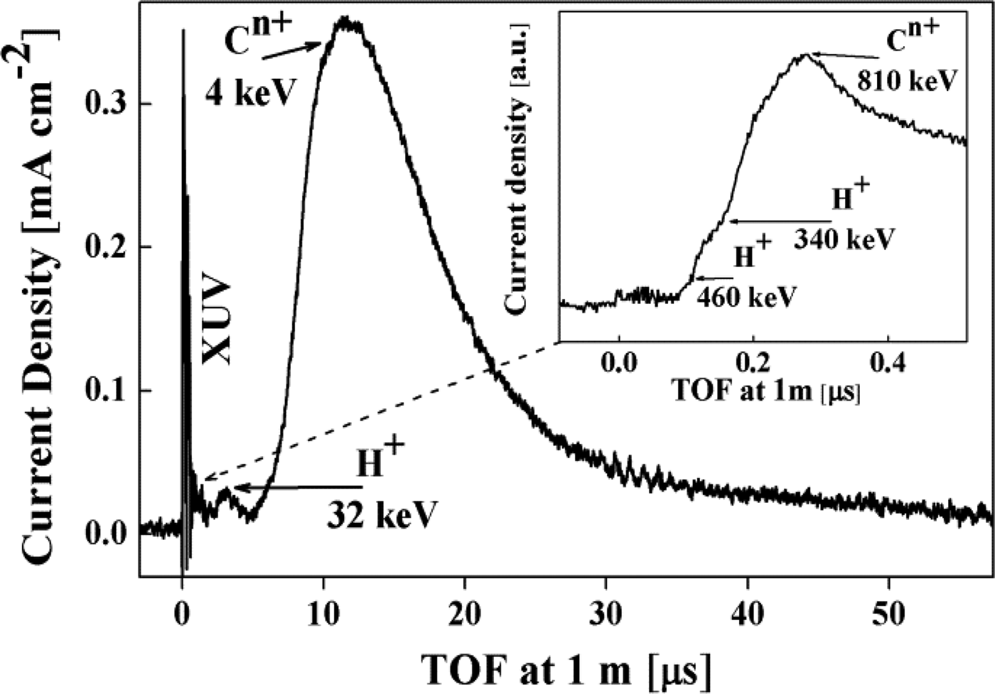
A 15 µm thick Mylar foil was laser irradiated under the same experimental conditions described for the LDPE irradiation. Ion time-of-flight (TOF) spectra (Fig. 2) referred to the plasma generated during Mylar irradiation on the rear side of the target were collected by a Faraday cup and by a fast response SiC detector to evaluate the low energy and the high energy ion components, respectively (Margarone et al., Reference Margarone, Krása, Giuffrida, Picciotto, Torrisi, Nowak, Musemeci, Velyhan, Prokůpek, Láska, Mocek, Ullschmied and Rus2011). For our purposes, the features of this spectrum can be considered analogous to those obtained from the LDPE-generated plasma. The TOF signals show the presence of an XUV photo-peak followed by H+ and Cn+ signals (after several microseconds) peaked at the energies of 32 and 4 keV, respectively. A proton maximum energy approaching 500 keV, and H+ and Cn+ peak energies of 340 and 810 keV were measured by a fast response SiC detector for proton and carbon ions, respectively. The slow and the fast ion components can be associated to the nanosecond laser generated plasma (due to the laser pedestal) and to the femtosecond laser interaction, respectively.

Fig. 2. TOF spectra referring to plasma generated during irradiation of a 15 µm Mylar foil, collected by a Faraday cup (PIC) and a SiC detector (inset). It is noteworthy that the high energy component intensity collected by the SiC detector (inset) is negligible, being far below that of the low energy component.
It is important to observe that the XUV photo-peak impinges on the sample surface much earlier than the low energy ion component and right before the high energy ion component, whose estimated number density is anyway negligible (about 1% of the total ion contribution). Therefore the SiC surface is first irradiated by the XUV component together with the 40-fs NIR laser beam, the intensity of the latter being reduced to about 8·1011 Wcm−2 due to the unfocusing condition, the LDPE absorption, the plasma mirror effect induced on the front side of the LDPE target, and the absorption by the expanding LDPE plasma on the rear side of the LDPE target (the estimated transmitted NIR beam that irradiates the sample surface after all the above mentioned processes is indeed less than 1%).
3. RESULTS AND DISCUSSION
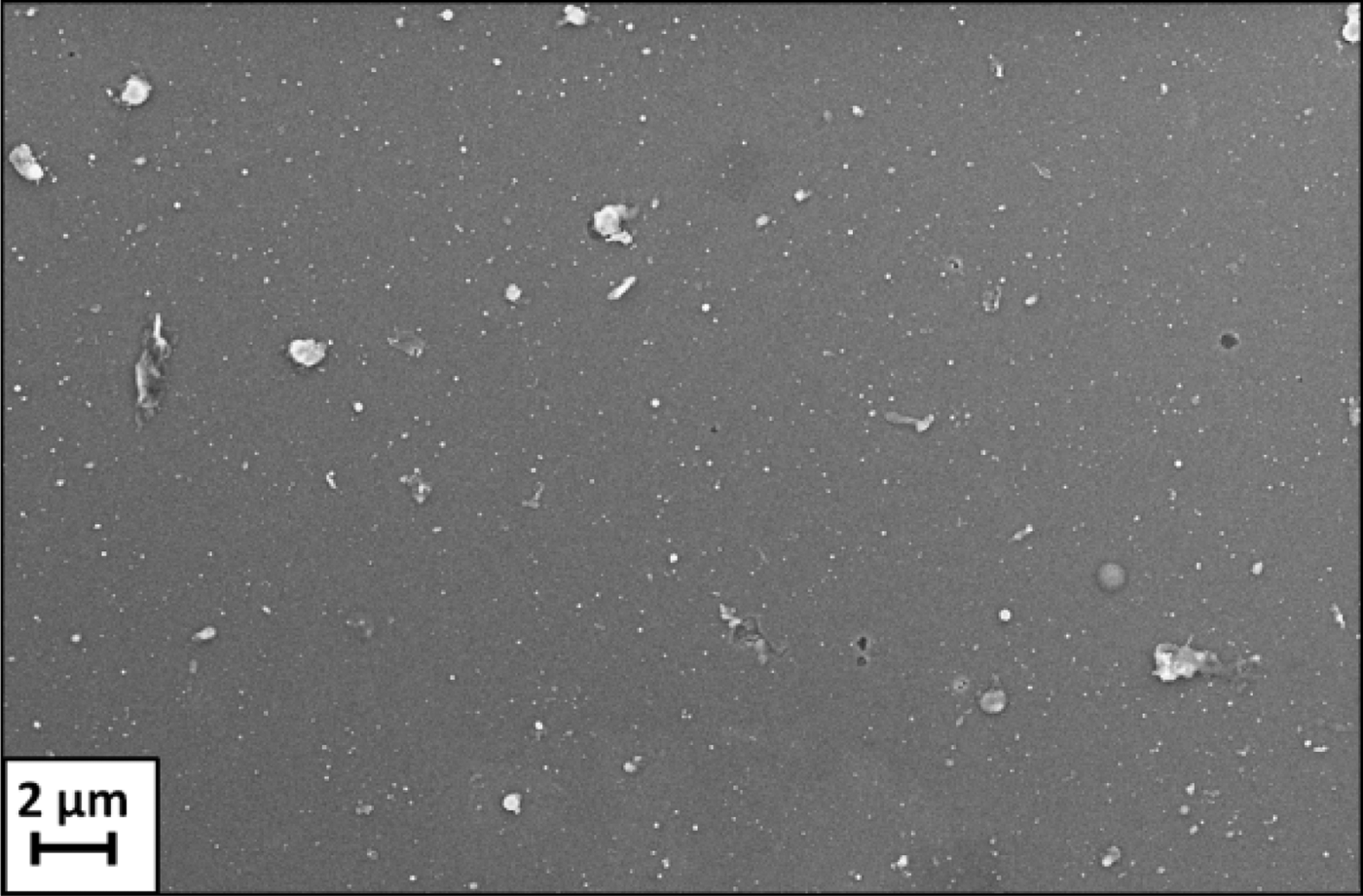
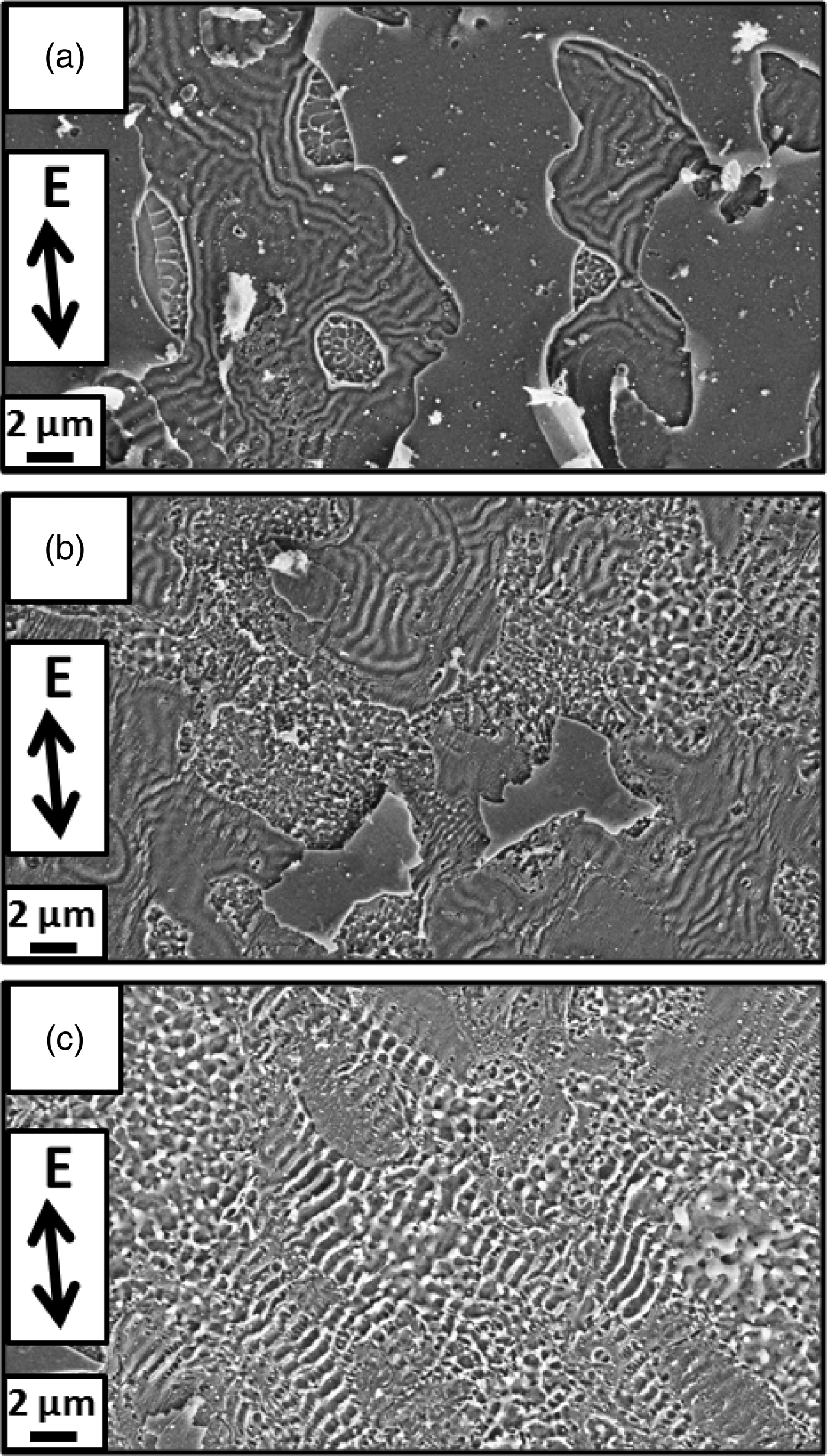
Different areas of the SiC sample were irradiated by a different number of LDPE plasma pulses, from 1 to 5, to observe the effects of the progressively cumulating plasma pulses on the SiC. Every irradiation produced a circularly shaped modification on the sample surface with a diameter of about 4 mm. For comparison, a reference SiC area was irradiated by five pulses of the unfocused femtosecond beam in the absence of LDPE. All the irradiated areas were analyzed by a scanning electron microscope (SEM Zeiss Supra 40 field ion instrument). Since the estimated NIR fluence at the sample surface (about 32 mJcm−2) was well below the ripple generation threshold of SiC in the femtosecond regime, the surface irradiated by unfocused femtosecond pulses in the absence of LDPE plasma appears unchanged with respect to the as-deposited material, as shown in Figure 3 (Dong & Molian, Reference Dong and Molian2003). Independently of the number of laser pulses, a wavy surface morphology is visible in all the areas irradiated by LDPE plasma pulses as shown in Figure 4: such ripples are most evident in the area irradiated by a single plasma pulse, and they lose definition with increasing pulse number. Energy dispersive X-ray spectroscopy (EDX, by SEM) indicates that all the plasma irradiated areas consist of SiC. It is noteworthy that the wavy structure extends across the entire areas irradiated by the laser induced plasma, without being limited to the center of a laser spot: indeed no laser spots are produced on the SiC surface, due to the indirect irradiation conditions.

Fig. 3. SiC surface irradiated by unfocused NIR light alone. The surface appears unmodified compared to the as deposited material.

Fig. 4. SEM images of the areas irradiated by LDPE plasma generated by single pulse (a), three pulses (b) and five pulses (c). The arrows indicate the direction of the laser electric field E polarization.
All modified areas were analyzed by an atomic force microscope (AFM Bruker Dimension Icon with ScanAsyst and AFM Ntegra Prima, both working in tapping mode in air). The AFM measurements were affected by the presence of dust and impurities on the sample surface. We preferred not to clean the sample in order to avoid possible surface contamination and alteration.
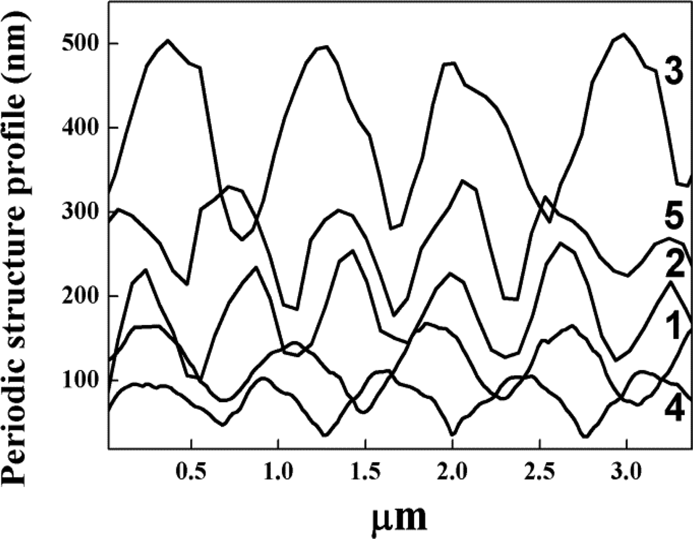
Representative profiles of the analyzed areas are shown in Figure 5. Independently of the number of laser pulses, the AFM profiles show that the wavy structure is characterized by a periodicity ranging from 638 nm to 826 nm with an average value of 710 nm, slightly smaller than the laser wavelength, and by a peak-to-valley depth ranging from 70 nm to 219 nm, leading to an average value of 123 nm. Every area, as related to a definite number of plasma pulses, is characterized by a specific, constant value of peak-to-valley depth and by a constant spatial periodicity.

Fig. 5. AFM periodic structure profiles of areas irradiated by LDPE plasma generated by 1 to 5 laser pulses. The numbers on the right side indicate the laser pulse number.
On the basis of these results, the formation of plasma induced ripples is likely to be associated to the coupling of the unfocused femtosecond laser beam with the incoherent XUV component of the laser-produced LDPE plasma. The experimental results may be explained in terms of a simplified physical picture. The strong coupling between the NIR laser beam and the XUV/NIR-irradiated solid is attributed to efficient free electron generation via linear absorption of highly energetic XUV photons. Such clouds of free electrons undergo free-carrier absorption of NIR light, in turn leading to an increase in the absorption coefficient of optical radiation and consequently to a fast, strong increase of both temperature and pressure in the near-surface region of the illuminated material (Bulgakova et al., Reference Bulgakova, Stoian, Rosenfeld, Hertel, Miotello and Ossi2010). The resulting highly unstable state relaxes via material melting and convective mass transport can then be taken into account (Siwick et al., Reference Siwick, Dwyer, Jordan and Miller2003; Ravindran et al., Reference Ravindran, Srinivasan and Marathe2004). The interference between waves scattered by defects in the material and the incident beam produces a spatial modulation of the light intensity in the molten material which leads to a consequent spatial modulation of the pressure, where the crests correspond to sites with higher enthalpy, intensity and pressure. In such a liquid material, a collective behavior in the form of convection currents arises and causes the transport of mass into the areas where the intensity, i.e., the pressure, is higher, ultimately leading to the generation of a periodic surface structure. Thus the formation of rippled structures corresponds to an efficient relaxation of local tension at the material surface. Nevertheless more detailed experiments are planned to validate the proposed model and to investigate the phenomena that take place during the interaction between the LDPE plasma and the SiC surface.
4. CONCLUSIONS
In summary, we have shown that the irradiation of a SiC surface with femtosecond laser-generated LDPE plasma leads to an efficient generation of periodic structures: such nanostructures grow over the entire area irradiated by the plasma, regardless of the propagation direction of the laser beam, only when the laser beam is coupled with an incoherent XUV source. We attribute the generation of these ripples to the effect of the dual action of the unfocused femtosecond laser beam with the incoherent XUV component of the laser-generated LDPE plasma. It is remarkable that the incoherent XUV radiation is here obtained directly from the plasma, removing the need for a directional XUV source such as high-order harmonics (Mocek et al., Reference Mocek, Jakubczak, Chalupsky, Park, Lee, Kim, Nam, Hajkova, Toufarova, Gemini, Margarone, Juha and Rus2012). This fact, together with the use of a NIR beam out from the focus position, permits obtaining an enhanced generation of a periodic surface structure with a very simple experimental setup. Remarkably, the surface areas affected by the highly repetitive self-organized periodic structuring are broad, homogeneous, and extend over regions of about 2 mm2 each, thus opening the way to large-scale applications of this surface treatment.
ACKNOWLEDGEMENTS
This work benefitted from the support of the Czech Science Foundation (Project P205/11/1165), the Czech Republic's Ministry of Education, Youth and Sports support of the HiLASE (CZ.1.05/2.1.00/01.0027), the ELI-Beamlines (CZ.1.05/1.1.00/ 483/02.0061), and the OPVK (CZ.1.07/2.3.00/20.0087) projects, and the Czech Technical University in Prague (RVO68407700).


