Introduction
Abdominal angiostrongyliasis (AA) is an infection caused by Angiostrongylus costaricensis Morera & Cespedes, 1971, a metastrongylid nematode that lives inside mesenteric arteries (Morera, Reference Morera1973). Infection with A. costaricensis occurs via ingestion of raw molluscs that harbour third-stage larvae (L3) or their mucus containing larvae released on fruit and vegetables (Bonetti and Graeff-Teixeira, Reference Bonetti and Graeff-Teixeira1998). L3 are able to penetrate the intestinal wall and migrate through blood or lymphatic vessels to become adult worms inside mesenteric arteries. First-stage larvae (L1) are released with rodent faeces, and may infect molluscs by penetrating their tegument or after ingestion (Morera, Reference Morera1973; Mota and Lenzi, Reference Mota and Lenzi2005). The L1 of molluscs molt twice to evolve to L3, with L3 representing the infective stage for vertebrate hosts (Morera, Reference Morera1973; Mota and Lenzi, Reference Mota and Lenzi2005).
Here we report the identification of a new mollusc host, the Chinese slug Meghimatium pictum (Stoliczka, 1873), which is a pest of vineyards in southern Brazil and is associated with human abdominal angiostrongyliasis, and we raise the hypothesis that A. costaricensis may infect humans through grape consumption.
Materials and methods
A 69-year-old female grape farmer living in a rural area of Marau (28°26′42″S, 52°12′35″W), Rio Grande do Sul, Brazil, presented with liver nodules and intestinal lesions. Following a histopathological examination, the farmer was diagnosed with abdominal angiostrongyliasis. When the area surrounding the farmer's house was searched at night, a large number of slugs with the same external characteristics were found climbing on grape plants in her backyard. Seven of these slugs were collected and digested with pepsin solution according to the method of Wallace and Rosen (Reference Wallace and Rosen1969) to recover larvae.
Larvae with a subterminal notch (suggestive of metastrongylid larvae) were subsequently inoculated per os in Swiss mice, which are susceptible to A. costaricensis infection (Mentz and Graeff-Teixeira, Reference Mentz and Graeff-Teixeira2010). The experimental infection of mice was conducted under ethical clearance, CEUA 15/00443. After 30 days, adult worms were obtained and identified based on their morphology and their localization in the mesenteric arteries (Morera, Reference Morera1973; Mota and Lenzi, Reference Mota and Lenzi2005; Spratt, Reference Spratt2015). After the initial identification of M. pictum infection, we surveyed the area around the farm to estimate the prevalence of infection in the slug population.
Two slugs with lengths/widths of 40.12/7.90 mm and 42.59/7.57 mm were deposited in the malacological collection of the Institute Oswaldo Cruz (CMIOC), Rio de Janeiro, Brazil (CMIOC 9.997). The slugs were dissected and examined under a stereomicroscope for morphological analysis.
Partial sequencing of the 5′ region of the cytochrome c oxidase I (COI) gene from one slug specimen was generated and deposited in GenBank (accession number: KX781994). In brief, fragments of COI were polymerase chain reaction (PCR) amplified with the primers LCO-1490 (5′GGTCAACAAATCATAAAGATATTGG) and HCO-2198 (5′TAAACTTCAGGGTGACCAAAAAATCA) (Folmer et al., Reference Folmer1994) according to the protocol of Hayes et al. (Reference Hayes2009) at the Laboratório de Referência Nacional em Esquistossomose – Malacologia (LRNEM) at IOC/FIOCRUZ. DNA sequencing was carried out at the Genomic Platform - RPT01A (Rede de Plataformas Tecnológicas Fiocruz).
Results and discussion
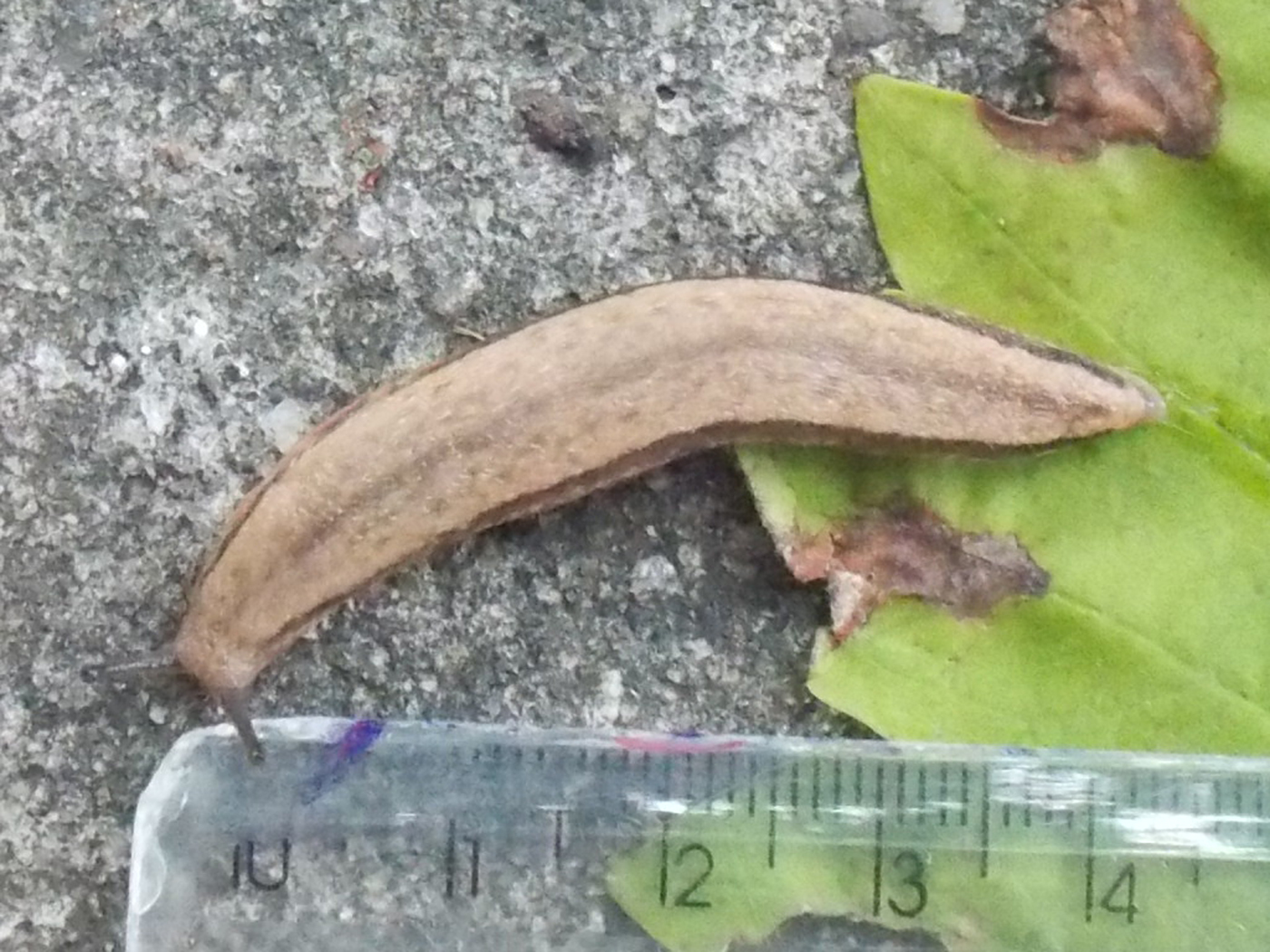
The bodies of the slugs were characterized by a beige background colour on the sides and dorsum, with two dark brown to black lateral stripes, and one medial stripe that was generally lighter than the lateral stripes (fig. 1). Below the lateral stripes and surrounding the central medial stripe there was a scattering of dark brown irregular spots or short lines. The foot of these slugs occupied the entire ventral extension and was cream coloured. The penis was claviform, short, thick, and had a constriction halfway along its length. The vas deferens was slightly longer than the penis. The bursa copulatrix had a spherical to oval form and both the penis and the bursa copulatrix opened in a large, barrel-shaped atrium. These external and internal characteristics are consistent with those previously described for Meghimatium pictum (Stoliczka, 1873) according to Tsai et al. (Reference Tsai, Lu and Kao2011) and Gomes et al. (Reference Gomes2011).

Fig. 1. The invasive slug Meghimatium pictum (Stoliczka, 1873).
A total of 655 base pairs were amplified and this DNA sequence was identical to the COI sequence deposited in GenBank (JQ712572) from an M. pictum collected in southern Brazil (Gregoric et al., Reference Gregoric2013).
After 30 days inoculating the larvae in Swiss mice, adult nematodes were found and collected from the mesenteric arteries of the mice. Based on the morphology and the localization of adult worms in the mesenteric arteries the adult worms were identified as A. costaricensis. The morphology and length of copulatory bursa rays were in accordance with the updated description of A. costaricensis (Morera, Reference Morera1973). Also, there is only one other species in the genus Angiostrongylus known to exhibit this type of localization, namely A. siamensis, which occurs in Asia (Spratt, Reference Spratt2015).
A total of 245 specimens of M. pictum (ranging from 0.03 g to 2.25 g in weight) were collected from the same area to estimate prevalence. Eleven slugs were infected with metastrongylid larvae, producing a prevalence of 4.5%. Two, three, four and eight larvae were detected in four individually digested molluscs. Another seven slugs were pooled for digestion, from which 200 L3 were recovered.
Many species of terrestrial molluscs have been reported as successful hosts of Angiostrongylus spp. larvae in various countries of Central and South America. Slugs from the Veronicellidae family are the best-adapted hosts in the southern region of South America, especially species belonging to the genera Phyllocaulis, Sarasinula and Belocaulus (Bonetti and Graeff-Teixeira, Reference Bonetti and Graeff-Teixeira1998). However, several other families of terrestrial molluscs are also naturally infected with A. costaricensis (Graeff-Teixeira et al., Reference Graeff-Teixeira1993; Maurer et al., Reference Maurer2002; Ohlweiler et al., Reference Ohlweiler2010).
Meghimatium pictum is a Stylommatophora slug belonging to the Philomycidae family. Stylommatophora encompass c. 95% of all terrestrial gastropods, whereas Systellommatophora, which contains Veronicellidae slugs, encompass < 1% (Thomé et al., Reference Thomé, Gomes and Picanço2006; Ponder and Lindberg, Reference Ponder and Lindberg2008). Both groups (Stylommatophora and Systellommatophora) are traditionally distinguished in orders or clades (Bouchet et al., Reference Bouchet2005; Ponder and Lindberg, Reference Ponder and Lindberg2008).
The slug M. pictum is considered endemic to China. However, it has also been found in Brazil and Argentina (Gomes et al., Reference Gomes2011; Gregoric et al., Reference Gregoric2013). The global dispersal of terrestrial gastropods has been facilitated by an expanding global economy and associated transportation opportunities (Robinson, Reference Robinson1999). In 2011, this slug was recorded for the first time in Brazil and quickly emerged as an agricultural pest in vineyards (Baronio et al., Reference Baronio2014). Terrestrial molluscs are considered one of the most significant and intractable threats to sustainable agriculture worldwide (Barker, Reference Barker2002).
Meghimatium pictum is found mainly in the south-eastern regions of China (Li et al., Reference Li2006). This region is also inhabited by M. bilineatum, which is a phylogenetically sister-species of M. pictum that is a confirmed carrier of another species of Angiostrongylus, A. cantonensis. This species is the primary causative agent of eosinophilic meningoencephalitis (EoM) in humans (Wang et al., Reference Wang2008).
Cases of EoM caused by A. cantonensis in Brazil have only recently been documented, with the parasite being detected in several mollusc species along the coast, including Achatina fulica, Bradybaena similaris, Sarasinula spp. and Subulina octona (Graeff-Teixeira et al., Reference Graeff-Teixeira1993; Morassutti et al., Reference Morassutti2014), but not in M. pictum. To our knowledge, there is also no previous report of M. pictum being infected with A. costaricensis.
The infective stage of angiostrongylid worms may be carried by the slime of molluscs (Bonetti and Graeff-Teixeira, Reference Bonetti and Graeff-Teixeira1998). Consequently, the risk of angiostrongyliasis infection may be associated with the consumption of vegetables and fruit, especially grapes, because molluscs can come in contact with the external surfaces of this fruit that is often consumed in nature. Furthermore, grape harvesters and garden keepers may be at a higher risk of infection, because their hands could be contaminated with L3 released from snails, with L3 remaining infective in the environment for three to 17 days (Richinitti et al., Reference Richinitti, Fonseca and Graeff-Teixeira1999). The low parasitic burden that was observed in this case (4.5%) supported previous findings that the natural infection of molluscs with A. costaricensis is characterized by very low parasitic burden (Rambo et al., Reference Rambo, Agostini and Graeff-Teixeira1997; Laitano et al., Reference Laitano2001).
This study confirmed M. pictum as a new intermediate host of A. costaricensis. We also suggest there is a risk of human infection through consuming grapes, because M. pictum slugs were frequently found on the fruit, both during our surveys and based on information provided by the infected grape farmer. Considering the low specificity of angiostrongylid worms for their intermediate hosts and the documented natural infection of M. bilineatum by A. cantonensis, which is a sister-species of M. pictum in China (Li et al., Reference Li2006), the present report should be considered an alert of the high potential risk for both abdominal and cerebral angiostrongyliasis transmission to humans in Brazil.
Financial support
Financial support was provided by the Brazilian National Council for Scientific and Technological Development (CNPq) (PVE 401904/2013-0), and the Coordination for the Improvement of Higher Education Personnel (CAPES) (Grant “PE-Parasitologia 1427/2011/Edital 32” and Grant 307005/2014-3).
Conflict of interest
None.
Ethical standards
Ethical standards are in accordance with Brazilian regulations.