Introduction
Granulomatosis with polyangiitis is a multisystemic granulomatous disease characterised by a triad of necrotising granulomata of the respiratory tract, glomerulonephritis and disseminated necrotising vasculitis. The disease is described as autoimmunity-driven inflammation of small blood vessels caused by antineutrophilic cytoplasmic antibodies (ANCAs) in the classification of vasculitis by the Chapel Hill Consensus Conference (Table I).
Table I Classification of cutaneous vasculitis*

* According to the Chapel Hill Consensus Conference, 1994
Granulomatosis with polyangiitis was previously known as Wegener's granulomatosis after Friedrich Wegener, who described the disease in 1936. More recently, the boards of directors of the American College of Rheumatology, American Society of Nephrology and the European League Against Rheumatism suggested that the name should be changed to ‘granulomatosis with polyangiitis’ so as to be more descriptive and to shift away from the use of eponyms.Reference Falk, Gross, Guillevin, Hoffman, Jayne and Jennette1
The disease commonly presents to otolaryngologists with involvement of the nose or paranasal sinuses (85 per cent of cases).Reference McDonald and DeRemee2 Primary otological symptoms alone are unusual: these often only become apparent with cranial nerve or meningeal involvement, and can lead to delayed diagnosis.Reference Ito, Shinogi, Yuta, Okada, Taki and Matsukage3 Otological symptoms forming part of a spectrum of presenting symptoms occur in up to 38 per cent of cases.Reference McCaffrey, McDonald, Facer and DeRemee4 We report a case series of 4 patients presenting within 12 months, initially with varied otological symptoms alone.
Case reports
Case one
A 58-year-old man presented with a 2-month history of gradual bilateral hearing loss, but no systemic symptoms. An examination showed that he had dull, thickened tympanic membranes, and a full ENT examination was otherwise unremarkable. Clinical investigations demonstrated bilateral conductive hearing loss of 30 dB and flat tympanograms. A diagnosis of otitis media with effusion was made and the patient opted for urgent grommet insertion. Intra-operatively, his tympanic membranes and middle-ear mucosa were noted to have an unusual thickened appearance. Thin serous fluid was removed by suction and grommets were inserted. Unfortunately, despite surgical management, his symptoms deteriorated, he developed sero-sanguinous otorrhoea and his hearing levels worsened. Serological specimens showed no microbiological growth, and a biopsy showed no granulations. Given these unusual findings, serological analysis for angiotensin-converting enzyme, antineutrophilic cytoplasmic antibodies (ANCAs) and autoantibodies was performed: the sample was positive for proteinase 3 ANCAs. The patient was promptly referred to a physician with suspected granulomatosis with polyangiitis; however, treatment was deferred so that a tissue diagnosis could be made. The patient was followed up three weeks after surgery in the otolaryngology clinic. He had developed nasal crusting, and an urgent biopsy was arranged. However, he became acutely unwell within 24 hours and was referred to a physician, who arranged for a renal biopsy to be taken. Histological analysis of the biopsy was consistent with granulomatosis with polyangiitis.
Case two
A systemically well 64-year-old man presented with a 1-month history of left-sided otalgia and discharge. Otomicroscopy revealed polypoidal granulations and debris within the external auditory canal. A further ENT examination was unremarkable. An audiogram confirmed left-sided conductive hearing loss. Suction clearance was performed and he was treated with topical steroids. Swabs showed no microbiological growth.
There was no improvement upon review a week later. A computed tomography (CT) scan of the temporal bones demonstrated soft tissue opacification of the middle-ear and mastoid air cells. Cortical mastoidectomy and grommet insertion were performed. The mastoid was full of granulations and histological analysis revealed inflammation of unknown origin. Suspicion was raised by the unusual findings and treatment resistance, and samples were taken for serological analysis. These proved to be positive for proteinase 3 ANCAs. The patient was referred to a physician and his symptoms improved after immunosuppressant therapy and steroid administration.
Case three
A 47-year-old woman with hearing loss attended an ENT clinic. A medical history and examination confirmed a diagnosis of bilateral otitis media. Swabs showed no microbiological growth. She was treated with antibiotics.
One month later, she presented as an emergency, with bilateral otalgia, worsening hearing loss, bilateral nasal obstruction and right-sided lower motor neuron facial nerve palsy. Upon examination, she was found to have bulging tympanic membranes and right-sided House–Brackmann grade V facial palsy (Figure 1). Rhinoscopy confirmed generalised, significant inflammation of the mucosal surfaces within both nasal cavities.

Fig. 1 Case three: clinical photograph showing right-sided grade V facial nerve palsy
Owing to a clinical suspicion of granulomatosis with polyangiitis, examination of the ears and nose was performed under anaesthetic as an emergency, along with bilateral grommet insertion and nasal biopsy. Serological analysis for ANCAs was undertaken. Granulations on both tympanic membranes and on the nasal mucosa were apparent (Figure 2). Initial serological analysis demonstrated a normal white cell count and mildly raised C-reactive protein (29 mg/l); ANCA tests were initially negative. The nasopharyngeal biopsy showed numerous multinucleated giant cells, well-formed granulomas and fibrinous degeneration in vessel walls, consistent with a diagnosis of granulomatosis with polyangiitis. She is currently under the care of the rheumatologists for the treatment of this disease. Repeated proteinase 3 ANCA tests have been positive.

Fig. 2 Case four: endoscopic photographs showing bilateral tympanic membrane granulations (a) & (b) and nasal mucosa granulations (c)
Case four
A 15-year-old girl presented as an emergency with left-sided otalgia, hearing loss and left lower motor neuron facial nerve palsy. She was otherwise well, except for a recent diagnosis of pyoderma presenting as facial lesions (Figure 3). An examination revealed grade VI facial nerve palsy on the left side, bulging tympanic membranes and left-sided crusted facial lesions, which had not responded to antibiotics given by the dermatologists. There were no other ENT symptoms or other findings upon examination. An audiogram confirmed severe to profound bilateral mixed hearing loss with thresholds of 90 dB.

Fig. 3 Case four: clinical photograph showing facial lesions resembling pyoderma
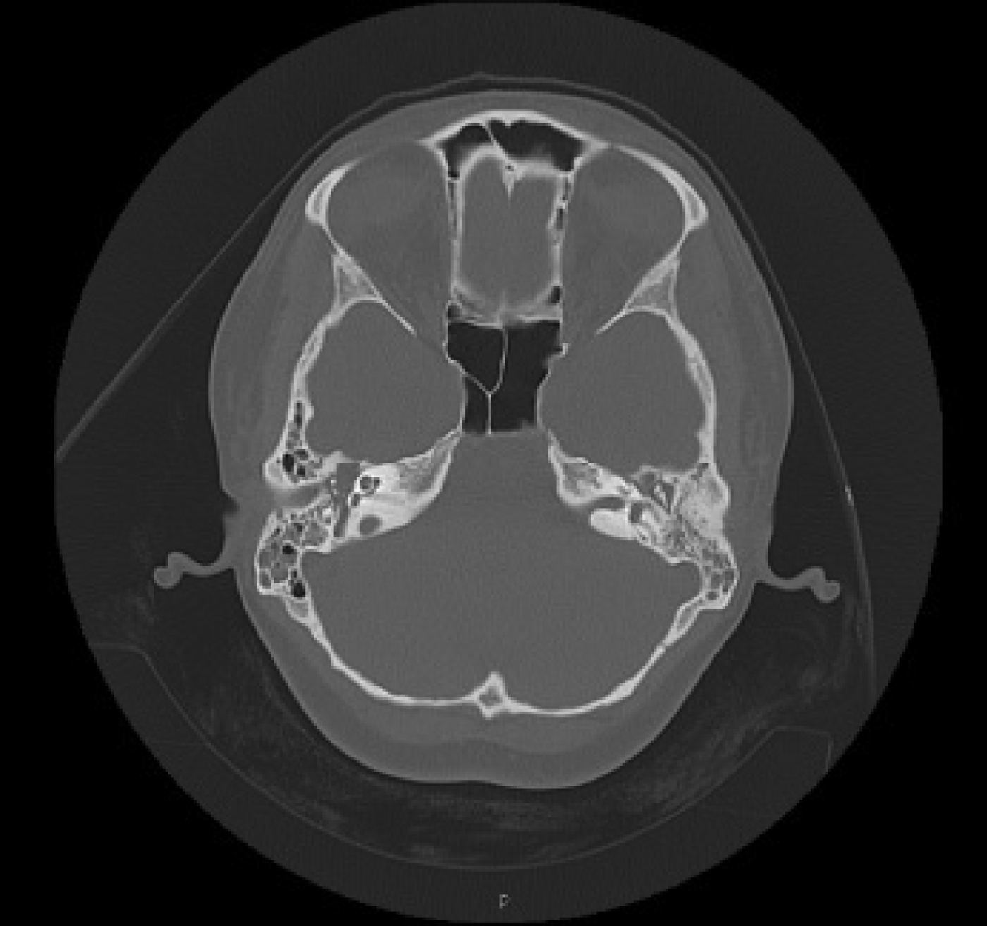
Bilateral myringotomy was performed, thin serous fluid was removed by suction and bilateral grommets were inserted. A biopsy showed no granulations. A one-week oral steroid treatment, topical combined gentamicin–hydrocortisone ear drops and eye care was commenced. An urgent CT scan demonstrated opacification in middle-ear and mastoid cells, but an intact tegmen (Figure 4). Initial serological analysis demonstrated a C-reactive protein level of 38 mg/l, a white cell count of 12.5 × 10⁹/l (neutrophils 8.3 × 10⁹/l) and a negative ANCA test; swabs were negative for bacteriological growth.

Fig. 4 Case four: axial computed tomography scan of the temporal bone showing opacification of mastoid air cells on the left
A review at the Otology Clinic after one month showed no improvement, with grommet extrusion and a recurrence of effusions and conductive hearing loss. Facial nerve palsy had not improved. Owing to a continued suspicion of granulomatosis with polyangiitis, ANCA testing was repeated, revealing proteinase 3 ANCA positivity. Facial lesions were biopsied and reported as non-specific inflammation. Despite this, the clinical picture overwhelmingly favoured a diagnosis of granulomatosis with polyangiitis; thus, treatment for this disease was commenced and symptoms significantly improved.
Discussion
Granulomatosis with polyangiitis commonly presents to the otolaryngologist with involvement of the nose or paranasal sinuses (in 85 per cent of cases).Reference McDonald and DeRemee2 This case series presents patients with initially localised otological granulomatosis with polyangiitis. As this is an unusual presentation, there were significant challenges to overcome before the clinician could make a correct diagnosis. The otological manifestations of granulomatosis with polyangiitis include serous otitis media, sensorineural hearing loss, chronic otitis media, vertigo and facial palsy.Reference Gubbels, Barkhuizen and Hwang5, Reference Dagnum and Robertson6 These symptoms may be secondary to rhinological disease; therefore, nasal endoscopy should always be performed to exclude this possibility.
A literature review identified three case series of patients suffering from granulomatosis with polyangiitis (shown in Table II).Reference Nikoloua, Vlachtsis, Daniilidis, Petridis and Daniilidis7–Reference Tsuzki, Fukazawa, Takebayashi, Hashimoto and Sakagami9 In all series, a primary otological presentation of granulomatosis with polyangiitis was rare. Our series of four patients with a primary otological presentation demonstrates that an increased clinical awareness has led to early diagnosis of this potentially lethal condition. Remission rates with treatment can be as high as 85 per cent,Reference Preuss, Stenner, Beutner, Laudes and Klussmann10 but there is still a possibility of systemic involvement and fatality. Granulomatosis with polyangiitis should be considered when clinical findings are unusual or extensive, or when there is no response to systemic antibiotic treatment. Further investigation for granulomatosis with polyangiitis and the differential diagnosis of other disease, including tuberculosis, Churg-Strauss syndrome, sarcoidosis, polyarteritis nodosa, Lyme disease, syphilis and malignancy should be undertaken.
Table II Published case series of granulomatosis with polyangiitis

GPA = Granulomatosis with polyangiitis
As demonstrated by patients in our series, both serological and pathological testing can be less sensitive in the early disease stages. Antineutrophilic cytoplasmic antibody (ANCA) sensitivity can be as low as 60 per cent in localised granulomatosis with polyangiitis.Reference Ferri, Armato, Capuzzo, Cavaleri and Ianniello11 Histological analysis often reveals non-specific inflammation, and key histological findings are often not apparent prior to systemic involvement. ANCA testing has become more reliable through the use of the enzyme linked immunosorbent assay, which uses proteinase 3 and myeloperoxidase as antigens to bind to ANCAs. Using this method, the sensitivity and specificity of granulomatosis with polyangiitis testing by proteinase 3 ANCA detection are greater than 90 per cent and 98 per cent, respectively. Current recommendations are that the test should be repeated every 3–6 months for 24 months if there is a high index of suspicion.Reference Gubbels, Barkhuizen and Hwang5
• Granulomatosis with polyangiitis (Wegener's granulomatosis) can present very rarely with otological symptoms
• In non-responding cases of acute ear infection, facial palsy and otitis media with effusion, systemic causes such as granulomatosis with polyangiitis should be excluded
• Serological testing for antineutrophilic cytoplasmic antibodies and histological analysis may be negative in the initial disease stages
Immunosuppressant and steroid therapies are the first-line management of granulomatosis with polyangiitis. Surgical management in the active stage of the disease should also be carefully considered. The literature review and our cases demonstrate that symptoms can worsen post-operatively. It is unclear whether this is caused by normal disease progression or by surgery.Reference Takagi, Nakamaru, Maguchi, Furuta and Fukada8 The consensus is that for patients with complete facial nerve palsy and otorrhoea not responding to medical treatment, surgical decompression may have to be considered.
Conclusion
Granulomatosis with polyangiitis should be considered in unusual or non-responsive cases of middle-ear infection or effusions, with or without facial palsy. In cases of granulomatosis with polyangiitis with limited localised symptoms, diagnosis can be problematic, and is made more difficult by the reduced reliability of serological and histological testing at this stage. However, clinical suspicion should be maintained so that granulomatosis with polyangiitis can be diagnosed and treatment can be commenced to minimise systemic involvement.


