


INTRODUCTION
Humes (Reference Humes1994) counted 47 species of copepods as associates of sponges. The number of copepods associated with sponges has since been almost doubled. The genus Asterocheres Boeck, 1859 is the most speciose within the family Asterocheridae, living in association with cnidarians, bryozoans, echinoderms, and ascidians, but most of them (about 56%) have been found associated with sponges (Ivanenko & Smurov, Reference Ivanenko and Smurov1997; Bandera et al., Reference Bandera, Conradi and López-González2005). There are more than 70 nominal species in Asterocheres; Kim (Reference Kim2010) recognized 45 as valid and remaining as incompletely described species or species inquirendae, and additionally described 14 new species of the genus.
Only three nominal species of Asterocheres have been reported from the Antarctic. Brady (Reference Brady1910) described A. tenuicornis collected at a depth of 177 m. Eiselt (Reference Eiselt1965) re-studied type specimens of A. tenuicornis and described a new species, A. alter, on the basis of males, but the latter was recognized by Kim (Reference Kim2010) as an incompletely described species. Bandera et al. (Reference Bandera, Conradi and López-González2005) reported a third species of the genus, A. hirsutus Bandera, Conradi and López-González, Reference Bandera, Conradi and López-González2005, associated with a hexactinellid sponge in the depth of 804–930 m off South Shetland in the Antarctic.
Recently, one of authors (G.S.M.) had a chance to visit the King George Island in the Antarctic, and collected some invertebrates from the shallow water during SCUBA diving sessions. The collected invertebrates include several species of sponges, and washings of these sponges yielded three species of Asterocheres, including two new species. This paper deals with these three species of Asterocheres.
MATERIALS AND METHODS
The copepods examined in the present paper were recovered from washing of sponges collected by SCUBA diving in the shallow water on the coast of the King George Island (62°S, 58°W), the largest island of the South Shetland Islands in the Antarctic. The collected copepods were fixed and preserved in 95% ethanol. Before microscopic observation and dissection, copepod specimens were immersed in lactic acid. Dissections were carried out using a reversed slide method. The dissected specimens were temporarily mounted in lactophenol and later sealed with Hoyer's medium. The intact type specimens have been deposited in the National Institute of Biological Resources, Incheon, Korea. Dissected paratypes are kept in the collection of I.-H. Kim. The descriptions are based on these dissected paratypes. In the descriptions of species the body lengths were measured from the anterior tip of the cephalothorax to the posterior margin of the caudal rami of a selected specimen. In the formula for the armature of legs 1–4, Roman numerals indicate spines and Arabic numerals represent setae.
RESULTS
SYSTEMATICS
Order SIPHONOSTOMATOIDA Thorell, 1859
Family ASTEROCHERIDAE, Giesbrecht, Reference Giesbrecht1899
Genus Asterocheres Boeck, 1859
Asterocheres spinosus sp. nov.
(Figures 1–3)
TYPE MATERIAL
Holotype: female (NIBRIV261851); washings of sponges; coast of King George Island, 62°14′19″S 58°46″36″W; collected 19 January 2012 during SCUBA diving by Seung-Gu Ra and Gi-Sik Min.
Allotype: male (NIBRVI0000261852). Same sampling data as holotype.
Paratypes: four females and two males (NIBRVI0000261853). Same sampling data as holotype.
All type materials have been deposited in the National Institute of Biological Resources, Incheon, Korea. Dissected paratypes (1 ♀, 1 ♂) are retained in the collection of I.-H. Kim.
DESCRIPTION
Female. Body (Figure 1A) with expanded prosome and narrow urosome. Body length 969 µm and maximum width 500 µm. Prosome 642 µm long. Cephalothorax 419 µm long, wider than long. Prosomal somites with rounded posterolateral corners. Urosome (Figure 1B) four-segmented. Fifth pedigerous somite 122 µm wide, with row of spinules along dorsal posterior margin. Genital double-somite 115 × 117 µm, almost as long as wide, widest across anterior quarter, with tapering posterior three-quarters, anterodorsal elevation on both sides, about 50 spinules on lateral margin posterior to genital area, and minute spines on posterior region of ventral surface; genital areas located dorsally slightly anterior to halfway length of somite. First free abdominal somite 54 × 63 µm, and ventrally covered with minute spinules. Anal somite and caudal rami covered with minute spinules on ventral and dorsal surfaces. Anal somite 53 × 55 µm. Caudal ramus (Figure 1C) subrectangular, 47 × 25 µm (length to width ratio 1.88:1), with two naked dorsal setae and four pinnate distal setae.
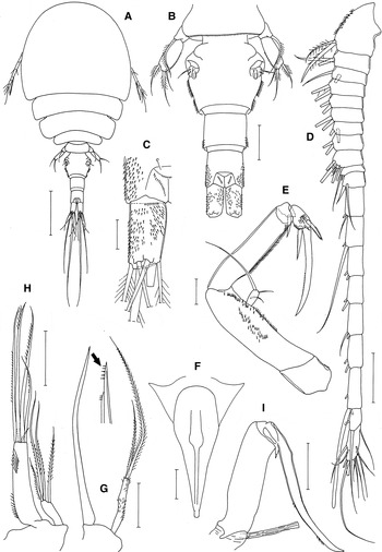
Fig. 1. Asterocheres spinosus sp. nov., female: (A) habitus, dorsal; (B) urosome, dorsal; (C) left caudal ramus, dorsal; (D) antennules; (E) antenna; (F) oral siphon; (G) mandible; (H) maxillule; (I) maxilla. Scale bars: A, 200 µm; B, D, F–I, 50 µm; C, E, 20 µm.
Rostrum as weak ventral prominence on cephalothorax. Antennule (Figure 1D) 465 µm long and 20-segmented; ninth segment with seven setae; eighteenth segment with two setae and one aesthetasc; terminal segment with 11 setae; other segments each with two setae; setae on first segment pinnate, other setae naked; setae fragile. Antenna (Figure 1E) consisting of coxa, basis, 1-segmented exopod, and 3-segmented endopod. Coxa short and unarmed. Basis 85 × 27 µm, with a number of spinules. Exopod 14 × 6 µm (ratio 2.33:1), with one proximal and two unequal distal setae. First endopodal segment 85 × 20 µm, as long as basis, with row of dense spinules along outer margin. Second endopodal segment short, with one spiniform seta. Third endopodal segment with spinules and one minute and two spiniform, barbed setae; terminal claw weakly curved and 72 µm long, with minute spinules on concave margin.
Oral siphon (Figure 1F) 225 µm long, extending to midway between maxilliped and leg 1, widest in proximal third (77 µm wide across this region), and terminated by hyaline extension. Mandible (Figure 1G) consisting of stylet and palp. Stylet 210 µm long, with acutely pointed distal apex bearing 5 + 3 spinule-like teeth. Palp 1-segmented, slender, 64 × 9 µm with spinules, and two pinnate distal setae; shorter seta 92 µm, and longer seta 177 µm, extending over tip of stylet. Maxillule (Figure 1H) bilobed. Inner lobe 77 × 18 µm with setules proximally and subdistally and five distal setae, including one small and four larger setae 130, 126, 126, and 83 µm, respectively. Outer lobe much smaller than inner lobe, 26 × 10 µm, with one subdistal and three distal, weakly pinnate setae, longest one 105 µm long, and shortest one 42 µm long. Maxilla (Figure 1I) two-segmented; proximal segment (syncoxa) with hyaline extension (tube of maxillary gland); a tapering process present between proximal and distal segment; distal segment (basis) elongate, claw-like, longer than proximal segment, with several spinules subdistally. Maxilliped (Figure 2A) consisting of syncoxa, basis and four-segmented endopod; syncoxa with one small inner distal seta and several outer distal spinules; basis 113 × 37 µm, with one minute inner seta and spinules on outer margin; four endopodal segments with two, one, one, and one setae, respectively; fourth endopodal segment 48 µm long; terminal claw 85 µm long and weakly curved.

Fig. 2. Asterocheres spinosus sp. nov., female: (A) maxilliped; (B) leg 1; (C) exopod of leg 1; (D) leg 2; (E) endopod of leg 3; (F) leg 4; (G) leg 5; (H) left side of genital double-somite, dorsal. Scale bars: A, B, D–F, 50 µm; C, G, H, 20 µm.
Legs 1–4 with three-segmented rami, spinules on mediodistal corner of basis, relatively large, naked outer seta on basis, and bicuspid outer distal corner of second endopodal segment (Figure 2B–F). Outer spine on first exopodal segment of leg l 32 µm long. Terminal spine on third endopodal segment of leg 3 51 µm long, that of leg 4 42 µm long. Armature formula of legs 1–4 as follows:
Leg 1: coxa 0-1 basis 1-1; exp. I-1; I-1; III, 2, 2; enp. 0-1; 0-2; 1, 2, 3
Leg 2: coxa 0-1 basis 1-0; exp. I-1; I-1; III, I, 4; enp. 0-1; 0-2; 1, 2, 3
Leg 3: coxa 0-1 basis 1-0; exp. I-1; I-1; III, I, 4; enp. 0-1; 0-2; 1, 1 + I, 3
Leg 4: coxa 0-1 basis 1-0; exp. I-1; I-1; III, I, 4; enp. 0-1; 0-2; 1, 1 + I, 2
Leg 5 (Figure 2G) 2-segmented; proximal segment fused with somite, with dorsolateral seta; distal segment (exopod) 53 × 23 µm (2.30:1), with spinules on outer margin and 1 seta inner side and 3 distal setae. Leg 6 represented by 1 seta and 1 spinule on genital operculum (Figure 2H).
Male
Body (Figure 3A) similar to that of female, but narrower. Body length 912 µm and maximum width 423 µm. Prosome 577 µm long. Cepahlothorax 400 µm long. Urosome (Figure 3B) five-segmented. Fifth pedigerous somite 115 µm wide. Genital double-somite large, 135 × 185 µm, much wider than long, with rounded anterolateral and posterolateral corners. Three free abdominal somites 21 × 62, 23 × 54, and 38 × 52 µm, respectively, with spinules on ventral surface. Caudal ramus 35 × 22 µm (ratio 1.59:1).

Fig. 3. Asterocheres spinosus sp. nov., male: (A) habitus, dorsal; (B) urosome, ventral; (C) antennules; (D) ninth segment of antennules; (E) tenth segment of antennules; (F) maxilliped; (G) endopod of leg 1; (H) third endopodal segment of leg 2; (I) exopod of leg 5. Scale bars: A, 0.2 mm; B, C, F, 50 µm; D, E, G–I, 20 µm.
Antennule (Figure 3C) 18-segmented, geniculate between segments 16 and 17 with two setae each on segments 1–8; armature formula of ninth to terminal segments 5 + 2 aesthetascs, 1 + aesthetasc, 2, 2, 2 + aesthetasc, 2, 2, 3, 3 + aesthetasc, and 11; penultimate segment with pointed distal process.
Rostrum, antenna, oral siphon, mandible, maxillule, and maxilla as for female. Maxilliped with tapering process on inner margin of basis (Figure 3F); fourth endopodal segment 51 µm and terminal claw 103 µm, much longer than that of female.
Third endopodal segment of leg 1 with patch of spinules on outer side and pronounced mid-terminal process (Figure 3G). Third endopodal segment of leg 2 with pectinate outer terminal process (Figure 3H). Legs 3 and 4 as for female.
Free segment of leg 5 (Figure 3I) 38 × 18 µm (ratio 2.11:1). Leg 6 represented by two small setae on genital operculum (Figure 3B)
ETYMOLOGY
The specific name spinosus (‘full of thorns’ in Latin) refers to the spinules-rich abdominal somites of the new species.
REMARKS
The caudal ramus in species of Asterocheres seldom exceeds 1.5 times as long as wide. Only seven species of the genus were described or figured to have an elongate caudal ramus which is at least 1.5 times as long as wide: A. fastigatus Kim, Reference Kim2010, A. hirsutus Bandera, Conradi and López-González, Reference Bandera, Conradi and López-González2005, A. kervillei Canu, 1898, A. latus (Brady, 1872), A. lilljeborgi Boeck, 1859, A. suberitis Giesbrecht, 1897, and A. tenuicornis Brady, Reference Brady1910. Of these, A. tenuicornis has a very long caudal ramus more than 5 times longer than wide, judging from the illustrations provided by Brady (Reference Brady1910) and Eiselt (Reference Eiselt1965) and, therefore, differs from A. spinosus sp. nov. The other six species differ also from A. spinosus sp. nov., because they all have a two-segmented mandibular palp. These six species may be further differentiated from A. spinosus by their following traits.
In A. fastigatus, the antennules is 17-segmented in the female and 14-segmented in the male, the cephalothorax is expanded, much larger than the remaining part of prosome, the exopod of the antenna is elongated, about half as long as the first endopodal segment, and the posterior half of the genital double-somite of the female is strongly tapering (Kim, Reference Kim2010).
In A. lilljeborgi Boeck, 1859, which is a circum-Arctic species associated with echinoderms, the body is large, 1.41–1.47 mm in length in the female, the prosomal somites are very broad, with angular posterolateral corners, and the apical setae on the outer lobe of the maxillule are longer than those of the inner lobe (Ivanenko & Ferrari, Reference Ivanenko and Ferrari2003).
The remaining four species have 21-segmented antennule in the female and reveal further differences as follows:
Stock (Reference Stock1967) treated A. kervillei Canu, 1898 as a junior synonym of A. echinicola, but Bandera & Conradi (Reference Bandera and Conradi2009b) redescribed and recognized it as a distinct species. In this species the antennule of the male is 17-segmented, the genital double-somite of the female bears setules (rather than spinules) on the lateral margins, and the larger one of distal setae on the mandibular palp is more than 6 times as long as the smaller one (Bandera & Conradi, Reference Bandera and Conradi2009b).
In A. latus (Brady, 1872) which was redescribed by Bandera & Conradi (Reference Bandera and Conradi2009b) the urosome is covered by epicuticular scales and the genital double-somite of the female is ornamented with setules on lateral margins (Bandera & Conradi, Reference Bandera and Conradi2009b).
In A. suberitis Giesbrecht, 1897, which is known in the Mediterranean Sea, the antennule of the male is 17-segmented, the lateral margin of genital double-somites of the female is ornamented with setules, one of apical seta on the inner lobe of the maxillule is spatulate at tip (Bandera & Conradi, Reference Bandera and Conradi2009a), and the larger one of distal setae on the mandibular palp is several times as long as the smaller one, as illustrated by Giesbrecht (Reference Giesbrecht1899).
Asterocheres hirsutus Bandera, Conradi and Lopez-Gonzalez, 2005 recorded from a sponge in the deep sea of the Antarctic is closely similar to A. spinosus in having spinules on the surface of the urosomal somites and especially on the lateral margin of the genital double-somite of the female and similar forms of the oral siphon, maxillule and leg 5. However, in addition to the difference in the segmentation of the mandibular palp which is 2-segmented in A. hirsutus, the latter species has 17-segmented male antennules bearing no aesthetasc on the ninth and tenth segments, a longer oral siphon which extends to the intercoxal sclerite of leg 1, and a longer caudal ramus which is 2.5 times as long as in the female (Bandera et al., Reference Bandera, Conradi and López-González2005).
Asterocheres rai sp. nov.
(Figures 4–6)
TYPE MATERIAL
Holotype: female (NIBRVI0000261854); washings of sponges; coast of King George Island, 62°14′19″S 58°46″36″W; collected 19 January 2012 by SCUBA diving by Seung-Gu Ra and Gi-Sik Min.
Paratype: 1 female (NIBRVI0000261855). Same sampling data as holotype.
All type materials have been deposited in the National Institute of Marine Biological Resources, Incheon, Korea. Dissected paratypes (1 ♀, 1 ♂) are retained in the collection of I.-H. Kim.
DESCRIPTION
Female. Body (Figure 4A) cyclopiform, fleshy and violet-tinted. Body length 1.61 mm and maximum width 800 µm. Prosome 1.11 mm long. Cephalothorax 644 µm long, expanded and sub-globular. Urosome (Figure 4B) four-segmented. Fifth pedigerous somite 169 µm wide. Genital double-somite 175 × 237 µm, much wider than long, expanded laterally in anterior half, and strongly tapering in distal half; lateral margins smooth, without setules or spinules; genital openings located dorsolaterally at midlength. First free abdominal somite 70 × 115 µm. Anal somite 81 × 96 µm, with minute spinules along ventral posterior margin. Caudal ramus (Figure 4C) sub-quadrate, 42 × 35 µm (length to width ratio 1.20:1), with six setae.
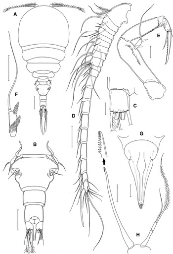
Fig. 4. Asterocheres rai sp. nov., female: (A) habitus, dorsal; (B) urosome, dorsal; (C) right caudal ramus, ventral; (D) antennules; (E) antenna; (F) exopod of antenna; (G) oral siphon; (H) mandible. Scale bars: A, 200 µm; B, 100 µm; C, E, F, 20 µm; D, G, H, 50 µm.
Rostrum absent. Antennule (Figure 4D) 465 µm long and 20-segmented. Armature formula: 2, 2, 2, 2, 2 (fifth segment), 2, 2, 2, 7, 2 (tenth), 2, 2, 2, 2, 2 (fifteenth), 2, 2, 2 + aesthetasc, 2, and 11; two setae on first segment pinnate but other setae naked. Antenna (Figure 4E) consisting of coxa, basis, one-segmented exopod, and three-segmented endopod. Coxa short and unarmed. Basis 96 × 33 µm, with a longitudinal row of minute spinules. Exopod 20 × 7 µm, with one long apical seta and characteristically one subapical and one proximal transformed seta covered by translucent mucilaginous substance (Figure 4F). First endopodal segment 77 × 23 µm, as long as basis, with row of minute spinules along outer margin. Second endopodal segment short, with 1 spiniform seta. Third endopodal segment with three setae (one of them minute); terminal claw spiniform, 59 µm long, with spinules along concave margin.
Oral siphon 298 µm long, extending to midway between maxilliped and leg 1, consisting of expanded proximal part and tapering distal part, with hyaline knob on distal end (Figure 4G). Mandible (Figure 4H) consisting of stylet and palp. Stylet 258 µm long, with about 14 teeth distally. Palp two-segmented, slender, 86 µm long, with longer pinnate seta (226 µm) extending over tip of stylet and smaller naked seta (107 µm). Maxillule (Figure 5A) bilobed. Inner lobe 94 × 34 µm with setules laterally and four setae distally, largest 168 µm. Outer lobe much smaller than inner lobe, 42 × 12 µm, with four naked distal setae, largest 92 µm. Maxilla (Figure 5B) stout and two-segmented; proximal segment (syncoxa) with hyaline extension (tube of maxillary gland); a small, tapering extension present between proximal and distal segments; distal segment (basis) claw-like, about as long as proximal segment, with two patches of minute spinules near halfway. Maxilliped (Figure 5C) consisting of syncoxa, basis and four-segmented endopod; syncoxa with one small inner distal seta and patch of minute spinules at outer distal corner; basis 114 × 54 µm, with few spinules on outer margin; four endopodal segment with two, one, one, and one setae, respectively; fourth endopodal segment 43 µm long; terminal claw 63 µm long, with small spinules along concave margin.
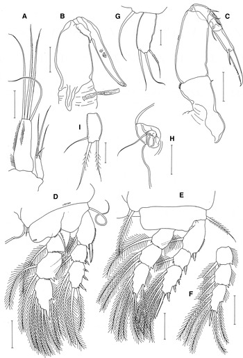
Fig. 5. Asterocheres rai sp. nov. Female: (A) maxillule; (B) maxilla; (C) maxilliped; (D) leg 1; (E) leg 2; (F) endopod of leg 3; (G) leg 5; (H) left side of genital double-somite. Male: (I) exopod of leg 5. Scale bars: A–F, H, 50 µm; G, I, 20 µm.
Legs 1–4 with three-segmented rami and bicuspid outer distal corner of second endopodal segment (Figures 5D–F, 6A). Inner coxal seta of leg 1 vestigial. Outer spine on first exopodal segment of leg l 35 µm long. Other spines on exopods of legs 1–4 small. Terminal spine on third endopodal segment of leg 3 28 µm long, that of leg 4 21 µm long. Armature formula of legs 1–4 as in preceding species.

Fig. 6. Asterocheres rai sp. nov. Female: (A) leg 4. Male: (B) habitus, dorsal; (C) urosome, ventral; (D) antennules; (E) maxilliped; (F) third endopodal segment of leg 1; (G) endopod of leg 2; (H) endopod of leg 4. Scale bars: A, C, D, H, 50 µm; B, 200 µm; E–G, 20 µm.
Leg 5 (Figure 5G) two-segmented; proximal segment fused with somite, with dorsolateral seta; distal segment (exopod) 54 × 23 µm (ratio 2.35:1), with smooth lateral margins and three distal setae. Leg 6 represented by one seta and one spinule on genital operculum (Figure 5H).
Male
Body (Figure 6B) narrower than that of female. Body length 790 µm and maximum width 375 µm. Prosome 537 µm long. Cepahlothorax 411 µm long, distinctly longer than wide. Urosome (Figure 6C) five-segmented. Fifth pedigerous somite 105 µm wide. Genital double-somite much wider than long, 115 × 169 µm, with rounded anterolateral and posterolateral corners. Three free abdominal somites 25 × 68, 31 × 59, and 26 × 57 µm, respectively. Caudal ramus 23 × 24 µm (ratio 0.96:1).
Antennule (Figure 6D) 17-segmented, geniculate between fifteenth and sixteenth segments; armature formula: 2, 2, 2, 2, 2 (fifth), 2, 2, 2, 6 + aesthetasc, 2 (tenth), 2, 4, 2, 2, 2 (fifteenth), 1 + aesthetasc, and 11; penultimate segment with pointed distal process.
Rostrum, antenna, oral sipon, mandible, maxillule, and maxilla as in female. Maxilliped (Figure 6E) with tapering process on inner margin of basis; fourth endopodal segment 35 µm, and terminal claw 52 µm.
Leg 1 without inner coxal seta; third endopodal segment with elongate and distally blunt outer distal process (Figure 6F). Outer margin of second and third exopodal segments of legs 2 and 3 with row of thick setules (Figure 6G). Leg 3 as in female. Second endopodal segment of Leg 4 with only one inner seta.
Free segment of leg 5 (Figure 5I) 29 × 15 µm (ratio 1.93:1), with slightly convex outer margin. Leg 6 represented by two unequal setae on genital operculum (Figure 6C).
ETYMOLOGY
The specific name ‘rai' is derived from the family name of our field assistant Seung-Gu Ra.
REMARKS
Asterocheres rai sp. nov. is easily recognizable by its four diagnostic features: (1) large body size; (2) absence of setae or spinules on the lateral margins of the genital double-somite in the female; (3) presence of only a single seta on the exopod of the antenna; and (4) presence of a distal prologation of the third endopodal segment of leg 1.
Species of Asterocheres generally have a small body, most of them being less than 1.0 mm in length. The body length of A. rai, 1.61 mm in the female, is probably the record of the longest length in the genus, followed by the next longest records in A. lilljeborgi Boeck, 1859 where the length of female is up to 1.47 mm (Ivanenko & Ferrari, Reference Ivanenko and Ferrari2003) and in A. simulans (T. Scott, 1898) where the length of female is up to 1.44 mm (Ivanenko, Reference Ivanenko1997) .
The genital double-somite of the female of Asterocheres generally bears setules, occasionally spinules, on the lateral margins, although this ornamentation is overlooked in many previous records. The smooth lateral magins of the somite are considered a significant feature of A. rai.
The exopod of the antennule of Asterocheres is generally armed with one apical, one subapical and one proximal seta, with few exceptional records of the presence of one or two setae. However, in none of the latter cases the presence of transformed setae covered by additional mucilaginous substance(s) on the exopod as in A. rai has been reported.
Asterocheres rai sp. nov. belongs to the ‘crinoidicola group’ (Kim, Reference Kim2010) having a distal prologation of the third endopodal segment of leg 1. Four species have previously been reported in this group: A. crinoidicola Humes, 2000, A. pilosus Kim, 2004, A. unioviger Kim, Reference Kim2010, and A. trisetatus Kim, Reference Kim2010 (Kim, Reference Kim2010). In these four species the caudal ramus is longer than wide, the genital double-somite of the female carries setae or spinules on the lateral margins, and the oral cone does not extend beyond the maxilliped, unlike in A. rai.
Asterocheres hirsutus Bandera, Conradi & López-González, Reference Bandera, Conradi and López-González2005
(Figure 7)
MATERIAL EXAMINED
Five females; washings of sponges; coast of King George Island, 62°14′19″S 58°46″36″W; collected 20 January 2012 by SCUBA diving by Seung-Gu Ra and Gi-Sik Min.
SUPPLEMENTARY DESCRIPTION
Female. Body length 1.02 mm (0.96–1.07 mm) in average, based on five specimens. Prosome rather expanded (Figure 7A). Urosome (Figure 7B) ornamented with scattered minute spinules dorsally and ventrally (spinules more dense on ventral surface). Genital double-somite 110 × 115 µm, slightly wider than long; ventral surface with tapering plate-like elevation (indicated by an arrowhead in Figure 7C) on both sides; posterior half of lateral margin with about 20 spinules (Figure 7I). Two free abdominal somites 69 × 62 and 62 × 54 µm, respectively. Caudal ramus (Figure 7D) 76 × 25 µm, 3.04 times as long as wide, with six setae.
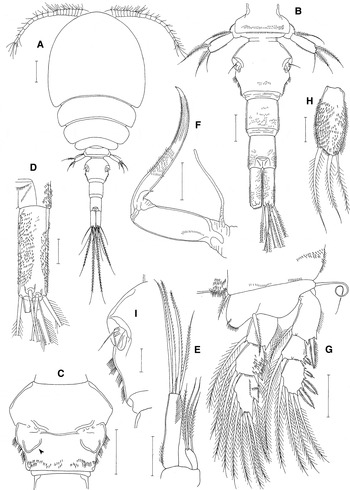
Fig. 7. Asterocheres hirsutus Bandera, Conradi and López-González, female: (A) habitus, dorsal; (B) urosome, dorsal; (C) Genital double-somite, ventral; (D) right caudal ramus, dorsal; (E) maxillule; (F) maxilla; (G) leg 1; (H) exopod of leg 5; (I) left side of genital double-somite, dorsal. Scale bars: A, 100 µm; B, C, E–G, 50 µm; D, H, I, 20 µm.
Rostrum indistinct, tapering, longer than wide, with ambiguous posterior tip. Antennule 21-segmented; suture line indistinct between last two segments. Antenna with exopod of 17 × 7 µm (ratio 2.43:1); terminal endopodal segment bearing one claw and three setae (including one minute seta); terminal claw weakly curved, 103 µm long, as long as three endopodal segments combined.
Oral siphon extending to proximal part of intercoxal sclerite of leg 1. Mandibular stylet armed with five minute teeth distally. Mandibular palp distinctly two-segmented, with one longer and one shorter setae distally; combined segments and longer seta shorter than stylet. Maxillule (Figure 7E) with five setae (including one small seta) on inner lobe and four setae on outer lobe. Maxilla (Figure 7F) and maxilliped as figured in original description.
Leg 1 (Figure 7G) and legs 2–4 also as in original description. Spinulation of these legs more developed on posterior surface than on anterior surface.
Free segment of leg 5 56 × 24 µm, 2.33 times as long as wide, with dense spinules on ventral surface (Figure 7H) and less dense spinules on dorsal surface; all of three distal setae pinnate. Leg 6 represented by one seta and one spinule on genital operculum (Figure 7I).
REMARKS
Type specimens of Asterocheres hirsutus Bandera, Conradi and López-González, Reference Bandera, Conradi and López-González2005 were derived from a depth of 804–930 m off South Shetland in the Antarctic (Bandera et al., 2005). In contrast, our specimens were sampled by SCUBA diving in the shallow water. Despite this great difference of sampling depths, our specimens are determined as A. hirsutus. The dense covering of spinules on the urosome and legs, which are more dense on the ventral surface (as shown in figure 4D of Bandera et al., Reference Bandera, Conradi and López-González2005) than on the dorsal surface, the presence of multiple rows of spinules on terminal claws of the antenna, maxilla and maxilliped (as shown in figures 1E, 2G, H, and 4C of Bandera et al., Reference Bandera, Conradi and López-González2005), the presense of a pair of plate-like elevations on the ventral surface of the female genital double-somite (see Figure 7C of this paper and figure 4G of Bandera et al., Reference Bandera, Conradi and López-González2005), and the rather elongate caudal rami led us to determine our specimens to be A. hirsutus.
Bandera et al. (Reference Bandera, Conradi and López-González2005) recorded body lengths of type specimens as 759–875 µm in the female (0.96–1.07 mm in our specimens) and the length to width ratio of the caudal ramus as 2.5:1 (3.04:1 in our specimens). These discrepancies are considered to be derived from variations within the species or observational errors.
ACKNOWLEDGEMENT
This work was supported by the Polar Academic Programme (PD11010), KOPRI (G.-S. Min)


