


Introduction
Acute mastoiditis is an uncommon complication of otitis media in the antibiotic era. However, it is still a serious condition owing to the possible extracranial and intracranial complications. The most frequent complication of acute mastoiditis is subperiosteal abscess.Reference Luntz, Brodsky, Nusem, Kronenberg, Keren and Migirov 1 , Reference Kangsanarak, Fooanant, Ruckphaopunt, Navacharoen and Teotrakul 2 Bilateral acute mastoiditis associated with bilateral subperiosteal abscesses is extremely rare, with only one previous report.Reference Oyarzabal, Patel and Tolley 3
This report documents bilateral acute mastoiditis associated with bilateral subperiosteal abscesses in a young child. A short video, available on The Journal of Laryngology & Otology website, demonstrates the key stages of the operation (Appendix 1).
Case report
A two-year-old boy was referred by his general practitioner with a three-day history of right-sided otalgia and a green, purulent discharge. One day after the right ear discharge commenced, the left ear also started discharging.
The child's past history included failure to thrive, for which he had required calorie supplements. Immune screening performed at this stage had been normal, other than marginally elevated immunoglobulin E levels consistent with an allergic enteropathy. Previous duodenal biopsies had confirmed the presence of enteritis, most likely to be allergic in nature.
On presentation, the child was febrile (38°C) and irritable with bilateral protruding ears. His external auditory canals were full of pus and granulation tissue. There were no focal neurological signs, and the remainder of his physical examination was normal.
The patient's white blood cell count was 14.1 × 10/l.
He was treated for 72 hours with intravenous co-amoxiclav (260 mg), which was subsequently changed to ceftriaxone (400 mg) and flucloxacillin (200 mg) following culture of Staphylococcus aureus and Streptococcus pneumoniae from his ear swabs. However, he failed to improve on the above management.
A computed tomography (CT) scan confirmed bilateral mastoid abscesses with destruction of the mastoid cortex (Figures 1 and 2).

Fig. 1 Coronal computed tomography scan showing opacified canal and mastoid cavity.
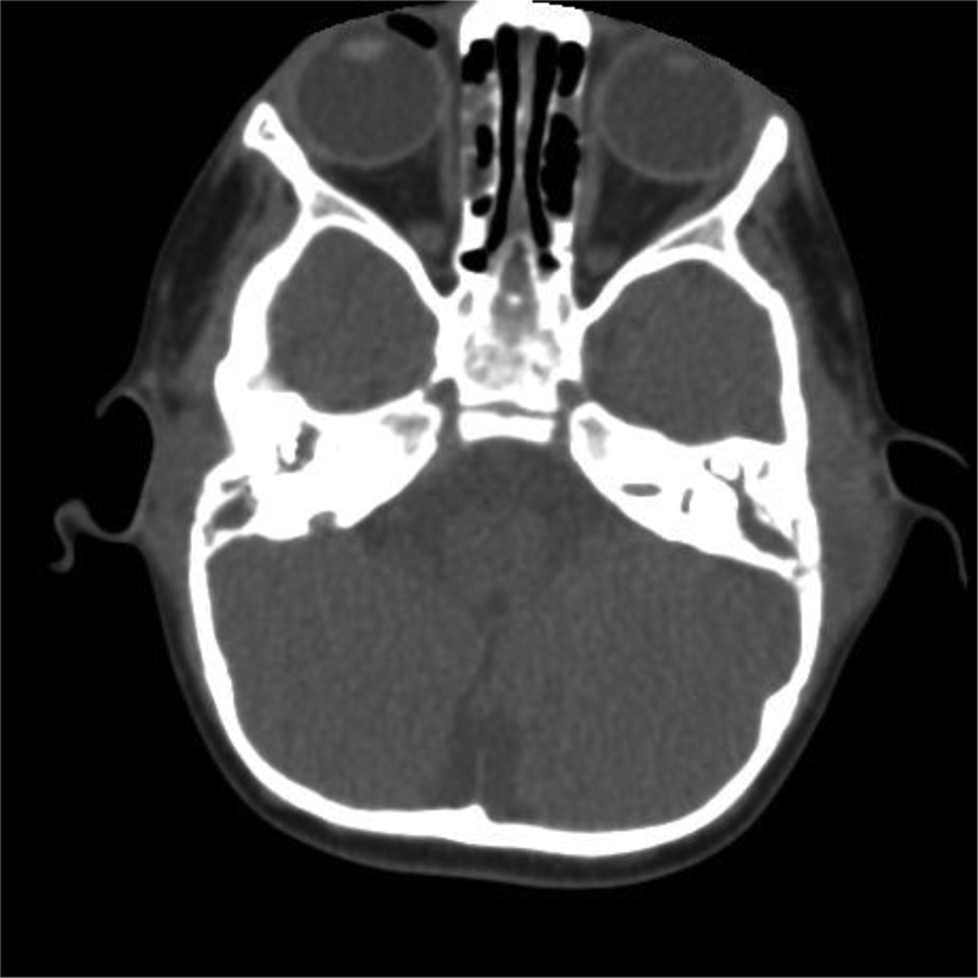
Fig. 2 Axial computed tomography scan showing bilateral subperiosteal abscesses.
Free field audiometry showed reduced hearing thresholds of 30–50 dB across all frequencies.
Bilateral surgical drainage of the subperiosteal abscesses and bilateral cortical mastoidectomies were carried out (see video, Appendix 1). Following incision through the skin and periosteum, a large amount of pus was released bilaterally (Figure 3). As might be expected, there was some bleeding during the cortical mastoidectomy due to the presence of infection and inflammation, and Surgicel (Ethicon, Summerville, New Jersey, USA) was used to aid haemostasis.

Fig. 3 Surgical photograph showing pus being released following incision through skin and periosteum.
Tissue sent intra-operatively for histopathological analysis showed bilateral ear polyps (consistent with acute mastoiditis) with inflamed metaplastic squamous mucosa and ulceration. There was no evidence of cholesteatoma.
Post-operatively, the child recovered well, and continued on intravenous antibiotics (ceftriaxone 400 mg and flucloxacillin 200 mg) for a further week. He had no fever post-operatively and his white blood cell count returned to normal. He was discharged home on oral penicillin V and flucloxacillin, following microbiology advice.
On review in the out-patient clinic two months later, the child was well. Examination of his ears revealed bilateral dry tympanic membrane perforations. Repeated conditioned free field audiometry showed normal hearing thresholds of 20 dB across all test frequencies (0.250–4 kHz).
Discussion
The introduction of antibiotics has significantly reduced the incidence of acute mastoiditis following acute otitis media, from 50 per cent in the pre-antibiotic era to 0.4 per cent recently. 4 Following the onset of otitis media, acute mastoiditis develops as a result of the connection between the middle ear and the mastoid cavity via the aditus and antrum. The resultant mucosal oedema can block the normal drainage pathways of the mastoid cavity, resulting in accumulation of pus. A subperiosteal abscess may form if the infection persists and extends outside the mastoid cavity. This spread may occur through the tympanomastoid suture or along vascular channels (emissary veins). Alternatively, the mastoid cortex can be eroded directly by the inflammatory process, causing subperiosteal abscess formation.
Bilateral acute mastoiditis is rare. Only a few cases have been described in recent decades, one of which was in a child with a rare leukocyte adhesion deficiency.Reference Andreasson and Fons 5 , Reference Martinez, McNellis, Weber and Adkins 6 Bilateral mastoiditis as a result of mycobacterial infection has also been reported, which presents a particular challenge to treat.Reference Avery, Eavey, Della Torre, Ramos and Pasternack 7 , Reference Munoz, Ruiz-Contreras, Jimenez, Jimenez, Mate and Calvo 8 In addition, there have been cases of bilateral ‘masked’ mastoiditis following multiple antibiotic therapy, presenting with bilateral facial nerve palsy.Reference Fukuda, Sugie, Ito and Kikawada 9 , Reference Tovi and Leiberman 10 It is therefore possible that bilateral mastoid involvement is much more common than previously thought, but that antibiotics control the disease and it remains subclinical.
Bilateral acute mastoiditis associated with bilateral subperiosteal abscesses is even rarer, and has only been reported once previously, by Oyarzabal et al. Reference Oyarzabal, Patel and Tolley 3 Interestingly, this case was also complicated by lateral sinus thrombosis. In most cases, subperiosteal abscess secondary to acute mastoiditis occurs in patients less than four years old.Reference Migirov, Yakirevitch and Kronenburg 11 Characteristic features include protruding ears, retroauricular oedema, erythema and tenderness over the mastoid area. This is associated with pyrexia, otalgia and otorrhoea.
Acute mastoiditis can occur in different stages. Acute mastoiditis with associated periostitis can result if infection spreads to the periosteum. Once the infection destroys the bone of the mastoid air cells, acute mastoid osteitis develops and a subperiosteal abscess may result. A different stage, referred to as subacute (‘masked’) mastoiditis, may develop from incompletely treated acute otitis media after 10–14 days of infection; signs may be minimal but serious complications may still occur.
Computed tomography is the imaging technique most commonly used. The majority of cases do not require imaging and resolve with antibiotics. However, CT is a useful diagnostic tool in determining the type of therapy required. It demonstrates mastoid cortical erosion, subperiosteal abscess and intracranial complications. Computed tomography has a sensitivity of 97 per cent in the diagnosis of complicated acute mastoiditis.Reference Migirov 12 However, CT can miss, and underestimate the extent of, subperiosteal abscess, and therefore it is important to base decisions about surgical intervention on clinical signs as well as imaging results.Reference Migirov, Yakirevitch and Kronenburg 11 , Reference Migirov 12 Magnetic resonance imaging can be performed if CT findings suggest intracranial complications, due to the former imaging modality's higher sensitivity for detecting fluid collections.Reference Vazquez, Castellote, Piqueras, Mauleon, Creixell and Pumarola 13
The mainstay of acute mastoiditis management is intravenous antibiotics. The commonest organism responsible is Streptococcus pneumoniae,Reference Luntz, Brodsky, Nusem, Kronenberg, Keren and Migirov 1 , Reference Vassbotn, Klausen, Lind and Moller 14 , Reference Gorphe, de Barros, Choussy, Dehesdin and Marie 15 although in some series S pyogenes ranks first.Reference Migirov, Yakirevitch and Kronenburg 11 , Reference Zanetti and Nassif 16 In the largest published series of subperiosteal abscesses, the commonest organism was S pyogenes followed by Staphylococcus aureus.Reference Migirov 12 In 17–44 per cent of cases, cultures are negative,Reference Luntz, Brodsky, Nusem, Kronenberg, Keren and Migirov 1 , Reference Migirov, Yakirevitch and Kronenburg 11 , Reference Vassbotn, Klausen, Lind and Moller 14 , Reference Gorphe, de Barros, Choussy, Dehesdin and Marie 15 probably reflecting the fact that many patients have already received multiple courses of antibiotics. Other causative organisms commonly identified include Pseudomonas aeruginosa, Haemophilus influenzae and Escherichia coli. Recently, antibiotic prescribing restrictions have been introduced due to more frequent development of antibiotic resistance, and this has contributed to an increase in the incidence of acute mastoiditis and associated complications.Reference Benito and Gorricho 17 It may also be the case that the development of antibiotic resistance and vaccination against S pneumoniae and H influenzae have resulted in a change in the pathogens involved. This has led to a change in antibacterial management, and some authors have suggested a reduction in the surgery rate.Reference Bakhos, Trijolet, Moriniere, Pondavan, Al Zahrani and Lescanne 18 In the case presented, the organisms cultured were S pneumoniae and Staphylococcus aureus, and appropriate intravenous antibiotics were given according to microbiology advice. Due to the variation in causative organisms and differences in local antibiotic resistance patterns, it is essential to discuss these cases with the microbiologist so that the most suitable antibiotic regimen can be used.
Surgery also has a role to play in the management of acute mastoiditis. However, the timing and extent of surgery required is controversial. Currently, some authors support the treatment of uncomplicated acute mastoiditis with myringotomy (if the eardrum is intact) and intravenous antibiotics, and this is effective in approximately 65 per cent of cases.Reference Luntz, Brodsky, Nusem, Kronenberg, Keren and Migirov 1 It has been suggested that mastoidectomy should be reserved for acute mastoiditis associated with subperiosteal abscess, cholesteatoma, intracranial complications and/or otorrhoea persistent for more than two weeks despite adequate antibiotic treatment.Reference Zanetti and Nassif 16 Surgery in these cases can be challenging due to the presence of acute infection and inflammation and the anatomy of the paediatric temporal bone.
A recent study by Gorphe et al. Reference Gorphe, de Barros, Choussy, Dehesdin and Marie 15 reviewed 36 cases of acute mastoiditis managed over a 10-year period; 97 per cent of patients underwent surgery, and there was a correlation between the length of stay and the time delay from admission to surgery. This would suggest that early surgery may be the optimum management, as all cases of acute mastoiditis resolved safely in Gorphe and colleagues' series. However, the extent of surgery is debatable, as mastoidectomy carries a higher risk of complication. It is traditional to treat subperiosteal abscess with drainage and mastoidectomy. However, in Gorphe and colleagues' study two patients with subperiosteal abscess were treated with intravenous antibiotics, a ventilation tube and drainage of the abscess, without mastoidectomy.
Bakhos et al. Reference Bakhos, Trijolet, Moriniere, Pondavan, Al Zahrani and Lescanne 18 have suggested that this conservative surgical approach is an effective alternative to mastoidectomy in the treatment of acute mastoiditis with subperiosteal abscess.Reference Bakhos, Trijolet, Moriniere, Pondavan, Al Zahrani and Lescanne 18 In their study, all patients with acute mastoiditis recovered fully whichever surgical treatment was used, but the length of stay was shorter if mastoidectomy was not performed.
Table I summarises the management of published cases of bilateral acute mastoiditis. With very few cases described, there is obviously no agreed standard management protocol adopted in all cases. In one previously reported case, the patient recovered with antibiotics alone,Reference Honner, Kudela and Handler 19 but in the other four previously reported cases surgery was performed. In one such case, it was not clear exactly what type of surgery was performed,Reference Munoz, Ruiz-Contreras, Jimenez, Jimenez, Mate and Calvo 8 but in the remaining three cases bilateral mastoidectomy was performed. In one case, there was a delay of one day between surgery on each side, while in another there was a delay of two and a half months.Reference Martinez, McNellis, Weber and Adkins 6 , Reference Avery, Eavey, Della Torre, Ramos and Pasternack 7 Interestingly, in the only other published case of bilateral acute mastoiditis and bilateral subperiosteal abscesses, Oyarzabal et al. Reference Oyarzabal, Patel and Tolley 3 performed simultaneous bilateral cortical mastoidectomy, bilateral abscess drainage with insertion of mastoid drains, and bilateral insertion of ventilation tubes, similar to our case. In our case, the patient's middle ear was open and discharging yet he did not improve on intravenous antibiotics. We believed that surgical intervention, in the form of simultaneous bilateral cortical mastoidectomy and abscess drainage, was the best option for our patient. This was supported by intra-operative identification of large amounts of pus and granulation tissue in the mastoid cavity. Performing simultaneous bilateral cortical mastoidectomies also had the obvious advantage that the patient only required a single general anaesthetic.
Table I Published cases of bilateral acute mastoiditis

Pt = patient; yr = years; bilat = bilateral; CHL = Conductive Hearing Loss; mth = months; HL = hearing loss; abs = antibiotics; R = right; L = left; post-op = post-operative; wk = weeks; IV = intravenous; op = operation
-
• Bilateral acute mastoiditis is extremely rare
-
• Computed tomography is the commonest imaging technique
-
• Initial treatment of uncomplicated acute mastoiditis is myringotomy (if needed) and intravenous antibiotics
-
• The reported case of bilateral mastoiditis and subperiosteal abscesses was managed successfully with simultaneous cortical mastoidectomy
There are increased risks when performing paediatric cortical mastoidectomy for acute mastoiditis, compared with simply inserting ventilation tubes and draining an abscess. However, with a sound knowledge of the relevant anatomy, and bearing in mind the fact that the facial nerve is more lateral and the mastoid process smaller in the paediatric temporal bone, the incision can be placed higher, avoiding the risk of damage to the facial nerve.
Conclusion
Cases of bilateral acute mastoiditis and associated subperiosteal abscesses are very unusual. The reported case illustrates the use of bilateral cortical mastoidectomy in the successful management of this condition following failed antibiotic therapy.
Appendix 1. Supplementary video material
A short video demonstrating the key stages of the operation is available online at The Journal of Laryngology & Otology website, at http://journals.cambridge.org/sup_S002221511200117Xsup001.


