


If a man is to shed the light of the sun upon other men, he must first of all have it within himself. Romain Rolland
We link through mentors. My senior mentor is Paul Lurie. His contributions to paediatric cardiology have been important, and his impact on me has been profound. Paul embarked on his career in 1948, working at Yale University with Ruth Whittemore, the first fellow of Helen Taussig. In this review, I summarise his life and contributions, indicating how he is justified in being inducted to the Hall of Fame.
Early years
Paul was born on 18 November, 1917, in Amsterdam, New York, 7 months after the United States entered the First World War. His grandfather founded the chain of Lurie Department Stores, which grew substantially before the Depression. Following the crash of the stock market in 1929, and loss of manufacturing jobs in the northeastern part of the United States, the chain had progressively dwindled by the time Paul’s father assumed the helm. Many years later, the last Lurie Department Store closed in Amsterdam, near Albany, where Paul and his wife Barbara live today.
At 12 years of age, Paul suffered a serious facial infection, successfully treated by the family doctor, Laverne Adelbert Bouton. Physicians often have motivating experiences compelling them to pursue medicine, and this event was an epiphany for Paul. He excelled in high school, and discovered an enthusiasm for some of the new, liberal ideas of the era. One of his pals at high school was Izzy Demsky, better known later as Kirk Douglas. Following graduation, Paul entered Harvard in 1934, where he majored in sociology.
In his second year at Harvard, the police arrested Paul and six other students for distributing handbills in support of labour unions at a factory gate without a permit. The judge deemed their action both legal and laudable, and proposed a plea of nolo contendere. These young idealists accepted the plea bargain, and the judge promptly released them. Paul grew up in a moderately conservative Jewish home. Although his run-in with the law startled his parents, they supported his actions.
Various distinguished young men made up the Harvard class into which Paul matriculated. Members voted Joseph P. Kennedy, Jr as class president after he memorised the first name of every classmate. Paul often saw Joe Kennedy, along with his gangly younger brother, John Fitzgerald, who joined the class in 1940, and subsequently became President of the United States, deep in conversation with John Kenneth Galbraith, then an instructor at Harvard in Economics. Other classmates included members of the Roosevelt family, John and Kermit, later historians Arthur Schlesinger, Jr, and Theodore H. White, and the future United States of America Secretary of Defense under President Ronald Reagan, Caspar Weinberger.1
Paul graduated from Harvard in the summer of 1938 during a period of American isolation, preceding the onset of the Second World War. In contrast, Nazi Germany marched as the world equivocated. Germany annexed Austria in March, and Jewish persecution began. France and Britain agreed to Germany taking over the Sudetenland, while British Prime Minister, Neville Chamberlain, claimed the dubious “peace in our time”. November brought the infamous Kristallnacht as Jewish synagogues and businesses suffered destruction. Simultaneously, the Nazis interned the first 26,000 inmates in the early death camps of Dachau, Buchenwald, and Sachsenhausen.2
When Paul applied to medical school, he elected Harvard as his first choice. An interviewer for admissions, probably with knowledge of his previous arrest, posed a question. He asked Paul to choose which system of government he preferred, fascism or communism. The interviewer failed to offer Paul a third alternative. With visions of swastikas etched in his mind from newsreels, Paul reluctantly answered communism, which led to him attending his second choice, the Columbia University College of Physicians and Surgeons.
In July 1938, when Paul joined in the medical school at Columbia, Europe teetered on the edge of an abyss. Maude Abbott was at the height of her fame. At Boston Children’s Hospital, John Hubbard asked Robert Gross to ligate a persistently patent arterial duct.Reference Gross and Hubbard3 At Johns Hopkins, Helen Taussig, still unknown, was organising clinical paediatric cardiology. In his third and fourth years at the College of Physicians and Surgeons, Paul attended an adult evening cardiology clinic at Beth Israel Hospital run by Harry Gold. This experience stirred his interest, because the patients with chronic rheumatic cardiac disease had a multiplicity of cardiac murmurs.
Four months into his fourth year of medical school, the attack of 7 December, 1941 on Pearl Harbor changed the world forever. Officials encouraged medical students to finish their studies and complete a 1-year internship before entering the medical corps. Similar to his colleagues, Paul believed that he would eventually enter general practice and planned to complete 1 year each of paediatric and adult medicine training. But, in his fourth year at Columbia, the Chairman of Paediatrics at Yale, Grover Powers, invited Paul to interview for internship, and later offered him a position.
In February, 1942, 4 months before medical school graduation, Paul met Barbara Samuel when she visited her cousin, a first-year medical student living in a dormitory room across from Paul’s. Barbara Samuel, born in Britain, was visiting her American relatives in 1939 when war broke in Europe. Although her father, Harry Samuel, stayed in London throughout the war, he urged Barbara to remain in the United States. To immigrate legally to America, Barbara first had to journey to Cuba, and remained there until the American embassy granted her a visa. Barbara re-entered the United States via Ellis Island in April, 1940.4 Following their meeting at Columbia University in February, Paul and Barbara married 3 months later, on 31 May, 1942.
Ongoing training
During his internship at Yale, Paul spent time at the Children’s Center, which had a convalescent home populated by patients with rheumatic fever. This additional experience hastened his interest in cardiology. Paul also took part in work by Daniel C. DarrowReference Darrow and Yannet5, which led to the groundbreaking discovery of loss of potassium in diarrhoea. Acting on a request by Darrow, Paul passed a Miller–Abbott tube through the intestinal tract of the patient for serial analysis of the electrolytes. Though not vascular access, inserting the Miller–Abbott tube became the first experience of “passing catheters” for Paul Lurie.
Following his year as an intern, the military assigned Paul to the Army Air Corps (Fig 1). He joined the 327th Airdrome Squadron in the second Air Commando Group, and was shipped to a remote section of India, Cox’s Bazaar, near the Burma border, now Bangladesh. Among his memories of this time, Paul relates that the fighter squadron of the Air Commando Group made a daring rescue flight to Japanese-held Thailand to free American prisoners of war held just outside Bangkok.

Figure 1 Paul Lurie in 1944.
In early 1945, Paul’s younger brother became gravely ill with kidney failure. His family requested the Army to send Paul back to the United States of America. Before Paul could arrive, his brother died, and within a few weeks, his father suffered a fatal cardiac attack. That same year, Alfred Blalock and Helen TaussigReference Blalock and Taussig6 reported the first construction of systemic-to-pulmonary arterial for palliation of patients with tetralogy of Fallot.
Following the end of the Second World War, Paul returned to Yale, where he completed a paediatric residency from 1946 to 1948. Paul saw more patients with rheumatic fever at the Children’s Center, providing him more experience with auscultation, and accelerating his enthusiasm for children with cardiac problems. After his residency, Paul still intended to enter private practice. When buying his first car, the salesman convinced Paul that a doctor needed an impressive automobile, specifically the large black Oldsmobile 98 he had on his lot. The crafty car salesman sold Paul the idea that such a car would provide him status with his patients and be a comfortable transport for house calls. Paul bought the pitch by the salesman and the car, but academics rather than private practice would be his passion.
Grover Powers suggested that Paul apply for a fellowship at the United States National Institutes of Health to study cardiac physiology. Funding by the National Institutes of Health was a way for Yale to pay his salary, because no funded fellowships in paediatric cardiology existed in the 1940s. The National Institutes of Health accepted his application, and in 1948, Paul began his career in the emerging specialty of paediatric cardiology. The same year, his fellow intern from the Yale class of 1942–1943, Ruth Whittemore (Fig 2), had returned to Yale from Johns Hopkins. Through the war years, Ruth had completed her paediatric residency, and had undertaken two additional years of training with Helen Taussig at Johns Hopkins.
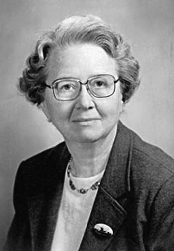
Figure 2 Ruth Wittemore. She was the first fellow of Helen Taussig and the first Director of Paediatric Cardiology at Yale University. From the Yale Bulletin and Calendar obituary in January, 2002.
Frank Gray also came to Yale then. Previously, Gray had been an internal medicine resident at Johns Hopkins, where he worked in the cardiac catheterisation laboratory with physiologist Richard Bing. Joining Frank Gray in the cardiac catheterisation laboratory at Yale, Paul Lurie was among the first individuals, if not the first individual, in the United States of America exclusively trained in paediatrics to perform cardiac catheterisations in children. Ruth Whittemore, Frank Gray, and Paul Lurie launched paediatric cardiology at Yale. Paul Lurie relays the following about Ruth Whittemore during their time at Yale: “I only cath’d two days a week. The rest of the time, I worked with Ruth seeing patients, reading electrocardiograms, writing reports, and so on. She had her own clinic space equipped with her own fluoroscope and good supply of barium. You can see the Taussig influence. We had a warm relationship with mutual respect, dating back to being interns together on a very small service under Dr Powers, who thought we were all great people, and we responded to that confidence. She was a much more compulsive physician than I was, but I went along with her because she knew so much more cardiology than I did. She was kind with patients and parents and spent a lot of time explaining and listening. The cath lab didn’t interest her much. She gowned up and came into the lab and helped in the early days but less and less as Frank and I appeared to be trustworthy”.
Paul provided me with a firsthand account about the early days of cardiac catheterisation and angiography based on his experiences at Yale between 1948 and 1950.
My recollections may not be perfect, but I am comforted by the thought, perhaps in error, that there is no one around to challenge them.
I knew that I was not a real pioneer. Nelson Ordway, former Powers [Grover Powers Chief of Paediatrics at Yale] resident a few years ahead of me was back at Yale for a short period before going on to run a Paediatric department at LSU [Louisiana State University in New Orleans]. He [Ordway] had worked very hard on getting a lab set up to perform blood oxygen analyses (Roughton-Scholander method, which I doubt is in use anywhere today) and respiratory gas analyses on the Van Slyke apparatus. We were simply allowed to walk in and take that over, the space, the able technician, and the apparatus and other equipment.
The actual lab in which the catheterizations were done had been initiated by the adult cardiologists, but after we got started we never saw them. I have no idea why Drs. Geiger and Goodyer [adult cardiologists] abandoned the scene so readily; probably they had more pressing interests and figured that Frank Gray would take care of any adults that needed caths. In any event, they had gotten a room set up with an old X-ray table contributed by Dr. Hugh Wilson of Radiology. It did not have a table-mounted fluoroscope. To visualize the catheter you held a twenty-pound gadget that had an eyepiece on one end and a small fluoroscopic screen at the far end of a foot-long, light-proof tunnel. In order to see anything you had to wear red goggles for about a half hour to get maximally dark-adapted. A couple of extremely heavy lead aprons were also provided by the radiologists. Exposure badges had not come into fashion. Of course, there was no air conditioning.
Another essential feature of the room was a very high ceiling. This allowed us to measure (I should say estimate) hydrostatic pressures with an infusion pole augmented with some yardsticks and lots of adhesive tape.
Our very first electrocardiograms were made on a string galvanometer directly descended from Einthoven. The catheters were woven, lacquered, designed for ureteral use. We rinsed them for hours and re-used them for months!
The fact that Frank [Gray] had done some caths with Richard Bing gave me a feeling of reasonable confidence. The other reassuring knowledge was that Cournand and Richards had been doing caths on patients in shock in the accident ward at Bellevue without harming them.
We started by doing a few adults before we moved down into kids but we did not get into really tiny infants. Any entrance into the left side of the heart or aorta was terra incognita and better get out fast. We did no contrast studies in the cath lab. Our emphasis was more on circulatory physiology than anatomic diagnosis. (This was Bing’s influence on Frank who continued to do pulmonary physiology in adults.) We tried valiantly to get reasonable results with the three-bag inhaled carbon dioxide equilibration indirect method for determining cardiac output comparing the results with direct measurements of blood oxygen content. That whole effort came to naught but I guess it made us look like scientists. Dan Darrow nominated me for Sigma Xi [founded in 1886 to honor excellence in scientific investigation] on the basis of such nonsense.
We did a few antecubital vein injections of a very irritating early contrast medium, Diodrast, through a large bore needle. These were done in the X-ray department with lots of cooperation. I remember holding the arm aloft so as to get the help of gravity to empty the vein. Filming was done without an X-ray film changer, as I recall on a table with a Bucky [X-ray equipment], which permitted two films about two seconds apart, single plane. If our timing was lucky, we could get a reasonable look at the right heart but nothing much at all if we were interested in the left side.
Sedation varied a lot as we kept trying various methods. My favorite was none at all, just local and sweet talk, which worked surprisingly well.
I look back on this early period and cannot think of a time that I was either terribly scared or just plain bored. I really was enjoying the whole thing. It was especially delightful to have the gradual acquisition of new equipment, which made our activities in the cath lab easier and more accurate. That first two-channel recorder with direct writing electrocardiography and pressure! Wow, what a change that made! The table-mounted fluoroscopic screen, another Wow! The ear oximeter from Earl Wood at the Mayo Clinic that I went there to get and be shown how to operate, another Wow! “I guess you get the idea of how it was from all this. In a word, GREAT.
Paul Lurie also relates that his good friend, Alexander Nadas, visited him at Yale in 1949 to gather firsthand information about setting up a paediatric catheterisation laboratory. Previously, internist Lewis Dexter had undertaken the earliest cardiac catheterisations at the Peter Bent Brigham Hospital. Nevertheless, the Children’s Hospital had no facility. Another internist, Walter Goodale, had an interest in congenital cardiac anomalies. Following the visit by Alexander Nadas with Paul Lurie, Goodale organised the first catheterisation laboratory at Boston Children’s Hospital, which a few years later Nadas placed under the leadership of Abraham Rudolph.
At Yale, Paul also worked with William Glenn, who in 1948 succeeded Harris Shumacker as the cardiac surgeon. Paul says of working with Glenn, “Collaboration with the surgeon was close because Bill Glenn was a pretty cautious guy. I remember getting several phone calls at home late in the evening before surgery…. But after all, what could he do? Just add a duct or subtract a duct. Usually the best diagnosis we could come up with before surgery was enough for that”. In 1958, Glenn published his results with the shunt created from the superior caval vein to the right pulmonary artery.Reference Glenn7
Riley Children’s Hospital in Indiana
In 1950, Paul and Barbara Lurie drove their oversized, black Oldsmobile 98 out West on a job-hunting trip. Paul eventually accepted the position as the first Chief of Paediatric Cardiology at Riley Children’s Hospital in Indianapolis, Indiana. Working at Riley, Lurie was the first to adapt the method of percutaneous entry, devised by Seldinger, for cardiac catheterisation in children (Fig 3).Reference Lurie, Armer and Klatte8 In 1953, Sven-Ivar Seldinger, a Swedish radiologist, building on previous work by a Harvard professor, E. Converse Peirce,Reference Peirce9 devised a percutaneous technique for adult angiography.Reference Seldinger10 Then, it was standard to enter a vessel by cutdown. Paul became enamoured with percutaneous entry in the late 1950s, and explains why he promoted it for children: “I was a poor surgeon and hated suturing vessels, never feeling sure they would recanalize. When I first heard about the Seldinger technique from Ross Jennings, Walter Judson’s technician down the hall, I went for it”. Paul further recounts that he presented his initial experience to a small group gathered at an early meeting of the cardiology section of the American Academy of Paediatrics. Presentations then were in the evening, informal, and after dinner. Paul was last and began by “Well for those who are still awake I will be presenting dessert”. Simultaneous to his experience with percutaneous entry, Paul and his colleagues were among the first to use an image intensifier and cinecardioangiography for congenital cardiac malformations.Reference Watson, Pickard, Lowe and Hill11–Reference Klatte, Campbell and Lurie13 Also at Riley, Paul Lurie studied the fascinating postural effects of tetralogy of Fallot, one of the only researchers to do such work.Reference Lurie14 He was also an early advocate of physical fitness and step-exercise testing in children with cardiac problems (Fig 4). Yet, after 17 years in Indiana, Paul decided it was time to move on.

Figure 3 Title page to Paul Lurie’s first report of using the Seldinger technique for vascular access in children during cardiac catheterisation. He signed this reprint to me during my fellowship at Childrens Hospital Los Angeles more than 30 years ago.

Figure 4 Paul Lurie in the early 1950s using an early ear oximeter during step exercise at Riley Children’s Hospital (courtesy of the Riley Children’s Hospital historical website).
Childrens Hospital Los Angeles in California
In 1967, the Chief of Paediatric Cardiology at Childrens Hospital of Los Angeles, Donald C. Fyler, returned to Boston Children’s Hospital. The same year, Childrens Hospital of Los Angeles recruited Paul Lurie to be the new Chief of Paediatric Cardiology. Childrens Hospital of Los Angeles is located on Sunset Boulevard, near Hollywood. Paul’s childhood friend, Izzy Demsky, had become a movie star, but Paul and Barbara Lurie had little time to socialise with Kirk Douglas. Paul continued to refine his technique for percutaneous entry for cardiac catheterisation in children. Manufactured dilators for small vessels were then uncommon, so Paul created his own by heating Teflon tubing over a Bunsen burner, pulling it out, allowing it to harden, and then cutting it at the right spot to make a deftly tapered dilator. Paul demonstrated this technique to me during my fellowship. Nonetheless, entry by cutdown remained popular. The tide began to turn when Abraham Rudolph visited Paul, and spent a day in the cardiac catheterisation laboratory observing his percutaneous techniques. With the interest of paediatric cardiologists like Abe Rudolph and others, percutaneous entry became the standard method in the catheterisation laboratories, intensive care units, and throughout hospitals.
In 1976, Paul undertook a sabbatical for 6 months at Hammersmith Hospital in London, where he studied with the acclaimed histochemist, Anthony Guy Everson Pearse.Reference Van Noorden15 Paul had an interest in myocardial disease. At Hammersmith, Paul worked with excised infundibular myocardium, which he was attempting to culture. Clinically, myocardial biopsy was in its infancy at the time. The technique was first reported in 1962 by Sakakibara and Konno in Japan.Reference Sakakibara and Konno16 During his experience at Hammersmith, Paul concluded that clinical myocardial biopsy could help unravel the classification of myocardial disease in infants. Paul learned that Peter J. Richardson, at King’s College Hospital in London, was then the only physician in Britain performing myocardial biopsies on adult patients with cardiomyopathies. Lurie requested to spend time with Richardson and discovered that he was using an Olympus biopsy forceps, appropriated from an endoscope. Promptly following his experience with Richardson, Lurie drove out to the Olympus Company located in Southend-on-Sea on the east coast of England, where he bought a biopsy forceps, packed in his suitcase with hastily purchased carpentry tools to divert the customs officials, and brought it back to Los Angeles.
On his return to Childrens Hospital of Los Angeles in 1977, Lurie set out to establish the clinical efficacy and safety of myocardial biopsy in infants and young children. In the United States, the use of myocardial biopsy was confined to the Adult Cardiac Transplantation Center at Stanford University. The administration and Department of Paediatrics at Childrens Hospital supported the efforts of Lurie. Paul applied for a grant from the National Institutes of Health that was swiftly and bluntly rejected. Not to be deterred, Paul launched experiments on dogs and rabbits, and became convinced of the safety of the procedure in small animals. With hospital approval, Paul began biopsies in infants and young children with cardiomyopathy, and presented his results in 1978.Reference Lurie, Fujita and Neustein17 Paul collaborated with pathologist Harry Neustein in these studies. They hoped that it might be possible to classify cardiomyopathies on the basis of ultrastructural primary lesions, as had been done in nephrology. As Paul admits now, in the cardiomyopathies, that might take many years and give indeterminate results. In contrast, the geneticists today, with their expertise and technology, are able to classify these diseases quickly down to fine biochemical aberrations. The team headed by Lurie and Neustein, however, was able to produce some useful reports; for example, the family with endocardial fibroelastosis and X-linked mitochondrial disease that was later expanded by Barth into the syndrome that properly bears his name.Reference Neustein, Lurie, Dahms and Takahashi18–Reference Barth, Scholte and Berden20 Paul retired 4 years later, and a grateful family donor supported trips by Lurie to more than 20 university medical centres for children in the United States to teach his technique for myocardial biopsy (Fig 5).

Figure 5 This picture was taken with the paediatric cardiology faculty in 1984 when Paul Lurie visited Childrens Hospital Los Angeles 2 years after his retirement. From left to right – Paul Lurie, Robert Stanton, Arno Hohn (Division Chief then), Masato (Mike) Takahashi, Tsun-Yee (Sunny) Lawrence, and Alan Lewis (courtesy of Paul Lurie).
An active “retirement”
Retired from the Childrens Hospital Los Angeles, Paul moved to Albany, New York, where in 1982 he was appointed as Professor of Paediatrics at Albany Medical College. In Albany, Paul founded the paediatric cardiomyopathy registry. The idea for the registry germinated during his trips to various medical centres while demonstrating his technique for cardiac biopsy. Paul Lurie had become acquainted with Bill Cook, of Cook Incorporated, during the time when Paul was at the Indiana University. Paul visited with Bill Cook in Bloomington, Indiana, around 1982 and Cook offered his help to support the fledgling cardiomyopathy registry with a grant of $25,000. With this money, Paul bought a computer and database programme, and painstakingly began entering data on the first patients himself. Early collection of data was slow, as Paul personally cajoled those working in paediatric cardiology to provide information on patients with cardiomyopathy. Some years later, nonetheless, several of our colleagues stepped up to help build the registry into a significant repository of information that aids medical research into the still vexing problem of paediatric cardiomyopathy.21 Paul especially credits Steven Lipshultz at the University of Miami, Lynn Sleeper at the New England Research Institutes, Jeff Towbin at Cincinnati Children’s Hospital, and Steve Colan at Boston Children’s Hospital for making the registry its current success.
Paul Lurie is now in his 90s, but he is still active academically. He recently published a commentary on ventricular non-compaction,Reference Lurie22 and he is currently working on a similar commentary on endocardial fibroelastosis. Paul Lurie is one of our innovators and founders of the field of paediatric cardiology. His contributions have been fundamental, and his influence on others is far-reaching. It gives me great pleasure to offer his story for his induction to the Paediatric Cardiology Hall of Fame.


