


Introduction
The Western honey bee (Apis mellifera L.) is considered the primary managed pollinator worldwide, and contributes significantly to the pollination of monoculture food crops (Potts et al., Reference Potts, Imperatriz-Fonseca, Ngo, Aizen, Biesmeijer, Breeze, Dicks, Garibaldi, Hill, Settele and Vanbergen2016). However, the on-going winter losses in the managed colonies of European-derived subspecies, particularly in North America, across Europe and the Middle East, represent a significant threat to healthy and sustainable human diet (Lee et al., Reference Lee, Steinhauer, Rennich, Wilson, Tarpy, Caron, Rose, Delaplane, Baylis, Lengerich and Pettis2015; Kulhanek et al., Reference Kulhanek, Steinhauer, Rennich, Caron, Sagili, Pettis, Ellis, Wilson, Wilkes, Tarpy and Rose2017; Gray et al., Reference Gray, Brodschneider, Adjlane, Ballis, Brusbardis, Charrière, Chlebo, Coffey, Cornelissen, Amaro da Costa and Csáki2019). Although evidence suggests that abiotic stressors such as exposure to agro-chemicals contribute to these colony losses, the effects of biotic stressors, chiefly, the invasive ecto-parasitic mite Varroa destructor and its associated viruses are most damaging (Francis et al., Reference Francis, Nielsen and Kryger2013). The genus Varroa (Acari: Varroidae) comprises of four species namely V. destructor, V. jacobsoni, V. rindereri and V. underwoodi (Anderson and Trueman, Reference Anderson and Trueman2000; Roberts et al., Reference Roberts, Anderson and Tay2015), of which V. destructor is considered the most widespread and economically damaging, especially the lineage of the Korean haplotype of this species (Lin et al., Reference Lin, Qin, Page, Wang, Li, Wen, Hu, Neumann, Zheng and Dietemann2018).
Varroa destructor has crippled the apicultural industry around the globe since it moved from its original host, the Eastern honey bee A. cerana to its new host, A. mellifera, which lacks the adaptive mechanisms that limit the reproductive capacity of the mite in its native host (Rosenkranz et al., Reference Rosenkranz, Aumeier and Ziegelmann2010). The mite feeds primarily on the bee's fat body but also on the haemolymph (Ramsey et al., Reference Ramsey, Ochoa, Bauchan, Gulbronson, Mowery, Cohen, Lim, Joklik, Cicero, Ellis and Hawthorne2019) and vectors pathogenic viruses, which have become more virulent due to the suppression of the honey bee immune response caused by Varroa parasitism (Francis et al., Reference Francis, Nielsen and Kryger2013; Annoscia et al., Reference Annoscia, Del Piccolo, Covre and Nazzi2015). Many strategies have been used to control this parasite to improve the health status of susceptible European honey bee subspecies including chemical treatment with synthetic acaricides, selective breeding of Varroa-tolerant stocks, biotechnical and biological intervention (Rosenkranz et al., Reference Rosenkranz, Aumeier and Ziegelmann2010; Plettner et al., Reference Plettner, Eliash, Singh, Pinnelli and Soroker2017). Among these, chemical treatment with synthetic acaricides remains the commonly used strategy to control the parasite population in the colonies to date (Rosenkranz et al., Reference Rosenkranz, Aumeier and Ziegelmann2010; Plettner et al., Reference Plettner, Eliash, Singh, Pinnelli and Soroker2017). Unfortunately, acaricide-based Varroa control measures are not sustainable because of their lethal effects on bees, the build-up of chemical residues in hive products and the development of mite-resistant populations (Rosenkranz et al., Reference Rosenkranz, Aumeier and Ziegelmann2010; Locke, Reference Locke2012). Therefore, other management options against the mites are required. Interestingly, the study of bee's cooperative behavioural defences called social immunity has revealed that propolis can mitigate, in part, the negative effects caused by Varroa parasitism and other biotic stressors on honey bee health (reviewed in Simone-Finstrom et al., Reference Simone-Finstrom, Borba, Wilson and Spivak2017).
Propolis is one of many hive products that has been extensively used in folk medicine since ancient times due to its pharmacological properties (Banskota et al., Reference Banskota, Tezuka and Kadota2001; Falcão et al., Reference Falcão, Vale, Gomes, Domingues, Freire, Cardoso and Vilas-Boas2013). Intensive research has demonstrated its antimicrobial, anti-inflammatory, anti-oxidative, anti-bacterial, anti-viral, anti-fungal, anti-tumour properties, among others (Banskota et al., Reference Banskota, Tezuka and Kadota2001). The biological properties of propolis are attributed to its sticky substance called resin or bee glue secreted by intact and wounded plants for defence against herbivores and pathogenic infection (Huang et al., Reference Huang, Zhang, Wang, Li and Hu2014). Although resin is primarily composed of flavonoid, terpene and phenolic compounds, its chemical composition is dependent on the phyto-geographical characteristic of the surrounding area, season and species of honey bee that collect it (reviewed in Anjum et al., Reference Anjum, Ullah, Khan, Attaullah, Khan, Ali, Bashir, Tahir, Ansari, Ghramh and Adgaba2019). Honey bees collect this resinous plant material and once in the hive, they mix it with varying amounts of wax and apparently their glandular secretions to generate propolis, which is then utilized for nest construction and protection against predators and microorganisms (Simone-Finstrom et al., Reference Simone-Finstrom, Borba, Wilson and Spivak2017). Initially, propolis was not selected in breeding programmes as a means of improving the health status of susceptible European honey bee subspecies due to its sticky nature (Simone-Finstrom et al., Reference Simone-Finstrom, Borba, Wilson and Spivak2017). However, it has become a subject of intensive research in the last decades as several studies have demonstrated the benefits of crude propolis and its ethanolic extracts on the health of Africanized and European honey bees. Their effects include reduced colony microorganism loads (Bastos et al., Reference Bastos, Simone, Jorge, Soares and Spivak2008; Simone-Finstrom and Spivak, Reference Simone-Finstrom and Spivak2012; Wilson et al., Reference Wilson, Brinkman, Spivak, Gardner and Cohen2015), reduced individual investment on costly immune activity (Simone et al., Reference Simone, Evans and Spivak2009; Borba et al., Reference Borba, Klyczek, Mogen and Spivak2015) and increased brood viability and worker lifespan (Nicodemo et al., Reference Nicodemo, De Jong, Couto and Malheiros2013, Reference Nicodemo, Malheiros, De Jong and Couto2014).
In relation to Varroa mite, previous studies have shown that the crude propolis envelope found in European honey bee colonies does not affect its survival and infestation levels (Borba et al., Reference Borba, Klyczek, Mogen and Spivak2015; Drescher et al., Reference Drescher, Klein, Neumann, Yañez and Leonhardt2017). In contrast, the crude propolis envelope does significantly affect the dynamics of Varroa-transmitted deformed wing virus in honey bees (Drescher et al., Reference Drescher, Klein, Neumann, Yañez and Leonhardt2017). Also, Drescher et al. demonstrated that the volatiles of propolis had no impact on the survival of the mite in laboratory assays, albeit the propolis used in their study was a year old (Drescher et al., Reference Drescher, Klein, Neumann, Yañez and Leonhardt2017). In contrast, the chemically enriched ethanolic extracts of propolis have been reported to have both narcotic and lethal effects on the mites in laboratory assays (Garedew et al., Reference Garedew, Lamprecht, Schmolz and Schricker2002, Reference Garedew, Schmolz and Lamprecht2003; Damiani et al., Reference Damiani, Maggi, Gende, Faverin, Eguaras and Marcangeli2010). Furthermore, Popova et al. reported that the chemical composition of propolis from Varroa-resistant honey bee colonies is distinct and had a significantly higher concentration of four biologically active compounds (caffeic acid, pentenyl caffeates, caffeic acid phenethyl ester and cinnamyl caffeate) than those of susceptible honey bee colonies (Popova et al., Reference Popova, Reyes, Le Conte and Bankova2014). While numerous field and laboratory studies have demonstrated the significant effects of propolis on the health of both European and Africanized honey bees, similar information remains scarce for the widely diverse African honey bee subspecies, which are known to incorporate huge amounts of propolis within their colonies compared to their European counterparts (Hepburn and Radloff, Reference Hepburn and Radloff1988). Previously, we demonstrated that the surviving African savannah honey bees A. m. scutellata found in Kenya have evolved cooperative defence behaviours such as grooming behaviour and suppression of mite reproduction to reduce Varroa's population build-up in their colonies without requiring any in-hive acaricide treatment (Nganso et al., Reference Nganso, Fombong, Yusuf, Pirk, Stuhl and Torto2017, Reference Nganso, Fombong, Yusuf, Pirk, Stuhl and Torto2018). In this study, we evaluated the effects of natural propolis found in A. m. scutellata colonies and its ethanolic extracts on the health status of this specific honey bee subspecies.
Materials and methods
Experimental apiary
The study was performed during the short rainy (from November to December 2017; temperature 20.9°C, average rainfall 2.0 mm) and cooler dry (from July to August 2018; temperature 18.5°C, average rainfall 0.59 mm) seasons at an apiary in Kithimani (1°8′S, 37°25E) located within the county of Machakos in Kenya. The short rainy season is characterized by the increased availability of flowering plants and consequently increased brood rearing in A. m. scutellata colonies, while the cooler dry season is characterized by the moderate availability of flowering plants and brood rearing (Nganso et al., Reference Nganso, Fombong, Yusuf, Pirk, Stuhl and Torto2017, Reference Nganso, Fombong, Yusuf, Pirk, Stuhl and Torto2018). Within the apiary, we selected randomly six queen right colonies of A. m. scutellata that originated from locally captured swarms for our study. All the colonies were housed in standard Langstroth hives and were subjected to standard beekeeping practices without acaricide treatment for Varroa mite. We previously found that the Varroa mite species in these colonies belonged to the Korean strain (K1 haplotype) (Nganso et al., Reference Nganso, Fombong, Yusuf, Pirk, Stuhl and Torto2017).
Collection of propolis
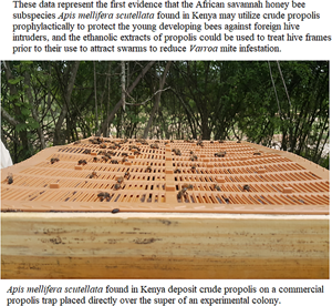
In November 2017 and July 2018, we placed a commercial propolis trap (see Fig. S1) directly over the top frames of the uppermost box (super) of each experimental colony and covered it with the colony lid to encourage the honey bees to seal the openings of the traps with propolis. A month later, December 2017 and August 2018, the traps were removed from the colonies, wrapped with aluminium foil before freezing at −80°C for 24 h in the laboratory located at the International Center of Insect Physiology and Ecology (icipe) Duduville campus, Nairobi (1°16′S; 36°49E). The propolis collected from an individual colony was knocked out from the trap, allowed to thaw at room temperature and weighed. All the propolis collected during each experimental period was mixed and grounded using an electric blender before storage at −80°C. The propolis sample was then used within 5 days in laboratory bioassays.
Assessment of the amount of worker brood and mite-infestation levels on worker honey bees in A. m. scutellata colonies
During each experimental period, the total amount of worker brood per colony was determined as described by Nganso et al. (Reference Nganso, Mani, Altman, Rafaeli and Soroker2020) while the mite-infestation level on approximately 100 worker adult honeybees in each colony was determined as previously described by Nganso et al. (Reference Nganso, Fombong, Yusuf, Pirk, Stuhl and Torto2017).
Assessment of the effects of crude propolis on Varroa mite's survival
Varroa mites were collected by the sugar shake method from six unrelated A. m. scutellata colonies in the experimental apiary (Nganso et al., Reference Nganso, Fombong, Yusuf, Pirk, Stuhl and Torto2017) and transferred in a container lined with moist filter paper with white-eyed pupae present. Only female mites with dark brown colour were used in all laboratory bioassays. The harvested mites were kept in an incubator at 34°C and 55% relative humidity (RH) placed in a dark room at icipe. They were used within 3 h from the time of collection in the field.
We used the method described by Drescher et al. (Reference Drescher, Klein, Neumann, Yañez and Leonhardt2017) to test the effect of propolis volatiles on the survival of mites but with the following modifications. Briefly, 1, 2, 4 and 6 g of grounded propolis were placed each on the lower side of the lid of Petri dishes (Fig. 1a). Fourteen mites were placed in the bottom of each Petri dish lined with moist filter paper. Propolis and mites were separated by a nylon mesh (<0.5 mm), which prevented direct contact but allowed the free release of propolis volatiles. To test the effects of propolis placed in direct contact with the mites, the above propolis weights (g) were placed each in the bottom of Petri dishes lined with moist filter paper and 14 mites were placed in direct contact with the propolis (Fig. 1b). In both experiments, Petri dishes without any propolis were used as control. Three biological replicates were performed for each treatment category. The Petri dishes were then placed in an incubator at 34°C and 55% RH and mite survival monitored under a dissecting microscope every 6 h for up to 36 h. During each observation time, the light was turned on briefly in the dark room. Mites were classified as ‘mobile’ when they were still active (able to move extremities); ‘immobile’ when they were inactive, but still alive (as validated by careful touching with a fine paintbrush); or ‘dead’ when they showed no movement after three subsequent stimulations with a fine paintbrush.

Fig. 1. Experimental setup of mites exposed to volatiles of propolis (a) and placed directly in contact with propolis (b) under laboratory conditions during the rainy and cooler dry seasons.
Assessment of the effects of ethanolic extracts of propolis on Varroa mite's survival
Extraction of propolis
Ethanolic extract bioassays were prepared and carried out using the method described by Damiani et al. (Reference Damiani, Maggi, Gende, Faverin, Eguaras and Marcangeli2010) but with the following modifications. Briefly, 10 g of propolis collected during each experimental period was grounded using an electric blender and extracted with 70% ethanol (1:30 w:v) by sonication for 20 min in an ultrasonic bath at 20°C (Bransonic ultrasonics cleaner, Branson Ultrasonics Corporation, Danbury, CT, USA). The resulting extract was filtered at room temperature using a Whatman grade 1 filter paper (Whatman International Ltd Maidstone England, England). The part trapped in the filter paper was re-extracted twice using the above procedure under the same conditions. From each of the three parallel extractions, 2 mL of the extract was evaporated to dryness in vacuo using a rotary evaporator (Heidolph, Germany) at 40°C. The percentage of balsam P (amount of matter soluble in 70% ethanol) in the propolis sample was calculated using the formula:

where g is the weight of the residue after evaporation of 2 mL of propolis 70% ethanol extract and M is the weight of the crude propolis (g) (Bankova et al., Reference Bankova, Bertelli, Borba, Conti, da Silva Cunha, Danert, Eberlin, Falcão, Isla, Moreno and Papotti2016). The mean of the three values was determined.
The three filtrates were combined and the solvent was evaporated in vacuo at 40°C. The dried propolis extract obtained was then weighed and the yield of extraction was calculated using the formula: Yield (%) = (P e/P m) × 100; where P e is the mass of the residue (g) after evaporation of propolis 70% ethanol extract and P m is the mass of the crude propolis sample (g) (Pujirahayu et al., Reference Pujirahayu, Ritonga and Uslinawaty2014). Acaricidal residues in the propolis sample, which may introduce false results, were not analysed because the colonies at the apiary in Kithimani were never treated with acaricides to control the mite-infestation levels and no subsistence farms were found at the vicinity of the apiary. The soft propolis extract obtained after evaporation was dissolved in 55% ethanol to reduce the effect of strong ethanol solution on the experimental mites. The following concentrations were prepared and used in the laboratory bioassay: 5, 7 and 10% (w/v).
Performance of bioassays
To test the effect of the ethanolic extract of propolis on the survival of the mites, the method described by Damiani et al. (Reference Damiani, Maggi, Gende, Faverin, Eguaras and Marcangeli2010) was used but with the following modifications. Briefly, 200 μL of each of the above propolis ethanolic concentrations were applied directly to the dorsal side of the idiosoma of six mites placed on a Petri dish lined with a piece of moist filter paper (3 × 3 cm). Similarly, control treatments were prepared by directly applying 200 μL of 55% ethanol solution to the dorsal side of the idiosoma of six mites placed on a Petri dish lined with a piece of moist filter paper (3 × 3 cm). Three replicates were used for each treatment concentration. All treatments were carried out at room temperature (22–24°C) and all the Petri dishes were incubated at 34°C and 55% RH. Mite survival was subsequently monitored under a dissecting microscope at 10, 20, 30, 40, 60, 120, 180, 240, 300 and 360 min (total = 10 observation points). Mites were classified as ‘mobile’ when they were still active (able to move extremities) or ‘dead’ when they showed no movement after three subsequent needle stimulations.
Statistical analysis
All statistical analyses were performed using R Software version 3.6.3 (R Development Core Team, 2020). We used Mann–Whitney–Wilcoxon test to compare the weight of propolis collected (g), balsam amount in the propolis (%), amount of worker brood and mite-infestation levels on adult workers between the hot and cooler dry seasons as the data were not normally distributed and their variance was not homogenous (Bartlett's test: P < 0.05) (Dalgaard, Reference Dalgaard2008). We also conducted spearman's rank-order correlation analysis to examine if there is a possible relationship between the weight of propolis collected and the amount of worker brood or mite-infestation levels on adult workers during each experimental period.
Using the Kaplan–Meier estimator in the ‘survival’ R package (Therneau, Reference Therneau2020), we assessed the survival probability of mites exposed to the volatiles of propolis and placed in direct contact with the propolis and test for differences between propolis samples and the control. For survival analyses, we combined data for the categories ‘immobile’ and ‘dead’ and compared to the number of mobile mites for each treatment group as described previously by Drescher et al. (Reference Drescher, Klein, Neumann, Yañez and Leonhardt2017).
The generalized linear mixed model (GLMM) assuming binomial distribution with logit link function was used to test the effects of concentration of ethanolic extracts of propolis, observation intervals and their interaction on the proportion of dead mites. The experimental units (mites) were considered as a random effect in the GLMM model to cater for possible correlation in the data due to observation intervals while the factors under study were considered as fixed effects. The Akaike's information criterion was used for assessing the quality of our model through comparison of related models. Statistical significance of pairwise differences was evaluated using Tukey's test in multcompView package (Piepho, Reference Piepho2004).
The generalized linear model (GLM) with negative binomial error distribution and logit link was also used to compare the total proportion of dead mites across all the observation intervals between the short rainy and cooler concentration of ethanolic extracts of propolis. The function ‘glm.binomial.disp’ in the package ‘MASS’ was used for this analysis (Scrucca, Reference Scrucca2018).
Results
Propolis intake and colony strength
The total amount of propolis collected from the experimental A. m. scutellata colonies during the short rainy season (mean ± s.e.: 41.4 ± 6.7 g) was significantly higher, approximately two and half-fold more than the amount of propolis collected during the cooler dry season (mean ± s.e.: 18.8 ± 1.4 g) (Wilcoxon rank-sum test: W = 36, P = 0.002). Also, the balsam amount in the propolis collected during the short rainy season (68 ± 1.7%) was higher than that obtained in the propolis collected during the cooler dry season (40 ± 12.6%), though this difference was not significant (Wilcoxon rank-sum test: W = 8.5, P = 0.12).
The total amount of worker brood recorded during the short rainy season (11 228 ± 1469 worker brood/colony) was higher than that recorded during the cooler dry season (8420 ± 1075 worker brood/colony), though this difference was not significant (Wilcoxon rank-sum test: W = 25, P = 0.31). In contrast, the mite-infestation levels on adult workers/colony recorded during the short rainy season (3.0 ± 0.7 mites/100 adult worker) were significantly lower, 2-fold less than that recorded during the cooler dry season (6.0 ± 1.2 mites/100 adult worker) (Wilcoxon rank-sum test: W = 29, P = 0.01).
During the short rainy season, the amount of propolis collected per colony ranged from 19.8 to 61.7 g and was correlated with neither the amount of worker brood (Spearman's rank correlation: r = −0.14, P = 0.80) nor mite-infestation level on adult workers (Spearman's rank correlation: r = 0.49, P = 0.32) per colony. Likewise, during the cooler dry season, the amount of propolis collected per colony ranged from 3.8 to 13.1 g and was correlated with neither the amount of worker brood (Spearman's rank correlation: r = −0.54, P = 0.30) nor mite-infestation level on adult workers (Spearman's rank correlation: r = 0.43, P = 0.39) per colony.
Effects of crude propolis on Varroa mite's survival
We found that mites exposed to propolis volatiles or placed in direct contact with the propolis lived as long as the untreated mites. In fact, the survival of Varroa mites exposed to propolis volatiles (P = 1, Fig. 2a) and placed in direct contact with the propolis (P = 0.51, Fig. 2b) was not significantly different between propolis samples and the control across times during the short rainy season. Likewise, during the cooler dry season, the survival of Varroa mites exposed to volatiles of propolis (P = 0.97, Fig. 2c) and placed in direct contact with the propolis (P = 1, Fig. 2d) was not significantly different between propolis samples and the control across times.

Fig. 2. Kaplan–Meyer survival curves of Varroa destructor exposed to volatiles of propolis (a and c) and placed directly in contact with propolis (b and d) under laboratory conditions during the short rainy and cooler dry season, respectively.
Effect of ethanolic extract of propolis on Varroa mite's survival
During the short rainy season, treatment concentration (GLMM: χ 2 = 36.59, d.f. = 3; P < 0.001), observation intervals (GLMM: χ 2 = 229.37, d.f. = 1, P < 0.001) and their interaction (GLMM: χ 2 = 30.90, d.f. = 3, P < 0.001) had a significant effect on mite's mortality (Fig. 3a). In the same vein, treatment concentration (GLMM: χ 2 = 45.94, d.f. = 3, P < 0.001), observation intervals (GLMM: χ 2 = 272.28, d.f. = 1, P < 0.001) and their interaction (GLMM: χ 2 = 24.55, d.f. = 3, P < 0.001) had a significant effect on mite's mortality during the cooler dry season (Fig. 3b). In fact, we found that mite's mortality generally increased with increasing propolis concentration while observation intervals had a negative quadratic relationship with mite's mortality across the two seasons. The mortality of the mites was high at the initial observation intervals but then reduced over time and increased again at higher observation intervals.

Fig. 3. Mean per cent mortality of Varroa mites at different treatment concentrations (control, 5, 7.5 and 10%) over ten progressive observation intervals (min) during the short rainy (a) and cooler dry season (b).
We also found that the total proportion of dead mites recorded after 360 min for each concentration of ethanolic extracts of propolis, 5% (glm.binomial.disp: χ 2 = 0.06, d.f. = 1; P = 0.80), 7.5% (glm.binomial.disp: χ 2 = 1.56, d.f. = 1; P = 0.21) and 10% (glm.binomial.disp: χ 2 = 0.54, d.f. = 1; P = 0.46) was not significantly different between the short rainy and cooler dry season.
Discussion
In this study, we anticipated that the African honey bees would deposit larger amounts of crude propolis in their colonies during the period of increased (short rainy season) vs reduced (cooler dry season) worker brood rearing to provide a prophylactic defensive barrier to the developing honey bees against microorganisms and hive intruders (Simone-Finstrom et al., Reference Simone-Finstrom, Borba, Wilson and Spivak2017). This is because previous studies have provided support of the adaptive significance of self-medication in honey bees as resin collection positively correlated with the levels of parasites and pathogens (Simone-Finstrom and Spivak, Reference Simone-Finstrom and Spivak2012; Drescher et al., Reference Drescher, Klein, Neumann, Yañez and Leonhardt2017). Moreover, the positive effects of crude propolis inside the hive on host fitness traits such as brood viability, worker lifespan, hygienic behaviour, honey production and pollen stores are well recognized (Nicodemo et al., Reference Nicodemo, De Jong, Couto and Malheiros2013, Reference Nicodemo, Malheiros, De Jong and Couto2014). As expected, we found that as brood density increased to approximately one and half-fold during the short rainy season, the quantity of propolis deposited by this honey bee subspecies in their colonies also increased to approximately two and half-fold, compared to typical cooler dry season density. Although the quantity of propolis did not correlate with brood density because of the small sample size of six colonies used in our study, our result suggests that this African honey bee subspecies may use propolis therapeutically to maintain a sterile environment within the nest, which is crucial for the proper development of their brood (Simone-Finstrom and Spivak, Reference Simone-Finstrom and Spivak2010).
In this study, we also observed no correlation between the quantity of propolis deposited in the nest and mite-infestation levels on adult African honey bees. Our results corroborate with those of previous studies in European honey bee colonies, which also reported no significant effects of the natural propolis envelope on mite loads using either our same or larger sample size of six colonies (Borba et al., Reference Borba, Klyczek, Mogen and Spivak2015; Drescher et al., Reference Drescher, Klein, Neumann, Yañez and Leonhardt2017). In contrast to this and previous findings, Pusceddu et al. found that the quantity of resin collected by the European honey bee, A. mellifera ligustica correlated with the levels of mite infestation (Pusceddu et al., Reference Pusceddu, Floris, Mura, Theodorou, Cirotto, Piluzza, Bullitta, Angioni and Satta2018). These contrasting effects of natural propolis on mite-infestation levels could be a result of the variable chemical composition of the resinous fraction of propolis in the differential geographical areas where these studies were conducted.
Our finding that propolis had no significant effect on Varroa mite loads at the colony level across the two seasons was further supported by our laboratory results, which showed that mites exposed directly to crude propolis via contact or exposed to the volatiles lived as long as the untreated mites (Fig. 1). We observed that mites placed directly on the crude propolis move relatively slow when compared to those exposed to propolis volatiles due to the sticky nature of the propolis. In general, Varroa mites may be unintentionally exposed to propolis volatiles inside the honey bee colonies but it still remains unclear if they can actually get in direct contact with crude propolis. It is important to note that these mites are mostly found on a bee: a forager bee, which carries them from one hive to another, or a nurse bee which carries them within a brood cell to reproduction, though they can sometimes move for a brief period on the surface of the comb (Nganso et al., Reference Nganso, Mani, Altman, Rafaeli and Soroker2020). Thus, it seems very likely that Varroa mites, unlike other external invaders, e.g. microorganisms, lizards, snakes, etc., may not come in close contact with the natural propolis envelope deposited by the honey bees on the hive entrance, holes and cracks. Our results agree with those of Drescher et al., who demonstrated no effects of propolis volatiles on the lifespan of V. destructor despite the fact that the propolis they tested in laboratory bioassay was not fresh but a year old (Drescher et al., Reference Drescher, Klein, Neumann, Yañez and Leonhardt2017).
In this study, the direct application of the propolis ethanol extract to the mites collected from the African savannah honey bee colonies caused significant and dose-dependent lethal effects on V. destructor across the two study periods (Fig. 2). In fact, approximately 40–65% of mites died within the first 10 min of the bioassay and only about 17–24% remained viable after 360 min. In contrast, practically none of the control mites died within the first 10 min of the bioassay and more than 80% remained viable at the end of the experiment. We also found that 5 and 10% ethanolic extract of propolis on average caused 80 and 100% mite mortality, respectively, after 360 min. This suggests that the bioactive compounds found in the resinous fraction of propolis are both non-volatile given the bath temperature and reduced pressure during rotatory evaporation, and relatively polar since extracted into ethanol. Pusceddu et al. (Reference Pusceddu, Floris, Mura, Theodorou, Cirotto, Piluzza, Bullitta, Angioni and Satta2018) noted narcotic effects on mites exposed to crude propolis samples collected from A. m. ligustica colonies, though as they suggested, these effects were minimal when compared to those caused by the ethanolic extracts of propolis samples collected from other A. mellifera colonies (Garedew et al., Reference Garedew, Lamprecht, Schmolz and Schricker2002; Damiani et al., Reference Damiani, Maggi, Gende, Faverin, Eguaras and Marcangeli2010).
Despite these results, we believe that the detrimental effects of the ethanolic extracts of propolis do not mirror the actual situation within the honey bee hive as practically no mites may get in direct contact with propolis in the hive as was suggested earlier by Drescher et al. (Reference Drescher, Klein, Neumann, Yañez and Leonhardt2017). More so, as a management tool for Varroa mites, the direct application of an ethanol extract is likely not practical. Thus, future studies should examine how crude propolis enhances colony longevity, productivity and bee's immune response against viruses vectored by the mites. Nevertheless, it would also be interesting to perform bioassay-directed fractionation of the ethanolic extract with the intent to identify the active ingredient(s) for the eventual control of mites and improvement of African honey bee health. Additionally, whilst our results showed similar acaricidal activity of the ethanolic extracts of propolis collected during the two distinct seasons on mite mortality, the origin, seasonal variation and plant extract composition should also be thoroughly explored to provide more robust data on the resinous plant species that best support the health of this African honey bee subspecies. In fact, there is some evidence that honey bees can discriminate among resinous plant species while foraging in the same landscape (Bastos et al., Reference Bastos, Simone, Jorge, Soares and Spivak2008; Wilson et al., Reference Wilson, Spivak, Hegeman, Rendahl and Cohen2013). In addition, variability in resin composition and antimicrobial activity between plant species in different regions and among closely related has been demonstrated (Bastos et al., Reference Bastos, Simone, Jorge, Soares and Spivak2008; Wilson et al., Reference Wilson, Spivak, Hegeman, Rendahl and Cohen2013, Reference Wilson, Brinkman, Spivak, Gardner and Cohen2015).
In conclusion, this study is the first to report on the contribution of propolis to the health of African honey bee colonies. Our finding suggests that this specific African honey bee subspecies may utilize crude propolis prophylactically to protect the young developing bees against foreign hive intruders. Furthermore, our result suggests that ethanolic extracts of propolis could be used to treat hive frames prior to their use to attract swarms to reduce Varroa mite infestation.
Supplementary material
The supplementary material for this article can be found at https://doi.org/10.1017/S0031182021000305
Acknowledgements
We gratefully thank C. Nzuki in Kenya for availing their apiaries for this study. We are also grateful to Muema Wilson, Onyimbo Nixon, Munyao Mutemwa, Gitari Macharia and Xavier Cheseto in Kenya for their assistance in the field and laboratory work. We also thank John J. Beck in USDA-ARS, CMAVE, Gainesville, Florida, USA for his critical review of the manuscript.
Author contributions
B.T. and B.N. conceived and designed the study. B.N. conducted data gathering and performed statistical analyses. B.T. and B.N. wrote the article.
Financial support
This research was supported financially by the following organizations and agencies: United States Department of Agriculture (USDA), ARS Grant #58-6615-3-011-F; UK's Department for International Development (DFID); Swedish International Development Cooperation Agency (Sida); the Swiss Agency for Development and Cooperation (SDC); and the Kenyan Government. Immense gratitude to the German Academic Exchange Service (DAAD) In-Region Scholarship for funding the research work through a PhD fellowship at the International Centre of Insect Physiology and Ecology (icipe) of Beatrice T. Nganso and the Office of International Research Programs at USDA-ARS for providing the financial support needed for the research conducted in the USA.
Conflict of interest
None.
Ethical standards
Not applicable.


