


Introduction
The World Health Organization defines an aneurysmal bone cyst as a ‘benign cystic lesion of bone composed of blood-filled spaces separated by connective tissue septa containing fibroblasts, osteoclast-type giant cells and reactive woven bone’.Reference Rosenberg, Nielsen, Fletcher, Fletcher, Unni and Mertens1
Aneurysmal bone cysts primarily occur in young females.Reference Hamilton, Voorhies, Wilkins and Rengachary2 While involvement of the tibia and femur is typical, presentation within the head and neck region is thought to occur in 12 per cent of cases.Reference Barnes3, Reference Kaffe, Naor, Calderon and Buchner4 The precise aetiology of aneurysmal bone cysts remains elusive. The condition had generally been conceptualised as ‘reactive’, until reports of clonal cytogenetic abnormalities indicated that the lesions were neoplastic. Lesions generally arise in isolation, although aneurysmal bone cyst like features may sometimes coexist with other bony lesions. A short history of pain and swelling in the affected area is the characteristic presentation, although symptoms and signs are dependent upon the site involved.Reference Martinez and Sessone5 The diagnosis of these benign but potentially very destructive lesions is evolving and improving as information about the cytogenetic features of giant cell rich bone lesions of the head and neck emerges.
The prominence of the cystic component of aneurysmal bone cyst is variable. When the cystic component is negligible, lesions histologically identical to aneurysmal bone cyst may, when occurring in the maxillofacial bones, be termed giant cell reparative granuloma or central ossifying fibroma. These terms, while essentially synonyms for ‘aneurymal bone cyst, solid variant’ are by convention used for lesions in the head and neck. Rarely, tumours histologically identical to aneurysmal bone cyst have been reported in soft tissue, sometimes termed ‘aneurysmal bone cyst of soft tissue’.Reference Nielsen, Fletcher, Smith, Rybak and Rosenberg6
The aim of this study was to examine the presentation, management and outcome of cases of head and neck aneurysmal bone cyst seen at Children's Hospital Boston; we also aimed to review current trends and theories regarding the diagnosis and treatment of aneurysmal bone cyst.
Materials and methods
A retrospective hospital approved chart review was undertaken of all patients presenting to Children's Hospital Boston between 1991 and 2006 with an aneurysmal bone cyst in the head and neck area. Patients were excluded from the study if: the lesion lacked a significant cystic component (i.e. befitting a diagnosis of giant cell reparative granuloma or central ossifying fibroma); the lesion occurred outside the head and neck region; the medical documentation was incomplete; or the patient was older than 18 years. Demographic details, radiological and pathological findings, management, and outcome were all evaluated.
Results
A total of nine patients were diagnosed with an aneurysmal bone cyst within the head and neck area during the study time period. Three patients were excluded from this group of 9: two patients with an aneurysmal bone cyst of the C7 vertebra, and a third patient whose documentation was incomplete. Patients' individual details are highlighted in Table I.
Table I Clinicopathological features of six patients with aneurysmal bone cysts of the head and neck

Pt no = patient number; mth = months; y = years; M = male; F = female
Of the six remaining patients, an equal number of males and females were affected, with a mean initial age at presentation of 9.6 years (range, eight months to 17 years). The mandible was the site most commonly involved (being involved in three patients). Individual cases of aneurysmal bone cyst of the maxilla, ethmoid and posterior neck soft tissue were also diagnosed. One patient had an asymptomatic presentation, with a lesion detected incidentally on computed tomography (CT) imaging requested for lip swelling following minor trauma. Three patients presented with an expanding mass with or without paraesthesia in the mandibular region. The patient with the ethmoidal aneurysmal bone cyst presented with a nasal mass, and a mass was noted on nasendoscopy. The final presentation was that of a swelling of the posterior neck, below the occipital prominence. Patients reported the presence of symptoms for a mean of 19 days (range, 10 days to one month).
All patients underwent pre-operative plain film, CT and magnetic resonance imaging (MRI) (see Figures 1, 2 and 3). Expansile soft tissue masses were identified in a left anterior ethmoid sinus and a right anterior maxilla (with an associated perforation of the floor of the nose). Expansile cystic lesions of the mandible were identified in three patients, while multiple fluid levels were visible on MRI of the occiptal lesion. Pre-operative embolisation was not performed for any case.
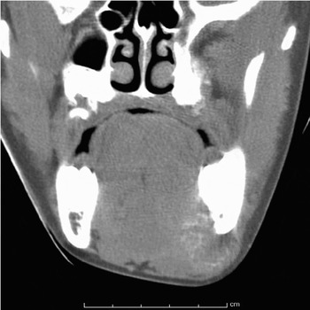
Fig. 1 Coronal, non-contrast, reformatted computed tomography image of aneurysmal bone cyst of the left lower mandible, showing the thin, bony septations of the expansile mandibular lesion.
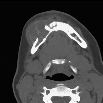
Fig. 2 Non-contrast, axial computed tomography image showing the ‘soap-bubble’ appearance of a right mandibular aneurysmal bone cyst.

Fig. 3 Coronal, non-contrast, reformatted computed tomography image of a left-sided aneurysmal bone cyst, showing an expansile lesion that occupies the left nasal cavity.
The three mandibular tumours were resected en bloc; the left condyle was reconstructed in one patient, while the remaining two had no cortical bone resected. The ethmoidal aneurysmal bone cyst required a midfacial degloving approach and medial maxillectomy. The maxillary aneurysmal bone cyst was enucleated and curetted, while the occipital lesion was also excised en bloc. Follow up, ranging from one to five years (mean, three years) showed that the ethmoidal lesion recurred eight months after initial treatment, then resolved with revision surgery. No other recurrences or complications were noted.
Gross examination of bone curettings in five patients revealed multiple tan-red soft bony tissue fragments. In the patient with the occipital soft tissue mass, the lesion was removed intact, and the specimen consisted of a 4.5 × 3.0 × 2.2 cm cyst with a diffusely calcified wall ranging in thickness from 0.3 to 0.9 cm. Microscopic examination in all cases showed blood-filled cystic spaces separated by variably thick septa composed of fibroblasts with admixed multinucleate giant cells and foci of ‘reactive’ woven bone (Figure 4). In one mandibular lesion (patient six), an incisional biopsy specimen lacked cysts, and the lesion was provisionally considered to be a solid-variant aneurysmal bone cyst, until excisional biopsy material revealed classic blood-filled cysts. In the ethmoidal lesion (patient one), areas of the lesion showed abundant woven bone, giving a pattern reminiscent of fibrous dysplasia in areas; however, the aneurysmal bone cyst was the overwhelmingly dominant pattern in this case. In five cases, fresh tissue was submitted for short-term culture and cytogenetic analysis. In one of these cases (patient one), tissue failed to grow in culture. In the four other cases, Giemsa-banding analysis revealed normal male (46,XY, patient three) and female (46,XX, patients four to six) karyotypes.

Fig. 4 (a) Photomicrograph of a maxillary aneurysmal bone cyst, showing fibroblasts and mononuclear cells with scattered multinucleate giant cells (arrows) most prominent along the lining of the cyst wall (H&E; original magnification ×60). (b) Photomicrograph of aneurysmal bone cyst of the mandible, showing calcification (white arrow), abundant mononuclear cells (arrowhead) and scattered giant cells (black arrow) (H&E; original magnification ×60).
Discussion
The term aneurysmal bone cyst was first coined by Jaffe and Lichtenstein in 1942 to describe two examples of erosive, expansile, blood-filled, cystic lesions in the vertebra of an 18-year-old male and in the pubic symphysis of a 17-year-old male.Reference Martinez and Sessone5 Reports of aneurysmal bone cysts of the head and neck region have been published as numerous individual case reports.Reference Struthers and Shear7–Reference David, Horvath, Horvath and Iles9 To the best of our knowledge, the current study represents the first series to evaluate paediatric aneurysmal bone cyst specifically in the head and neck region.
Our study identified six paediatric patients, aged eight months to 17 years, with aneurysmal bone cysts of the head and neck. We confirmed the propensity of head and neck aneurysmal bone cysts to affect the mandible; they occurred in this location in 50 per cent of our patients. Our series also included a soft tissue aneurysmal bone cyst occurring in the occiput of an eight-year-old. We believe this to be the second soft tissue aneurysmal bone cyst reported in the head and neck; the first was described as a common carotid artery mass in a seven-year-old boy.Reference Petrik, Findlay and Sherlock10 Surgical treatment, tailored among our patients to suit the anatomical regions involved, resulted in a 20 per cent recurrence rate, typical of aneurysmal bone cysts at other sites.
Clinical picture
Females may be at increased risk of developing aneurysmal bone cysts.Reference Struthers and Shear7 Between 80 and 85 per cent of these benign but potentially destructive, rapidly growing lesions are diagnosed in patients younger than 20 years.Reference Hamilton, Voorhies, Wilkins and Rengachary2, Reference Helliwel11 Aneurysmal bone cysts also show a tendency to be more aggressive in older compared with younger children;Reference Cottalorda and Bourella12 one study found that the recurrence rate was 32 per cent in patients younger than 15 years, 12 per cent in those older than 15 years and 0 per cent in those older than 25 years.Reference Szendroi, Cser, Konya and Renyi-Vamos8 Virtually any bone may be involved.Reference Mirra and Mirra13 The tibia and the femur are affected most commonly, but up to 12 per cent of aneurysmal bone cysts occur in the head and neck region, most commonly affecting the mandible.Reference Barnes3, Reference Kaffe, Naor, Calderon and Buchner4, Reference Simon and Springfield14
Typically, aneurysmal bone cysts in the head and neck region present with a rapid onset of pain and swelling.Reference Fechner and Mills15 Depending on location, symptoms may also include headache, diplopia, altered vision, proptosis, teeth loosening, conductive hearing loss, lip paraesthesia, abducens and facial nerve palsies, and even signs of raised intracranial pressure.Reference Barnes3, Reference Fechner and Mills15, Reference Kransdorf and Sweet16
The majority of aneurysmal bone cysts arise de novo and are referred to as primary aneurysmal bone cysts. However, up to 30 per cent of aneurysmal bone cysts are referred to as secondary aneurysmal bone cysts – this refers to areas histologically resembling aneurysmal bone cyst which may be seen adjacent to other bone lesions.Reference Dormans, Hanna, Johnston and Khurana17
Radiology
The classical radiological appearance of an aneurysmal bone cyst on plain films is that of an eccentric, ballooned, cystic expansion surrounded by a rim of sclerosis.Reference Schajowicz and Schajowicz18 The periosteum may be raised by new bone formation occurring between the margin of the aneurysmal bone cyst and the adjacent, unaffected bony cortices. The three stages of change have been grouped into a lytic phase, followed by a mature phase (with a peripheral bony shell and septa) and a late calcified stage. Although typically radiolucent (87 per cent), aneurysmal bone cysts may be radiopaque (2 per cent) or mixed (11 per cent).Reference Kaffe, Naor, Calderon and Buchner4 A ‘soap-bubble’ or ‘blow-out’ appearance of single or multiple cysts is also commonly seen (Figure 2).
Computed tomographic imaging shows a unicystic or multilocular lesion, and highlights any bone destruction.Reference Kaffe, Naor, Calderon and Buchner4 The multilocular lesion consists of several cavities of different densities. The vascular stroma and septa may enhance with intravenous contrast.Reference Barnes3, Reference Calliauw, Roels and Caemaert19 Magnetic resonance imaging demonstrates the cystic component of the lesion. Both T1- and T2-weighted sequences will demonstrate cysts with internal fluid–fluid levels. The cyst contents consist of blood degradation products in various stages of evolution and, as such, the signal intensity of the blood products will vary from hypointense to isointense to hyperintense.Reference Kransdorf and Sweet16 On angiography, a persistent venous circulation is typically demonstrated, with occasional arteriovenous shunting.Reference Lindbom, Soderberg, Spjut and Sunnqvist20
Radiologically, aneurysmal bone cysts may be staged according to the Enneking classification, as follows: in stage I (latent) the aneurysmal bone cyst remains static or heals spontaneously; in stage II (active) its growth is progressive but without cortical destruction; while in stage III (aggressive) there is progressive growth with cortical destruction.Reference Enneking21
The radiological differential diagnosis for aneurysmal bone cyst includes giant cell tumour, telangiectatic osteosarcoma, unicameral bone cyst, nonossifying fibroma, ossifying fibroma, enchondroma, fibrous dysplasia, myxoma, bone fracture, fibroblastic osteosarcoma and reparative giant cell granuloma of hyperparathyroidism.Reference Struthers and Shear7, Reference Fechner and Mills15, Reference Casadei, Ruggeri, Moscato, Ferraro and Picci22 The radiological differential diagnoses vary for classic aneurysmal bone cyst, solid-variant aneurysmal bone cyst and soft tissue aneurysmal bone cyst.
Pathology
The diagnosis of aneurysmal bone cyst is based on the combination of radiological and histological features. In the appropriate clinicoradiological setting, histological features can be diagnostic. As with most childhood bone lesions, it is unwise for a pathologist to render a diagnosis without thorough inspection of the radiology films. When aneurysmal bone cyst is suspected radiologically, an intra-operative radiology consultation with frozen section is often sought at the time of curettage. This is done in particular to rule out telangiectatic osteosarcoma, usually the primary concern in the radiological differential diagnosis.
Grossly, aneurysmal bone cyst consists of friable, haemorrhagic material that is often gritty. In en-bloc resections, cysts and cortical destruction can be appreciated. Microscopically, aneurysmal bone cyst shows a stroma composed of fibroblasts, multinucleate giant cells and bone, as well as cystic spaces often filled with blood and showing an increased number of giant cells lining the cavity.
The prominence of the cystic component in aneurysmal bone cyst can vary. Accordingly, aneurysmal bone cyst may be divided into two variants: classic (i.e. containing cysts; 95 per cent) and solid (5 per cent). In the head and neck, the solid variant of aneurysmal bone cyst is conventionally referred to as giant cell reparative granuloma, central giant cell granuloma or central ossifying fibroma; this type of lesion was excluded from our study because of lack of uniform consensus regarding its terminology.
Aneurysmal bone cysts may be seen in isolation (i.e. primary aneurysmal bone cyst); alternatively, aneurysmal bone cyst like histological features may appear in conjunction with another bone lesion (i.e. secondary aneurysmal bone cyst). The most common coexisting lesions associated with aneurysmal bone cyst are said to be chondroblastoma and giant cell tumour.Reference Kransdorf and Sweet23 Aneurysmal bone cysts have also been associated with a wide spectrum of other lesions, including osteoblastoma, fibromyxoma, fibrous dysplasia, haemangioma and osteosarcoma.Reference Fechner and Mills15, Reference Kransdorf and Sweet23–Reference Brindley, Greene and Frankel25 The literature on aneurysmal bone cysts is confounded by rare reports of ‘malignant transformation’, which may represent undersampling or misinterpretation of malignant tumour with secondary aneurysmal bone cyst like change.Reference deMello, Archer and Blair26
The histological classification of giant cell rich lesions of bone is often problematic, particularly in the head and neck region.Reference Panoutsakopoulos, Pandis, Kyriazogloe, Gustafson, Mertens and Mandahl27 Our traditional concepts are currently being refined as cytogenetic characterisation of the various lesions progresses. Aneurysmal bone cysts have in the past been generally thought to be ‘reactive’ lesions, particularly as they were seen as ‘degenerative’ phenomena adjacent to other bone lesions. However, it has become recognised that the chromosomal translocation t(16;Reference Dormans, Hanna, Johnston and Khurana17) (q22;p13) is a recurring abnormality in primary aneurysmal bone cysts, indicating that these lesions represent a clonal proliferative process.Reference Oliveira, His, Weremowicz, Rosenberg, Dal Cin and Joeseph28, Reference Dal Cin, Kozakewich, Goumnerova, Mankin, Rosenberg and Fletcher29 Further study has identified that these translocations involve the genes USP6 (an oncogene) and CDH11 (a promoter); their rearrangement is found in a majority of cases of primary aneurysmal bone cyst but not in secondary aneurysmal bone cyst.Reference Dal Cin, Kozakewich, Goumnerova, Mankin, Rosenberg and Fletcher29 Cytogenetic analysis has also confirmed that soft tissue lesions histologically identical to aneurysmal bone cyst are characterised by the same chromosomal rearrangement and thus appear to represent the same biological process.Reference Gleason, Kleinman, Debelenko, Rahbar, Gebhardt and Perez-Atayde30 Additional cytogenetic characterisation of bone lesions showing clinicoradiological and histological overlap with head and neck aneurysmal bone cyst is expected to further refine the field and may be of diagnostic use in the future.Reference Docquier and Delloye31
Treatment
Injection agents
Demineralised bone plus autologous bone marrow has been used as an ossification-inducing agent within an aneurysmal bone cyst, specifically targeting the cells of the aneurysmal bone cyst septae.Reference Glorion, Brunelle, Ghazai and Chotel32 This technique enables a minimally invasive approach and negates the need for extensive surgery and blood loss, particularly in cosmetically sensitive areas and poorly accessible lesions. Although this particular study only evaluated aneurysmal bone cysts of the long bones, pelvis, scapula and calcaneus, successful healing was observed in 11 cases, followed up to a mean of 3.9 years.
Intracystic injection of a pure alcohol solution has been used to successfully treat six of seven aneurysmal bone cysts in one series.Reference Szendroi, Antal, Liszka and Konya33 However, this mode of treatment still remains under evaluation.
Calcitonin is hypothesised to block osteoclast activity and to induce bony trabeculae formation in the fibrous septae of aneurysmal bone cysts. Intralesional injection of calcitonin into hypovascular aneurysmal bone cysts has shown good provisional results; three aneurysmal bone cysts healed completely and three incompletely, out of a series of seven, but follow up was only for 14 months.Reference Cottalorda, Kohler, Chotel, de Gauzy, Lefort and Louahem34 The calcitonin was administered three times a week for five weeks, under local anaesthesia.
Steroid injection has not shown benefit in the treatment of aneurysmal bone cyst.Reference Falappa, Fassaria, Fanelli, Genovese, Ascani and Crostelli35
Overall, the role of injection and adjunctive therapies in the treatment of aneurysmal bone cysts of the head and neck remains ill-defined, because the agents used have only been evaluated in cases of aneurysmal bone cyst outside this region. In addition, because of the low incidence of aneurysmal bone cyst (particularly in the head and neck area), prospective studies are more difficult, and so the standard of care has remained unaltered.
Embolisation
Alcoholic zein (Ethibloc; Ethnor Laboratories/Ethicon, Noderstedt, Germany) is a biodegradable, radiopaque, alcoholic solution of corn protein that may be injected into aneurysmal bone cysts. It causes thrombosis, inflammation and a fibrotic reaction by obstructing multiple afferent arterioles of the aneurysmal bone cyst at their venous end.Reference Topouchian, Mazda, Hamze, Laredo and Pennecot36 Injection is performed under CT or fluoroscopic guidance. What makes this controversial mode of treatment impractical in the head and neck area is that compression is required to prevent distal embolisation of the venous drainage system. Complications encountered have ranged from cellulitis to cutaneous fistulisation and pulmonary embolism.Reference Green, Bellemore and Marsden37
Selective arterial embolisation is used to reduce the blood supply to the lesion while not interfering with the supply to the surrounding structures. It is useful in lesions in inaccessible sites or where the use of tourniquets is not possible. When successful, progressive ossification of an aneurysmal bone cyst will typically occur within two to four months.Reference Cottalorda and Bourelle38 The use of selective arterial embolisation does not preclude surgery at a later stage. Ischaemia to nerves and viscera remains a concern, but this is typically unusual in the hands of an experienced radiologist.
Surgery
Eradication of the aneurysmal bone cyst by complete surgical excision remains the objective. En-bloc resection is the treatment with the lowest risk of recurrence. However, its role is more clearly defined in cases of aneurysmal bone cyst of the extremities; the majority of patients with head and neck lesions undergo curettage with or without bone grafting and cementation. Reported recurrence rates vary from 20 to 70 per cent.Reference Fechner and Mills15 It would appear that the type of curettage performed has an enormous influence upon these statistics; curettage via a small hole has a higher recurrence rate than that performed through a larger cortical window. This in turn is less effective than curettage via saucerisation (in which new subperiosteal bone is also removed), which has been shown in one study to have a 100 per cent success rate, albeit outside the head and neck area.Reference Schreuder, Veth, Pruszczynski, Lemmens, Koops and Molenaar39
Several authors have used adjunctive therapies in addition to curettage in the treatment of aneurysmal bone cysts. Selective arterial embolisation, as discussed earlier, may be used to reduce the vascularity of the lesion and to facilitate its excision.Reference Cottalorda and Bourelle38 Phenol is a cytotoxic agent that nonselectively destroys tumour and normal cells when applied directly to the surface of curetted tumours. It is unable to penetrate deeper than the surgical margin.Reference Marcove, Sheth, Takemoto and Healey40 Liquid nitrogen cryotherapy successfully treated all but one of 29 patients in one study, while a 96 per cent cure rate was achieved after a second cryotherapy treatment in another study.Reference Marcove, Sheth, Takemoto and Healey40, Reference Ozaki, Hillmann, Linder and Winkelmann41 The major concerns with this form of treatment include superficial skin necrosis, deep wound infection, flap necrosis, osteonecrosis, neurovascular injury and growth arrest. The heating effect of polymethylmethacrylate administers a cytotoxic 50°C heat to the surrounding tissue to a depth of 2.5 mm in cancellous bone and 0.5 mm in cortical bone.Reference Kumar, Singh, Phadke, Diwakar, Agarawal and Jain42 Cementation has the added benefits of preventing fracturing in weight-bearing bones and allowing early radiological evidence of localised recurrence to be identified. In cases of extremity aneurysmal bone cyst, curettage with a mechanical burr has been reported to control almost 90 per cent of lesions.
Radiation therapy
Aneurysmal bone cyst is amenable to radiation therapy, by a mechanism that has never been entirely elucidated.Reference Fajardo43 Using megavoltage therapy, the vascular lesions probably thrombose, with thickening of the vascular wall; the radiation is also thought to induce fibrosis and necrosis of the mesenchymal tissues.Reference Mendenhall, Zlotecki, Gibbs, Reith, Scarborough and Mendenhall44 Radiation therapy has been used successfully in cases of aneurysmal bone cyst recurrence and in sites that are anatomically inaccessible, e.g. within the temporal bone.Reference Fajardo43 Mendenhall et al. used low dose (26–30 Gy) radiotherapy in patients with incompletely resected, aggressive and/or recurrent aneurysmal bone cysts, and approximately 90 per cent were controlled by this method.Reference Mendenhall, Zlotecki, Gibbs, Reith, Scarborough and Mendenhall44
• The World Health Organization defines an aneurysmal bone cyst as a ‘benign cystic lesion of bone composed of blood-filled spaces separated by connective tissue septa containing fibroblasts, osteoclast-type giant cells and reactive woven bone’
• While involvement of the tibia and femur is typical, presentation within the head and neck region is thought to occur in 12 per cent of cases
• Aneurysmal bone cysts may be seen and managed by otolaryngologists. In the head and neck, they typically involve the mandible but may occur in unusual sites such as soft tissue
• Eradication of aneurysmal bone cysts by complete surgical excision remains the objective. En-bloc resection is the treatment with the lowest risk of recurrence
The emergence of osteogenic sarcoma or fibrosarcoma following radiation therapy for aneurysmal bone cyst has been reported.Reference Cawson, Binnie, Speight and Wright45 The other major side effect is arrest of bony growth in children. Accordingly, the importance of an accurate and thorough histological evaluation, and the need for routine follow up for all aneurysmal bone cyst patients, cannot be over-stated.
Chemotherapy has no role to play in the management of aneurysmal bone cysts.
Conclusion
Aneurysmal bone cysts may be seen and managed by otolaryngologists. In the head and neck, they typically involve the mandible but may occur in unusual sites such as soft tissue. They are benign but have a capacity for local destruction and recurrence. Computed tomography imaging shows a characteristic unicystic or multilocular lesion with contrast enhancement of the vascular stroma and septa. Surgical excision, where feasible, remains the ‘gold standard’ of care. Identification of the chromosomal rearrangement characteristic of aneurysmal bone cyst represents the most striking recent advance in the understanding of this disease; it has led to recognition of the genes involved in aneurysmal bone cyst pathogenesis, and it is emerging as a helpful adjunct to histological diagnosis. While adjunctive therapies have been utilised elsewhere in the body, their role in head and neck aneurysmal bone cyst remains ill-defined.


