


INTRODUCTION
Anthropogenic impacts and climate change are threatening the survival of coral reefs worldwide (Hughes et al., Reference Hughes, Baird, Bellwood, Card, Connolly, Folke, Grosberg, Hoegh-Guldberg, Jackson, Kleypas, Lough, Marshall, Nystroem, Palumbi, Pandolfi, Rosen and Roughgarden2003; Hoegh-Guldberg et al., Reference Hoegh-Guldberg, Mumby, Hooten, Steneck, Greenfield, Gomez, Harvell, Sale, Edwards, Caldeira, Knowlton, Eakin, Iglesias-Prieto, Muthiga, Bradbury, Dubi and Hatziolos2007). The rapid decline of reefs requires urgent reform of scientific approaches in order to understand the processes underlying the degradation (Hughes et al., Reference Hughes, Graham, Jackson, Mumby and Steneck2010). Studies of corals in mesocosms or tank systems under controlled conditions allow sophisticated experiments that hold a great promise to yield new insights in coral biology and are ideally suited to complement traditional field studies. The impact of ex situ experiments is strongly dependent on how close to natural corals behave in captivity. Tremendous progress has been made over the last 20 years in improving techniques for coral culturing that allow a sustainable propagation of corals not only by natural or induced fragmentation (many species), but also by sexual reproduction (e.g. Pocillopora and Tubastrea) (Petersen et al., Reference Petersen, Falcato, Gilles and Jones2007). Long term culture of corals offers the opportunity to establish distinct model strains for which information can be accumulated over time. Despite the obvious benefits of working with established models (Weis et al., Reference Weis, Davy, Hoegh-Guldberg, Rodriguez-Lanetty and Pringle2008), questions are often raised over the extent to which aquarium corals still show natural physiological responses in experimental treatments or if their responses are distorted by the acclimatization to life in captivity. The fact that corals can thrive for decades in experimental systems (Smith et al., Reference Smith, Pinzón and LaJeunesse2009) already suggests that basic physiological processes must function well. However, the relationship between corals and their symbiotic algae (zooxanthellae) shows a degree of flexibility and corals have been shown to change their dominant symbiont type at both the population and individual levels in response to changes in their specific environment (Baker, Reference Baker2003; Van Oppen et al., Reference Van Oppen, Baker, Coffroth, Willis, Van Oppen and Lough2009). These changes might result from ‘symbiont shuffling’, where there is a shift in dominance of existing algal types within the host, or by ‘symbiont switching’, where the host acquires new algal strains from the environment (Baker, Reference Baker2003; Van Oppen et al., Reference Van Oppen, Baker, Coffroth, Willis, Van Oppen and Lough2009). The degree to which corals are capable of changing their algal partners in nature is still controversially discussed (Goulet, Reference Goulet2006). Evaluating the diversity of zooxanthellae strains in aquarium corals promises insights into the general stability of the coral–algal relationship (Smith et al., Reference Smith, Pinzón and LaJeunesse2009). Moreover, the precise knowledge of the two partners of the symbiotic association facilitates the interpretation of the responses exhibited by experimental models to imposed stress conditions. The present study surveyed the zooxanthellae diversity in the experimental aquarium of the Coral Reef Laboratory at the National Oceanography Centre, Southampton. Where available, we selected different colour morphs of the species for the study in order to evaluate the potential influence of the host pigments on the zooxanthellae complement. The striking differences in the visual appearance of the colour morphs result from the differential expression of green fluorescent protein (GFP)-like proteins (Wiedenmann, Reference Wiedenmann1997; Wiedenmann et al., Reference Wiedenmann, Schenk, Rocker, Girod, Spindler and Nienhaus2002; Leutenegger et al., Reference Leutenegger, D'Angelo, Matz, Denzel, Oswald, Salih, Nienhaus and Wiedenmann2007a, Reference Leutenegger, Kredel, Gundel, D'Angelo, Salih and Wiedenmannb; Oswald et al., Reference Oswald, Schmitt, Leutenegger, Ivanchenko, D'Angelo, Salih, Maslakova, Bulina, Schirmbeck, Nienhaus, Matz and Wiedenmann2007; D'Angelo et al., Reference D'Angelo, Denzel, Vogt, Matz, Oswald, Salih, Nienhaus and Wiedenmann2008). Green and red tones are caused by green and red fluorescent proteins, whereas blue morphs derive their colour from a dominant expression of purple-blue non-fluorescent chromoproteins (Alieva et al., Reference Alieva, Konzen, Field, Meleshkevitch, Hunt, Beltran-Ramirez, Miller, Wiedenmann, Salih and Matz2008; D'Angelo et al., Reference D'Angelo, Denzel, Vogt, Matz, Oswald, Salih, Nienhaus and Wiedenmann2008). In many corals, the expression of the GFP-like proteins is regulated by the intensity of the incident blue light (D'Angelo et al., Reference D'Angelo, Denzel, Vogt, Matz, Oswald, Salih, Nienhaus and Wiedenmann2008). In contrast, the expression of these pigments in sea anemones and at least in some corals is largely independent of the light climate (Leutenegger et al., Reference Leutenegger, D'Angelo, Matz, Denzel, Oswald, Salih, Nienhaus and Wiedenmann2007a, Reference Leutenegger, Kredel, Gundel, D'Angelo, Salih and Wiedenmannb).
Zooxanthellae clades were determined by diagnostic restriction digest. For this purpose, the DNA region encoding the small subunit (SSU) ribosomal RNA of zooxanthellae was amplified and digested with TaqI and DpnII (Rowan & Powers, Reference Rowan and Powers1991; Bythell et al., Reference Bythell, Douglas, Sharp, Searle and Brown1997; Savage et al., Reference Savage, Goodson, Visram, Trapido-Rosenthal, Wiedenmann and Douglas2002). The analyses were supported by cloning and sequencing of the amplified SSU rDNA fragments.
MATERIALS AND METHODS
DNA isolation
DNA was isolated from zooxanthellae using a modification of a previously described method (Doyle & Doyle, Reference Doyle and Doyle1990). Tissue was homogenized in sterile filtered seawater. The homogenate was filtered through a 0.1 mm mesh to remove large cellular or skeletal debris. Zooxanthellae were precipitated from the solution by centrifugation (5 minutes/500×g/4°C). The zooxanthellae pellet was re-suspended in 1 ml lysis buffer containing: 2% cetyl-trimethylammonium-bromide (CTAB); 1.4 M NaCl; 0.5% 2-mercaptoethanol; 2% polyvinylpyrrolidone (PVP); 20 mM EDTA; 10 mM Tris–HCl, (pH 8.0); and incubated at 60°C for 30 minutes in a water bath. Subsequently, the DNA solution was thoroughly mixed with 1 ml chloroform: isoamyl alcohol (24:1) followed by centrifugation (5 minutes/20,800 × g/4°C). The aqueous phase was removed and the extraction was repeated twice using 1 ml phenol:chloroform:IAA (25:24:1) and 1 ml chloroform. The DNA was then precipitated from the solution by adding an equal volume of isopropanol and centrifugation (5 minutes/20,800 × g/4°C). The supernatant was removed and the DNA pellet was washed with 1 ml 70% ethanol followed by a further centrifugation step (5 minutes/20,800 × g/4°C). After removal of the supernatant, the DNA pellet was dried, subsequently dissolved in 50 µl TE and stored at –20°C for further analyses. DNA concentration and purity were determined with a Nanodrop–ND-1000 Spectrophotometer (Thermo Scientific, Wilmington, USA).
Amplification of zooxanthellae SSU rDNA
The SSU rDNA gene was amplified using zooxanthellae-specific polymerase chain reaction (PCR) primers ss5z and ss3z (Rowan & Powers, Reference Rowan and Powers1991). Reaction mixtures with a final volume of 50 µl contained 1mM dNTPs, 0.5 µM of each oligonucleotide, 2.5 U Taq Polymerase (Promega, Madison, WI, USA) and 3 mM MgCl2. Fragments were amplified in a MyCycler Thermal Cycler (Biorad, Hertfordshire, UK) using the following cycling steps: initial denaturation: 2 minutes/94°C, 40 cycles (denaturation 30 seconds/94°C; annealing 30 seconds/59°C; elongation 2 minutes/72°C) and final elongation: 5 minutes/72°C. The PCR products were separated on ethidium bromide stained agarose gels. Bands of the expected size of ~1.6 kB were purified from the gel using the JETQuick PCR Product Purification kit (Genomed, Löhne, Germany).
Restriction fragment length polymorphisms
The purified PCR products were split in two aliquots which were digested with the restriction enzymes TaqI and DpnII, respectively, following the manufacturer's protocol (New England Biolabs, Ipswich, MA, USA). Digested fragments were analysed by electrophoresis on ethidium bromide-stained agarose gels. The length of the restriction fragments were determined relative to a size standard and used for the identification of zooxanthellae phylotypes as described previously (Rowan & Powers, Reference Rowan and Powers1991; Bythell et al., Reference Bythell, Douglas, Sharp, Searle and Brown1997; Savage et al., Reference Savage, Goodson, Visram, Trapido-Rosenthal, Wiedenmann and Douglas2002).
Sequence analyses
For selected samples, the purified PCR fragments were cloned using the StrataClone kit (Agilent Technologies, Berkshire, UK). After purification of plasmids containing inserts of the expected size, two clones were sequenced for each species by a commercial provider (Macrogen, Seoul, Korea). Sequences were deposited at Genbank under the accession numbers (JN255733–JN255743). SSU sequences of Symbiodinium clades B, C, D and E were downloaded from GenBank and used for comparison. Sequences were aligned using MUSCLE (Edgar, Reference Edgar2004) and a phylogenetic tree was constructed applying the maximum likelihood method implemented in PHYML (Guindon et al., Reference Guindon, Lethiec, Duroux and Gascuel2004). Bootstrap analyses were performed using 100 replicates to evaluate the reliability of the tree topology (Felsenstein, Reference Felsenstein1985).
RESULTS AND DISCUSSION
Scleractinian corals and sea anemones are kept and propagated for experimental purposes in the Coral Reef Laboratory at the National Oceanography Centre of Southampton (NOCS) (http://www.soton.ac.uk/soes/research/staff/jwlw07.page?). Individuals have been co-cultured with each other in this and preceding aquarium systems together with numerous other hexacoral and octocoral taxa for periods between 2.5 and 10 years (Table 1). During this time, the organisms experienced comparable environmental conditions and were exposed to the same recirculated water body. This gave us the opportunity to evaluate if life in captivity affects the dominant zooxanthellae strains hosted by the experimental animals, i.e. via symbiont shuffling or exchange (Baker, Reference Baker2003; Van Oppen et al., Reference Van Oppen, Baker, Coffroth, Willis, Van Oppen and Lough2009). DNA was isolated and parts of the Symbiodinium SSU rDNA gene were amplified using PCR primers ss5z and ss3z (Rowan & Powers, Reference Rowan and Powers1991). The PCR products were subjected to a diagnostic restriction digest using TaqI and DpnII and the zooxanthellae clades were deduced from the restriction pattern obtained from agarose gel electrophoresis (Bythell et al., Reference Bythell, Douglas, Sharp, Searle and Brown1997; Savage et al., Reference Savage, Goodson, Visram, Trapido-Rosenthal, Wiedenmann and Douglas2002). One sample of zooxanthellae from Acropora clathrata which was prepared immediately after collection from Abu Dhabi (UAE) was analysed as control. The TaqI and DpnII restriction patterns place the zooxanthellae from A. clathrata within clade C (Table 2; Figure 1). The presence of this clade in corals from the Arabian Gulf is somewhat surprising since a dominance of clade D was suggested for animals in this particular environment (Baker et al., Reference Baker, Starger, McClanahan and Glynn2004). Moreover, a recent study found clade D zooxanthellae on A. clathrata colonies from the Arabian Gulf off the Iranian coast (Ghavam Mostafavi et al., Reference Ghavam Mostafavi, Fatemi, Shahhosseiny, Hoegh-Guldberg and Loh2007). This finding underlines that at least some coral species maintain variability in their association with zooxanthellae even in habitats experiencing extreme temperatures. Restriction analyses revealed that Montipora and Acropora species were found to harbour clade C symbionts. In contrast, clade A zooxanthellae were found in two Anemonia sulcata specimens. One of the individuals, A. sulcata from Elba, Italy, was already shown 9 years ago to contain clade A symbionts which are found in most cnidarians of the Mediterranean Sea and the European Atlantic coast (Savage et al., Reference Savage, Goodson, Visram, Trapido-Rosenthal, Wiedenmann and Douglas2002; Visram et al., Reference Visram, Wiedenmann and Douglas2006). The present study reveals that the symbionts have not changed despite living for a decade under tropical temperature conditions (constant water temperatures between 23 and 26°C) and in proximity to cnidarians containing mostly clade C symbionts. The red colour morph of Entacmaea quadricolor (Wiedenmann et al., Reference Wiedenmann, Schenk, Rocker, Girod, Spindler and Nienhaus2002) which was co-cultured with the two Anemonia specimens for nearly ten years harboured clade C zooxanthellae. One representative of the green morph of Entacmaea quadricolor showed an unusual restriction pattern. Whereas the TaqI digest yielded fragments with the lengths of ~880 and 700 base pairs (bp) indicative of clade C symbionts, the smaller fragment of the DpnII restriction was significantly longer than expected (Figure 1A).

Fig. 1. Determination of zooxanthellae clades. (A) Ethidium bromide stained agarose gels show the restriction patterns of amplified Symbiodinium small subunit (SSU) fragments. Numbers indicate the species/colour morph under study. The DNA fragments of each individual were digested with TaqI (left lane) and DpnII (right lane). Acropora microphthalma (1), Acropora millepora pink morph (2), A. millepora green morph (3), A. millepora blue morph (4), Acropora pulchra (5), Acropora clathrata (6), Montipora sp. brown morph (7), Montipora sp. green-purple morph (8), Montipora sp. (red morph Ulm) (9), Montipora sp. (red morph Wilhelma) (10), Montipora digitata (11), Entacmae quadricolor red morph (12), E. quadricolor green morph (13), and Anemonia sulcata collection 2000 (14), collection 2006 (15); (B) phylogenetic relationship of SSU sequences. Bootstrap values (% of 100 replicates) are shown next to the nodes. GenBank accession numbers are given for sequences that were not determined in the present study. Sequences derived from on host individual are numbered (seq. 1–seq. 2). The years (y) spent in aquarium culture are indicated at the end of the branch labels.
Table 1. Origin of samples and their predominant zooxanthellae clades.

a, by December 2010.
Table 2. Fragment lengths of small subunit rDNA restriction with TaqI and DpnII.
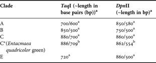
a, data from Rowan & Powers (Reference Rowan and Powers1991), Brown et al. (Reference Brown, Dunne, Goodson and Douglas2002) and Savage et al. (Reference Savage, Goodson, Visram, Trapido-Rosenthal, Wiedenmann and Douglas2002); b, data deduced from sequencing results of the present study.
To resolve the uncertainty about the nature of these zooxanthellae, SSU rDNA sequences were again amplified using the ss5z and ss3z primers, cloned and sequenced. Selected Acropora and Montipora sequences were determined for comparison. The sequences were aligned and a phylogenetic tree was constructed applying the maximum likelihood method (Figure 1B). Sequence analysis revealed that the zooxanthellae of the green morph of Entacmaea quadricolor also belonged to clade C, however, a ‘shift’ in one DpnII restriction site resulted in an increase in the length of the smaller fragment from 499 bp to 554 bp, explaining the observed change in the restriction pattern (Table 2).
Although SSU sequences have a limited power to identify subclades in comparison to internal transcribed spacer (ITS) regions (LaJeunesse, Reference LaJeunesse2001), the sequence analysis reveals well supported differences between the zooxanthellae from different individuals that suggest that each of them is associated with different clade C subtypes (Figure 1). The fact that the sequences could not be matched with known subclades of clade C is most probably due to the lack of appropriate SSU reference sequences in Genbank. A deeper classification at the level of subclades using ITS1/2 sequencing is currently underway.
Sequences of zooxanthellae derived from the same individual are always most similar to each other (Figure 1B). Zooxanthellae of the red individual of Entacmaea quadricolor yielded sequences which are similar to those from the green individual, suggesting the presence of E. quadricolor-typical symbionts even after 27 years of aquarium culture. The divergence of sequences from representatives of different species with the same colour does not indicate an obvious connection between host pigmentation and the preference for a certain clade C subtype.
A high abundance of clade C symbionts in aquarium corals was also observed most recently by Smith et al. (Reference Smith, Pinzón and LaJeunesse2009). This correlation reflects the prevalence of clade C in Indo-Pacific reefs (Smith et al., Reference Smith, Pinzón and LaJeunesse2009; Van Oppen et al., Reference Van Oppen, Baker, Coffroth, Willis, Van Oppen and Lough2009) from which most of the studied corals originate, rather than a specific adaptation to aquarium conditions (Smith et al., Reference Smith, Pinzón and LaJeunesse2009). Importantly, no shift of the zooxanthellae complement to clade D is observed that could be interpreted as response to stressful conditions (Baker et al., Reference Baker, Starger, McClanahan and Glynn2004) during transport or culture.
The results show that a diversity of zooxanthellae is maintained in corals and sea anemones over a long period of aquaculture. Therefore, well managed aquarium systems do not appear to lead to a selection for different symbiont types adapted to aquarium conditions, or a mixing of symbionts between hosts within the same aquarium. Such specimens may therefore be suitable for studying the physiological responses of hermatypic corals and allies to various environmental parameters over long periods of time. Moreover, the stability of symbiotic associations suggests that aquarium systems can be helpful in ex situ conservation of threatened coral species.
ACKNOWLEDGEMENTS
We thank J. Gittins for technical support and B. Goessele (Aquarium Ulm) and I. Koch (Wilhelma, Stuttgart) for samples. Our appreciation is extended to A. Al-Hemeri of the UAE Federal Environment Agency and A. Al-Cibahy of the Environment Agency of Abu Dhabi for provision of CITES export permits (No. 09FEA555) for UAE corals. The study was funded by the German research foundation (DFG Wi1990/2-1) and NERC (NE/H012303/1). We acknowledge the Tropical Marine Centre London for sponsoring the Coral Reef Laboratory at NOCS.


