


Introduction
The parapharyngeal space is a potential space that lies lateral to the pharynx and deep to the palatine tonsils. This space is divided into the pre-styloid and post-styloid spaces by the fascia running from the styloid process to the tensor veli palatini muscle.Reference Riffat, Dwivedi, Palme, Fish and Jani1, Reference Shabab, Heliwell and Jones2 Lesions in this area have proven difficult to diagnose.Reference Arnason, Hart, Taylor, Trites, Nasser and Bullock3–Reference Yousem, Sack and Scanlan5 This is because of their deep location, inaccessibility and concerns about damaging important adjacent anatomical structures.Reference Su, Zhao, Li, Deng, Zeng and Cui4, Reference Yousem, Sack and Scanlan5
The parapharyngeal space biopsy is an important investigation in the management of parapharyngeal space tumours. These tumours are relatively rare and the surgeon is often faced with a wide range of differential diagnoses. There are a number of ways to access the parapharyngeal space, with some degree of associated morbidity, including needle biopsy through the mandibular or maxillofacial areas,Reference Su, Zhao, Li, Deng, Zeng and Cui4 biopsy via the buccal space,Reference Tu, Geyer, Mancall and Baker6 and access directly through the skin or oral mucosa if the lesion is palpable.Reference Yousem, Sack and Scanlan5
We present a novel, less invasive, image-guided transnasal endoscopic approach to parapharyngeal space biopsy that has been successfully used by the senior author for years without any significant morbidity.
Materials and methods
This technique is performed under general anaesthetic. We use the Medtronic Stealth LandmarX™ computed tomography (CT) image guidance system; the pre-operative scans need to be compatible with the LandmarX navigation system. The procedural steps are outlined below.
Step one
The patient is positioned supine with their head up, similar to the positioning for functional endoscopic sinus surgery.
Step two
The LandmarX image guidance system is set up as per protocol. The patient is registered and the equipment is calibrated. The pre-operative films have been carefully studied with a radiologist (Figure 1), and the relevant side is pre-operatively marked.
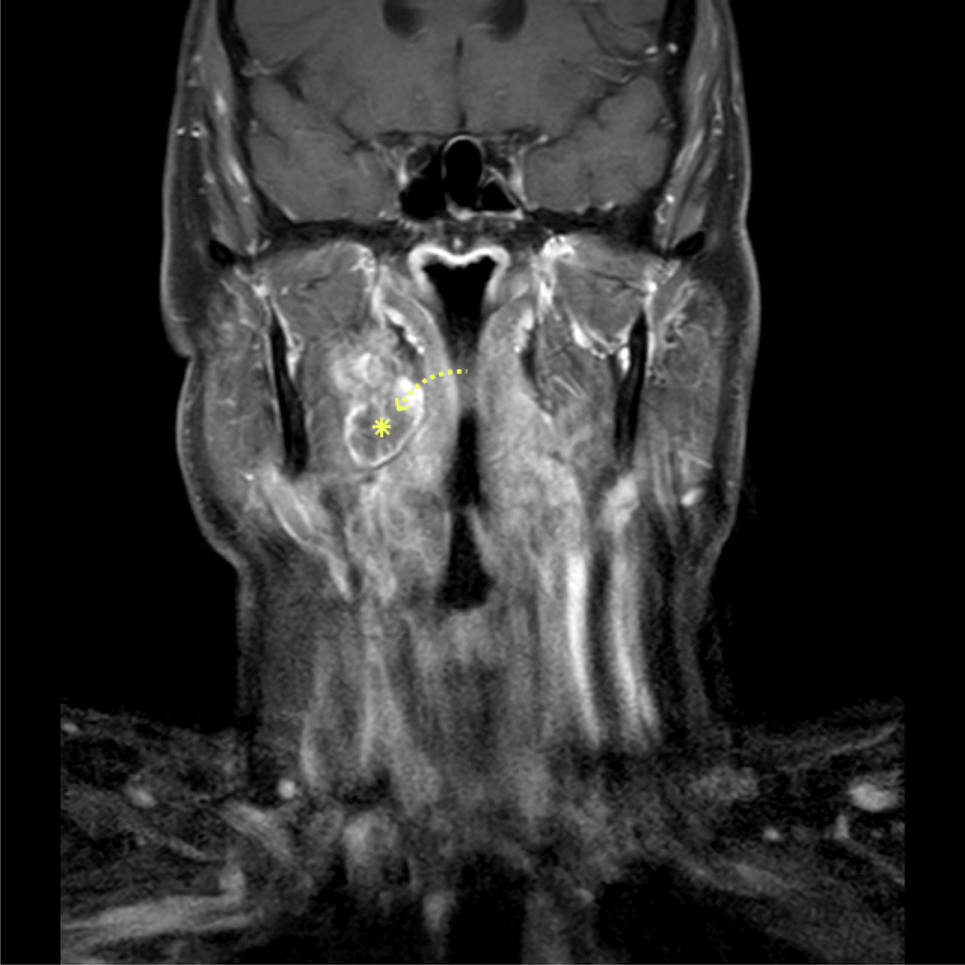
Fig. 1. Coronal, T1-weighted, post-gadolinium magnetic resonance imaging scan of the neck, performed pre-operatively for diagnosis and staging. The arrowhead indicates the biopsy route for a right parapharyngeal space mass, whilst the star highlights the target mass.
Step three
The lesion is identified using the curved LandmarX probe. The probe is placed into the nose and angled laterally, penetrating the mucosa and entering the parapharyngeal space. The image guidance system assists the surgeon in deciding the trajectory and depth of penetration into the parapharyngeal space (Figure 2). Care must be taken to determine the correct direction in which to sample the tumour without damaging any critical structures.

Fig. 2. Axial computed tomography scan of the neck acquired using the Medtronic LandmarX navigation system. The arrowhead indicates the biopsy route for a right parapharyngeal space mass, whilst the star highlights the target mass.
Step four
The sampling needle is usually a 22 gauge × 90 mm black spinal needle that is bent to exactly mirror the curve on the guidance probe. This is mounted onto a 10 ml syringe.
Step five
The LandmarX probe is removed and the needle is inserted into the location that has been pre-determined using the LandmarX equipment. The syringe plunger is withdrawn to create a negative pressure. The needle is moved in and out, whilst maintaining the negative pressure, in order to gain a satisfactory cytology sample.
Step six
The sample is injected into a pot of 10 per cent formalin and checked for adequacy. If more cells need to be obtained, step five may be repeated, usually with a fresh needle. Once a satisfactory sample has been obtained it can be sent to the laboratory for cytological assessment.
Step seven
The same procedure can be repeated using curved ‘giraffe’ forceps from the endoscopic sinus surgery set. The forceps are inserted in the closed position and opened within the tumour to obtain tissue samples for histology. A tissue sample would be superior to a cytological sample; however, this is not always achievable, as this depends on the proximity of the tumour to the pharyngeal wall. The surgeon checks for and controls any bleeding prior to finishing the procedure.
Discussion
Masses in the parapharyngeal space are relatively rare, but do account for 0.5 per cent of all masses arising in the head and neck.Reference Riffat, Dwivedi, Palme, Fish and Jani1 It is important to histologically type these lesions. The most common pre-styloid primary mass is a parotid tumour and the most common post-styloid mass is of neurogenic origin.Reference Riffat, Dwivedi, Palme, Fish and Jani1
Histological identification of the parapharyngeal space tumour allows the surgeon to make informed decisions on management and can also indicate prognosis. The 5- and 10-year survival rate for a benign lesion is 100 per cent. Conversely, in malignancy, the 5-year survival rate drops to 82–100 per cent and the 10-year survival rate decreases to 40–74 per cent. This dramatic fall in survival demonstrates a clear role for establishing histology in parapharyngeal space tumour management.
Biopsy of parapharyngeal space masses can be performed under image guidance. Su et al. describe a needle biopsy performed under CT image guidance through the skin of the ipsilateral submandibular region, above the hyoid and approximately 2 cm from the midline.Reference Su, Zhao, Li, Deng, Zeng and Cui4 Other than peri-procedural discomfort, there were no observed complications, and in seven patients a positive diagnostic result was obtained.Reference Su, Zhao, Li, Deng, Zeng and Cui4 This procedure is, however, more invasive. In addition, given the increased distance from the puncture site to the lesion, it does carry an increased theoretical risk of damage to important anatomical structures.
Anterior maxillofacial approaches have been described by numerous authors.Reference Yousem, Sack and Scanlan5, Reference Abrahams7–Reference Gupta, Henningsen and Wallace9 Unlike our novel technique, the maxillofacial approach may be limited by the osseous structures of the facial skeleton. This may limit the position of the needle and therefore the ability to access the lesion adequately.Reference Su, Zhao, Li, Deng, Zeng and Cui4 In addition, it may become difficult to avoid important structures. For this reason, this technique is often reserved for lesions on the superficial edge of the parapharyngeal space.Reference Su, Zhao, Li, Deng, Zeng and Cui4
Some lesions of the parapharyngeal space may bulge through the pharynx, becoming visible and palpable intra-orally. Yousem et al. describe a blind transoral biopsy.Reference Yousem, Sack and Scanlan5 The main drawbacks include the blind nature of the procedure, which therefore put the great vessels of the neck at risk, and there is potential for contamination from oral commensal organisms.Reference Yousem, Sack and Scanlan5 Fine needle aspiration cytology and Tru-cut® biopsies can be performed intra-orally. Needle aspiration has been demonstrated to be more accurate.Reference Castelli, Gattuso, Reyes and Solens10
We believe our procedure is relatively simple, safe and reproducible. The main disadvantage is the requirement of the LandmarX navigation system and the expertise required to use this equipment before attempting the procedure. All the fine needle aspiration or biopsy samples that have been collected via this technique so far have been adequate for cytological or histological analysis. There have been no complications experienced in any of the patients undergoing this technique.
Conclusion
Transnasal image-guided endoscopic aspiration or biopsy of the parapharyngeal space is a novel technique that has not been described previously. In trained hands, it has been shown to gain accurate results, without complication. The technique should be considered when faced with the difficult task of obtaining a tissue diagnosis of parapharyngeal space masses.
Competing interests
None declared


