Introduction
In 1870, Heinrich Waldeyer, a German anatomist–embryologist, established the central dogma of reproductive biology by introducing the idea that the ovary possesses a limited number of follicles at birth. He claimed that in the beginning of life all oogonia (the female germline stem cells) reserves are depleted as a result of their differentiation into oocytes and that the number of follicles diminished constantly with age, leading to menopause (Waldeyer-Hartz, Reference Waldeyer-Hartz1870). Despite some arguments that criticized Waldeyer's dogma (Kingery, Reference Kingery1917; Allen & Creadick, Reference Allen and Creadick1937), in 1951 Zuckerman put an end to the controversy in favour of a fixed number of oocytes at birth; during the lifetime there is no increase in the number of primary oocyte reserve cells beyond those initially present when the ovary developed (Zuckerman, Reference Zuckerman1951). Although this dogma has been established and supported by other scientists (Borum, Reference Borum1961; Peters et al., Reference Peters, Levy and Crone1962; Franchi et al., Reference Franchi, Mandl, Zuckerman and Zuckerman1962; Peters, Reference Peters1970; Faddy et al., Reference Faddy, Jones and Edwards1976; McLaren, Reference McLaren1984; Richardson et al., Reference Richardson, Senikas and Nelson1987; Anderson & Hirshfield, Reference Anderson and Hirshfield1992; Faddy, Reference Faddy2000), some studies have challenged this doctrine (Pansky & Mossman, Reference Pansky and Mossman1953; Vermandevaneck, Reference Vermandevaneck1956; Artem'eva, Reference Artem'eva1961; Johnson et al., Reference Johnson, Canning, Kaneko, Pru and Tilly2004; Zou et al., Reference Zou, Yuan, Yang, Luo, Sun, Zhou, Xiang, Shi, Yu and Zhang2009; Esmaeilian et al., Reference Esmaeilian, Gur Dedeoglu, Atalay and Erdemli2012; Sriraman et al., Reference Sriraman, Bhartiya, Anand and Bhutda2015; Woods & Tilly, Reference Woods and Tilly2012; Gong et al., Reference Gong, Lee, Lee, Kim, Lee, Chi, Ryu, Lee, Yum and Lee2010). However, since 2004, increasing numbers of scientists have questioned this central dogma of mammalian reproductive biology. In 2004, results of considerable study revealed that mitotically active germ cells exist in the ovaries of both young and adult mice and, depending on the number of oocyte atresia and reduction, these cells are required to constantly rejuvenate the follicle pool (Johnson et al., Reference Johnson, Canning, Kaneko, Pru and Tilly2004). Bukovsky's group revealed the possibility of neo-oogenesis and granulosa cell formation from surface epithelium of adult human ovaries (Bukovsky et al., Reference Bukovsky, Svetlikova and Caudle2005). In 2009, Mouse Vasa Homologue (MVH)-positive germline stem cells (GSCs) were isolated from the surface epithelium of neonatal mouse ovaries. Transplantation of these cells into the busulphan-treated mouse ovaries led to neo-oogenesis and offspring generation (Zou et al., Reference Zou, Yuan, Yang, Luo, Sun, Zhou, Xiang, Shi, Yu and Zhang2009). To trace GSCs in the surface epithelium of adult mammalian ovaries, Virant-Klun and collaborators scraped the ovarian surface epithelium (OSE) in postmenopausal women or young women with premature ovarian failure (POF). They reported the isolation of putative GSCs that spontaneously generated oocyte-like cells (OLCs) with the capacity to undergo parthenogenesis and development of blastocyst-like structures in vitro (Virant-Klun et al., Reference Virant-Klun, Zech, Rozman, Vogler, Cvjeticanin, Klemenc, Malicev and Meden-Vrtovec2008, Reference Virant-Klun, Rozman, Cvjeticanin, Vrtacnik-Bokal, Novakovic, Rulicke, Dovc and Meden-Vrtovec2009). In another study, a rare population of mitotically active oogonial stem cells was isolated by fluorescence-activated cell sorting (FACS) with DDX4 expression on the cell surface of adult mouse ovaries and human ovarian cortical tissues. These cells were cultured for several months on mouse embryonic fibroblast (MEF) feeders and differentiated spontaneously into OLCs, based upon morphology, gene expression (expression of DDX4, KIT, NOBOX, LHX8, GDF9, ZP1, ZP2, and ZP3) and haploid status (White et al., Reference White, Woods, Takai, Ishihara, Seki and Tilly2012). Scraping of OSE from post-natal human and sheep ovaries demonstrated the presence of two groups of stem cells including nuclear Oct-4-positive very small embryonic-like stem cells (VSELs) and slightly bigger cytoplasmic Oct-4-positive GSCs (Parte et al., Reference Parte, Bhartiya, Patel, Daithankar, Chauhan, Zaveri and Hinduja2014). The VSELs in chemoablated ovaries sustain the capacity to initiate germ cell cluster formation and spontaneous differentiation into OLCs (Sriraman et al., Reference Sriraman, Bhartiya, Anand and Bhutda2015). In addition to GSCs, somatic (pluripotent/multipotent) stem cells (SSCs) were also investigated in the post-natal ovaries; in the cortex of adult human ovaries Stimpfel et al. observed the small round SSEA4-positive cells with a high degree of plasticity and differentiation into various types of somatic cells of three germ layers in vitro: neuronal-like cells (ectoderm), adipogenic and osteogenic cells (mesoderm), and pancreatic-like cells (endoderm) (Stimpfel et al., Reference Stimpfel, Skutella, Cvjeticanin, Meznaric, Dovc, Novakovic, Cerkovnik, Vrtacnik-Bokal and Virant-Klun2013). Most recently, lineage-tracing methods to label a population of small germ cells that expressed the pluripotency factor (Oct-4) revealed persistent meiosis entry and primordial follicle replenishment under physiological conditions in post-natal mouse ovaries (Guo et al., Reference Guo, Li, Wang, He and Zheng2016). The establishment of female GSC lines from scarce ovarian cortical tissues that exist in follicular aspirates and in vitro differentiation of these cells into germinal vesicle (GV) stage oocytes have been performed successfully (Ding et al., Reference Ding, Liu, Xu, Wu, Hui, Ni, Wang, Du, Teng and Wu2016). Furthermore, Lee et al. demonstrated that putative stem cells existed in the ovarian cortex and exhibited differentiation potential into to develop into OLCs and for folliculogenesis in vivo (Lee et al., Reference Lee, Kim, Lee, Lee, Jeon, Jang, Ock, Lee, Park and Rho2016).
Although study results in favour of the possibility of neo-oogenesis in post-natal ovaries have intensified during the last decade, some investigators have refused this possibility. Liu and collaborators have reported the absence of early meiotic-specific (SPO11, PRDM9, SCP1, TERT and NOBOX) or proliferation germ cell markers (SCP3, Oct4/3 and C-KIT) in adult human ovaries (Liu et al., Reference Liu, Wu, Lyu, Yang, Albertini, Keefe and Liu2007). Zhang et al. failed to find large ovoid germ cells that expressed the VASA gene, meiotic-specific genes SCP1, SCP3, SPO11 and evidence for translation of DMC1, STRA8 and SCP3 in the surface epithelium of adult rat ovaries (Zhang et al., Reference Zhang, Lv and Xing2010; Zhang et al., Reference Zhang, Zheng, Shen, Adhikari, Ueno and Liu2012). A study on human ovary revealed that, after final elimination of GSCs, during the first 2 years post-natal the presence of pluripotent premeiotic germ cell markers (SSEA4, Oct-4, Nanog or MAGE-A4) and oogonia (by morphology) was not found (Byskov et al., Reference Byskov, Hoyer, Andersen, Kristensen, Jespersen and Mollgard2011). The presence of proliferative GSCs or new in vivo oocyte formation was not observed in adult mouse (Lei & Spradling, Reference Lei and Spradling2013) or monkey ovaries (Yuan et al., Reference Yuan, Zhang, Wang, Liu, Mao, Yin, Ye, Liu, Han and Gao2013). Recent life-long in vivo lineage-tracing experiments have indicated that the primordial follicle pool formed prenatally in mammalian ovaries is the unique source of developing follicles and oocytes throughout the reproductive life period (Zhang et al., Reference Zhang, Liu, Li, Busayavalasa, Shen, Hovatta, Gustafsson and Liu2014; Zhang et al., Reference Zhang, Panula, Petropoulos, Edsgard, Busayavalasa, Liu, Li, Risal, Shen, Shao, Liu, Li, Zhang, Zhang, Gerner, Sheikhi, Damdimopoulou, Sandberg, Douagi, Gustafsson, Liu, Lanner, Hovatta and Liu2015).
As mentioned above and summarized in some review articles (Gheorghisan-Galateanu et al., Reference Gheorghisan-Galateanu, Hinescu and Enciu2014; Esmaeilian et al., Reference Esmaeilian, Atalay and Erdemli2015; Pan et al., Reference Pan, Sun, Liang, Li, Zhou, Zhong and Zheng2016), the possibility of post-natal oogenesis in the adult mammalian ovaries has led to controversy that has progressively intensified over the last 10 years, and confirmation of this possibility would necessitate the revision of the current classic doctrine of reproductive biology. In this study we investigated the presence of putative germline and pluripotent stem cells in adult mouse ovary and the differentiation potential of these cells into oocytes, neural cells, osteoblasts and chondrocytes.
Materials and methods
Reagents and equipment
Reagents and equipment suppliers’ information are listed in Supporting Information.
Animals
In total, 40 wild type BALB/c adult (8-week-old) female mice were purchased from the Experimental Animal Facility of Ankara University and all procedures were conducted in accordance with Ankara University Animal Experiments Ethics Committee approval No. 2014–22–152.
Cell culture
Pre-differentiation process (Passages 0, 1, 2 and 3)
Both ovaries from female mice were dissected, rinsed gently in phosphate-buffered saline (PBS) and completely cleaned out from surrounding tissues under a stereomicroscope. Under laminar flow hood, ovaries were minced by sterile disposable surgical scalpel in Dulbecco's Modified Eagle's Medium/Ham's Nutrient Mixture F-12 (DMEM/F12) and specimens were incubated initially in 800 units/ml collagenase I at 37°C with gentle agitation for 15 min. The lysates was then incubated with 0.25% trypsin–EDTA at 37°C for 10 min. After neutralization of enzymatic reaction with 20% FBS, the suspension was centrifuged at 400 g for 5 min and the supernatant was carefully removed from the pellet. The pellet was re-suspended and the cells were passed through a 40 μm cell strainer to remove all of the remaining oocytes or follicles and cultured in DMEM/F12 supplemented with 5% fetal bovine serum (FBS), 1% penicillin–streptomycin–amphotericin B (PSA), 1.25 mg/ml bovine serum albumin (BSA), 0.6 mg/ml albumax I, 0.05 mM β-mercaptoethanol and 0.25 mM sodium pyruvate (SP) in a 5% CO2 incubator at 37°C (Passage 0 or P0); 10 ng/ml leukemia inhibitory factor (LIF) were used for self-renewal maintenance without spontaneous differentiation of ovarian stem cells (OSCs) from P1 to P3 steps. Two mouse ovaries were cultured in two T25 flasks. Medium was changed completely every 48 h. Passages 0, 1 2 and 3 lasted 15 days, 10 days, 8 days and 6 days respectively. During cell culture passage, attached cells were washed with PBS and detached from flasks with 0.25% trypsin–EDTA solution at 37°C for 3 min. Cell suspensions were centrifuged at 600 g for 8 min and then the pellet was re-suspended in DMEM/F12 and re-cultured.
Elimination of maternal oocytes and follicles
The purpose of using the whole ovary tissue instead of special parts, such as surface epithelium, was to conserve the in vivo conditions of the ovary as much as possible by retaining the stromal cells and by preservation of the natural OSCs microenvironment (niche). Maternal oocytes and follicles were cleared totally using the cell strainer (40 μm), at the P3 step (just before differentiation), the RT-PCR was carried out for detection of the ZP2 and ZP3 genes (oocyte-specific genes) to confirm complete removal of maternal oocytes.
Differentiation process (Passage 4)
Differentiation of germline and pluripotent stem cells of ovary into oocyte, osteoblast, chondrocyte and neural cells was performed during P4. Plates and flasks that were used for differentiation into oocytes and neural cells were coated with 0.5% gelatin, based on well established gelatin-coating protocols. During the differentiation process half of the medium was changed every 48 h. Cultures were observed carefully daily under an inverted microscope that was equipped with a heated stage and digital camera.
In vitro differentiation into oocytes
Differentiation of germline or pluripotent stem cells into oocytes was performed starting from the third day of P4, when cell confluency reached around 60%. Cells were grown in DMEM/F12 medium supplemented with 5% FBS, 1% PSA, 1.25 mg/ml BSA, 0.6 mg/ml albumax I, 0.05 mM β-mercaptoethanol, 0.25 mM SP, 1% ITS (insulin, transferrin and sodium selenite), 100 μg/ml β-estradiol, 0.05 IU/ml follicle stimulating hormone (FSH), 5 µM retinoic acid (RA), 5% follicular fluid (preparation method is given in Supporting Information) and 2 mM glutamine at 37°C in a 5% CO2 in air atmosphere for 4 weeks. At the end of 4 weeks floating OLCs were collected for analysis.
In vitro differentiation into neural cells
Differentiation of OSCs into neural cells was performed beginning from third day of P4, when cell confluency reached around 40%. Cells were grown in DMEM/F12 supplemented with 2% FBS, 1% PSA, 0.05 mM β-mercaptoethanol, 0.25 mM SP, 1% ITS, 10 µM RA, 30 ng/ml epidermal growth factor (EGF), 40 ng/ml basic fibroblast growth factor (bFGF), 0.1% dimethyl sulfoxide (DMSO), 1× N-2 supplement, 1× B-27 supplement and 2 mM glutamine at 37°C in a 5% CO2 in air atmosphere for about 2 weeks.
In vitro differentiation into osteoblasts
Differentiation of OSCs into osteoblasts was performed beginning from third day of P4, when cell confluency reached around 80%. Cells were grown in low glucose (1 g/l) DMEM (DMEM/LG) supplemented with 10% FBS, 1% PSA, 0.05 mM β-mercaptoethanol, 10 ng/ml bone morphogenetic protein 7 (BMP7), 10 mM glycerophosphate, 200 mM ascorbic acid, 0.1 µM dexamethasone and 2 mM glutamine at 37°C in a 5% CO2 in air atmosphere for about 3 weeks.
In vitro differentiation into chondrocytes
Differentiation of OSCs into chondrocytes was performed from first day of P4. In this assay 3 × 10⁶ cell aliquots suspended in 5 ml chondrogenic medium and centrifuged at 600 g for 5 min in 15 ml polypropylene centrifuge tubes. This medium contained high glucose (4.5 g/l) DMEM (DMEM/HG) supplemented with 2% FBS, 1% PSA, 1.25 mg/ml BSA, 0.05 mM β-mercaptoethanol, 0.25 mM SP, 1% ITS, 10 ng/ml transforming growth factor beta 1 (TGF-β1), 200 mM ascorbic acid, 0.1 µM dexamethasone and 2 mM glutamine. Pelleted cells were incubated 3 weeks at 37°C under 5% CO2 in air with loosened caps to permit gas exchange. Within 24–48 h of incubation, the precipitated cells formed a spherical aggregate at the bottom of each tube. Half of medium was changed carefully (without pellet distributing) every 2 days and aggregates were harvested on day 21 for further analysis.
Immunofluorescence staining
Cells that were cultured on 0.5% gelatin-coated glass coverslips in 24 well plate dishes were washed with Dulbecco's phosphate-buffered saline (DPBS) and fixed with 3.5% paraformaldehyde (PFA) for 30 min at room temperature (RT). Coverslips were washed with PBS and permeabilized with 0.3% Triton X-100 containing PBS (PBS-T) for 20 min at RT. Blocking of non-specific epitopes was carried out by incubation in 5% goat serum in 1% BSA–PBS-T medium for 30 min at RT. Thereafter, the cells were incubated with primary antibodies for 1 h at 37°C. Cells were washed three times with PBS-T, then incubated with secondary antibodies for 1 h at 37°C. Coverslips were washed three times, then covered with mounting medium and 24 h later images were taken using the fluorescence microscope. All experiments were conducted in triplicates and conditions were adjusted using respective negative controls (same samples treated identically except for the addition of primary antibody). The catalogue numbers and working concentrations of all primary and secondary antibodies used in this study are listed in Table S1.
Transmission electron microscopy (TEM) analysis
The embryoid bodies (EBs) were fixed with 2.5% gluteraldehyde in 0.1 M phosphate buffer (pH 7.4) at room temperature (RT) for 1 h and post-fixed in 1% phosphate-buffered osmium tetroxide for 1 h. Bodies were dehydrated by passage through a graded ethanol series and embedded in araldite. Ultrathin sections were contrast stained with uranyl acetate and lead citrate and observed using the TEM.
Haematoxylin and eosin (HE) histochemistry
Cells that were cultured on 0.5% gelatin-coated coverslips in 24 well plates, were stained with HE method at the pre-differentiation step. After fixation with 3.5% PFA, cells were re-hydrated using a decreasing alcohol series (100%, 96% and 75%) for 10 s each and washed with PBS. Following staining with haematoxylin for 30 s, cells were washed three times with PBS, stained with eosin for 1 min, washed again and then observed under a light microscope.
Alizarin red S histochemistry
Alizarin red S is a dye that binds to calcium salts and is utilized for detection of calcium mineralization in osteoblasts. At the end of the osteogenic differentiation process, medium was removed and the cells were washed with DPBS. Following fixation of the cells with 10% formalin for 20 min at RT, cells were washed with PBS. Then the cells were stained with 2% alizarin red S and incubated at RT in the dark for 45 min. Cells were washed again with PBS and observed under a light microscope.
Toluidine blue histochemistry
Toluidine blue is a cationic dye that stains sulfated glycosaminoglycans with negative charge metachromatically. At the end of the chondrogenic differentiation process, the presence of sulfated glycosaminoglycans in the extracellular matrix of the cell pellet was detected using toluidine blue staining. Medium was removed from the 15 ml polypropylene centrifuge tubes, the cells were washed two times with PBS and centrifuged at 600 g for 5 min. Thereafter, PBS was replaced with 4 ml 10% formalin, and cells were centrifuged again and incubated for 24 h at RT. Following fixation, formalin was removed, cells were washed with distilled water and centrifuged at 1300 g for 3 min. The pellet was then embedded in paraffin and 24 h later was cut into a series of 4-μm thin sections. Next, sections were deparaffinized with xylene, and hydrated using a decreasing alcohol series (100%, 96% and 75%) and washed with distilled water. Afterwards, the pellet stained with 1% toluidine blue for 3 min, washed again, dehydrated using increasing alcohol series (75%, 96% and 100%) and finally visualized under a light microscope.
RNA extraction and cDNA synthesis
Total RNA was extracted by TRIzol reagent according to manufacturer's instructions. The concentration of the isolated RNA and the ratio of absorbance at 260 nm to 280 nm were measured using a NanoDrop spectrophotometer. cDNA was synthesized from 1 µg of total RNA using the Transcriptor First Strand cDNA Synthesis Kit and Anchored-oligo[dT]18 primer following the manufacturer's recommended protocol. cDNA was synthesized for 30 min at 55°C and the reaction was stopped by incubation at 85°C for 5 min.
RT-PCR
Ovary tissues, undifferentiated cells of P3 step and differentiated cells were analysed by RT-PCR. Using Taq DNA Polymerase dNTPack Kit and primers, cDNAs were subjected to PCR amplification using a Thermal Gradient Cycler instrument in 20-µl reaction volumes. The amplification conditions included initial denaturation at 94°C for 2 min, followed by 45 cycles of denaturation at 94°C for 30 s, annealing at 58°C to 63°C for 30 s, elongation at 72°C for 45 s, and a 5-min final extension at 72°C. PCR products were examined by 2% agarose gel electrophoresis. Negative and positive controls were run for each pair of primers (data not shown) and results were representative of independent biological triplicates. The primers were designed to include intronic sequences between the forward and reverse pair or designed from exon–exon boundaries to avoid genomic DNA contamination if any remained in the RNA samples. Efficiencies of the each primer pair were found to be higher than 98% and no inhibitory effect was observed in amplification efficiencies (E = 2.0). All primers that were used in this study were designed by PerlPrimer v.1.1.21 software and listed in Table S2.
Quantitative Real-Time PCR (qRT-PCR)
Gene expression values in the differentiated cells were also analysed by qRT-PCR. Six independent repeats for each experiment were used. qRT-PCR reactions and analyses were performed using a LightCycler® 480/384 system using the LightCycler® 480 SYBR Green I Master in 20 µl reaction volume. Reactions were performed as technical triplicates and HPRT was used as a reference gene for normalization. The amplification conditions were initial denaturation at 95°C for 5 min, followed by 45 cycles of denaturation at 95°C for 10 s, annealing at 58−63°C for 20 s, and extension at 72°C for 10 s. The homogeneity of PCR amplicons was verified by running the products on 2% agarose gel and also by observing the amplification curves and melting peaks. 2−ΔΔCt method was used for the evaluation of the reaction results and statistical analyses were performed using GraphPad Prism v.6.0 software. The significance of the gene expression profiles was tested with t-test analysis and P-values less than 0.05 (t-test: P < 0.05) were considered as statistically significant.
Western blotting (WB)
The presence of germline- and pluripotent stem cell-specific proteins in the ovary tissue and the cells that were harvested from P3 step was determined by WB. Total proteins were extracted from samples with Complete Lysis-M solution and the lysates were centrifuged at 14,000 g for 10 min at 4°C. Protein concentration was evaluated by Bradford Assay. Equal amounts of proteins for every sample (40 µg) were separated by SDS-PAGE and transferred onto a PVDF membrane using the iBlot Dry Blotting System. After blocking with BSA–TBS-T (5% BSA-1× Tris-buffered saline-0.2% Tween 20) for 90 min at RT, membranes were incubated with anti-Oct-4 (1:500), anti-Sox2 (3 µg/ml), anti-Nanog (1:250), anti-DDX4 (2 µg/ml) and anti-DAZL (2 µg/ml) antibodies overnight at 4°C. Following washes with 0.2% TBS-T, membranes were incubated with horseradish peroxidase-conjugated secondary antibody (1:10,000) for 90 min at RT. Following final washes and incubation using an ECL (Enhanced Chemiluminescence) kit, the signals on the blots were visualized using a Kodak Image Station. Anti-beta actin (42 kDa) horseradish peroxidase-conjugated antibody (1:50,000) was used as the loading control. Results are representative of three experiments that were independently repeated and negative controls were evaluated with test samples on each run.
Results
Pre-differentiation step of ovarian cell culture
In the pre-differentiation step, putative germline and pluripotent stem cells were observed in spherical form that were 5–50 µm diameter (Fig. 1 A). A novel population of these cells with a 5–10 µm diameter is depicted here as ‘VSELs’ (arrows in Fig. 1 A); larger cells (arrowhead in Fig. 1 A) possibly are oocyte precursor cells that may develop from germline or pluripotent stem cells. The vast majority of OSCs was resting on the fibroblast cells and had features similar to mouse embryonic stem cells (ESCs), taking advantage of them as a feeder layer. The expression of germline (DDX4 and DAZL) and pluripotency-specific proteins (Oct-4, Nanog, Sox2 and SSEA1) was observed to be positive in the spherical cells (Fig. 1 B). A high nuclear/cytoplasm ratio, which is one of the characteristics of pluripotent stem cells, was observed in the stem cells with the 5–10 µm diameter (arrows in Fig. 2). In some stem cells that were larger than 15 µm, the nucleus was positioned just under the ooplasm, which is a major feature of oocyte precursor cells (arrows in Fig. 3). Spontaneous formation of EBs by migration and gathering of undifferentiated stem cells was observed during the P2 and P3 steps (Fig. 4 A). The presence of Oct-4, Nanog, Sox2 and SSEA1 proteins in EBs was detected by immunofluorescence staining (Fig. 4 B). TEM analysis of EBs revealed that cell–cell connections via cytoplasmic extensions, cell junctions; zonula occludens, zonula adherens and gap junctions, led to the formation of well organized and complex spheroid structures (Fig. 4 C). Indented nuclei with one or more nucleolus, plenty of free ribosomes in cytoplasm, enlarged granular endoplasmic reticulum sacs and large mitochondria, which are signs of active cells, were observed (Fig. 4 C). The formation of cells with the capacity to produce collagen and lipid was also identified (Fig. 4 C). We noticed continuous pulsing in one of the EBs that lasted 8 days (until next passage step), which might be due to spontaneous myogenic differentiation of stem cells (Movie S1).

Figure 1 Putative germline and pluripotent stem cells in the pre-differentiation step. (A) The vast majority of the spherical cells with 5–50 µm diameter were grown on fibroblast cells. Some of these cells with 5–10 µm diameter are VSELs (arrows) and the larger ones (arrowhead) possibly are oocyte precursor cells that may have originated from germline or pluripotent stem cells. (B) Expression of germline and pluripotent stem cell proteins in the cells of the pre-differentiation step. Most of the cells that are negative for these markers with big and oval nucleus are fibroblast cells. (C) HE histochemistry. Spherical OSCs were grown on fibroblast cells. Other cells with a large and oval nucleus are fibroblast cells.

Figure 2 High nuclear/cytoplasm ratio of OSCs with 5–10 µm diameter (VSELs) was observed by HE and fluorescence staining (arrows). These cells express pluripotent stem cells-specific proteins.

Figure 3 Periphery location of nucleus in putative oocyte precursor cells (arrows). These cells express germline stem cell-specific proteins.

Figure 4 EBs at the pre-differentiation step. (A) EBs development process that lasted about 3–5 days (I, II). Matured EBs (III). (B) Expression of pluripotent-specific proteins in the EBs. NC: negative control. (C) Ultrastructural analysis of EBs. Nucleus (N), nucleolus (n), mitochondria (M), granular endoplasmic reticulum (GER), free ribosomes (R), zonula adherens (black arrowheads), zonula occludens (white arrowheads), gap junctions (white arrows), collagen bundles (black arrows), intracellular lipid droplets (black asterisks), extracellular lipid droplets (white asterisks).
Differentiation into oocyte
At the start of the differentiation process, putative oocyte precursor cells (or primordial germ cells), which were attached to the culture dish (≤50) (arrows in Fig. 5 A), detached after about 2 weeks and started floating in the medium. Floating OLCs (50–120 µm) (black arrows in Fig. 5 B) were observed in the third week of differentiation and development of GV-like (white arrowhead in Fig. 5 B), polar body-like (white arrows in Fig. 5 B) and zona pellucida-like structures (black arrowhead in Fig. 5 B) were demonstrated in some of these cells. The expression of DDX4, DAZL and ZP3 proteins in the OLCs was demonstrated using immunofluorescence staining (Fig. 5 C). We found out that the formation of zona pellucida-like structures occurred only in the floating OLCs through the later days of differentiation, but not in the cells attached to the culture dish. At the end of fourth week of differentiation, nucleus division or binuclear form of OLCs (arrows in Fig. 5 D) and the zona pellucida hatching-like degeneration process (arrowhead in Fig. 5 D) were observed in a few OLCs only.

Figure 5 Differentiation into oocyte. (A) Putative primordial germ cells at the early days of oocyte differentiation process that attached to the culture dish (arrows). (B) Floating OLCs at the end of the third week of the oocyte differentiation process (black arrows). In some of these cells GV-like (white arrowheads), polar body-like (white arrows) and zona pellucida-like structures (black arrowheads) were observed. (C) Expression of DDX4, DAZL and ZP3 in the OLCs. (D) At the end of the forth week of the oocyte differentiation timeline, a few numbers of OLCs underwent nucleus division (arrows) and zona pellucida hatching degeneration process (arrowheads).
Neurogenic differentiation
Neurosphere formation (arrow in Fig. 6 A), neural cells (neurons (b–f), Fig. 6 A) and glial cells (arrowhead Fig. 6 A) were observed morphologically between days 8 to 14 of the neurogenic differentiation process. The tendency to make contacts between cytoplasmic processes of neural cells body and formation of neural nest-like structures was distinguishable (b–f, Fig. 6 A). Immunofluorescence staining revealed the presence of nestin (neurons and neural precursor cells marker) and GFAP (astrocyte cells marker) proteins in the differentiated cells (Fig. 6 B).

Figure 6 Neurogenic differentiation. (A) Formation of neural cells was observed morphologically. (a) Neurosphere (arrow). (b–f) Contacts between cytoplasmic processes of neural cells body and formation of neural nest-like structures. (g–i) Glial cells (arrowheads). (B) Expression of nestin and GFAP proteins in the neural differentiated cells.
Osteogenic differentiation
A calcified extracellular matrix was visualized as black spots in the cell monolayer that were induced into the osteogenic lineage for 21 days. The calcified matrix, which is the typical indication of mature osteoblasts, increased between days 10 to 21 (Fig. 7 A). Calcium deposits were specifically stained bright orange-red using the alizarin red S dye, which binds to calcium salts (Fig. 7 A).

Figure 7 Osteogenic and chondrogenic differentiation. (A) Osteogenic differentiation process from day 6 to 21. Black spots that appeared beginning from day 10 through 21 are calcified matrix. Alizarin red (AR) staining of this matrix led to orange-red metachromasia. (B) Toluidine blue and immunofluorescence staining of chondrogenic differentiated cells aggregate. Sulfated glycosaminoglycans deposition (blue metachromasia) in the extracellular matrix was assessed by toluidine blue (TB) staining. Collagen II deposition in the extracellular matrix was observed by immunofluorescence staining.
Chondrogenic differentiation
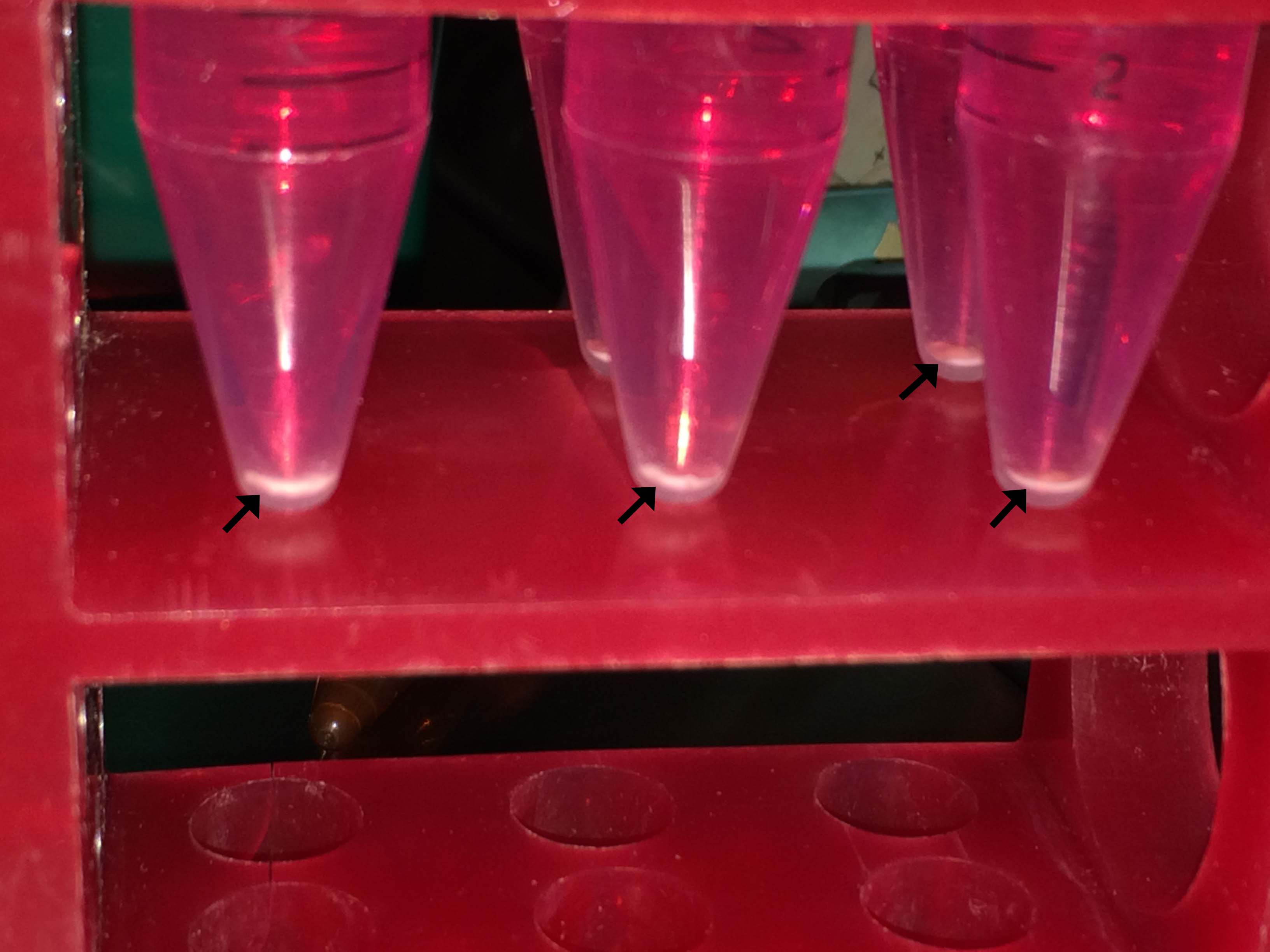
Differentiation into chondrocytes was performed in 15-ml polypropylene centrifuge tubes (Fig. S1) and aggregates (indicated by arrows) were harvested on day 21 for further analysis. The presence of sulfated glycosaminoglycans and collagen II protein in the extracellular matrix of the differentiated cells pellet was demonstrated by toluidine blue and immunofluorescence staining respectively (Fig. 7 B).
Analysis of gene expression by RT-PCR
In the ovary tissues and the cells that were harvested from P3 step, expression of the pluripotent stem cells markers (Oct-4, Sox2, Nanog and SSEA1) and germline stem cells/primordial germ cells markers (DDX4, DAZL, STRA8, DMC1, PRDM1 and SCP3) were detected by RT-PCR (Fig. 8 A). The expression of oocyte-specific (ZP2, ZP3 and NOBOX), neural cell-specific (Nestin and GFAP), osteoblast-specific (Osteocalcin, Osteopontin and ALP) and chondrocyte-specific (Aggrecan and Collagen II) transcripts in the differentiated cells was assessed by RT-PCR (b–e, Fig. 8 B). Conversely, RT-PCR results revealed the absence of oocyte-specific gene (ZP2 and ZP3) expression in the cells that were harvested from the P3 step (a, Fig. 8 B).

Figure 8 RT-PCR, qRT-PCR and western blot (WB) results. (A) Expression of pluripotent and germline stem cell markers in ovary tissues and the cells that harvested from the P3 step. (B) There is no expression of oocyte-specific genes in the P3 step (a). Expression of differentiated cells related markers (b–e). (C) qRT-PCR results in the differentiated cells. In the OLCs, expression of ZP3 was higher than ZP2 and NOBOX. In the osteoblasts, expression of Osteopontin was greater than Osteocalcin and ALP. In the chondrocytes, expression of Collagen II was higher than Aggrecan. In the neural cells, expression of Nestin was higher than GFAP. All results were statistically significant (t-test: P < 0.05). (D) Expression of pluripotent and germline stem cells proteins in ovary tissues and the cells that were harvested from the P3 step. OT: ovary tissues.
Analysis of gene expression in the differentiated cells by qRT-PCR
Results from qRT-PCR showed that ZP3 expression was higher than ZP2 and NOBOX in OLCs, Osteopontin expression was upregulated compared with Osteocalcin and ALP in the osteoblasts, Nestin expression was upregulated compared with GFAP in the neural cells and expression of the Collagen II was upregulated compared with Aggrecan in the chondrocytes (Fig. 8 C). All qRT-PCR results were statistically significant (t-test: P < 0.05).
Analysis of protein expression by WB
WB analysis revealed the presence of germline-specific (DDX4 and DAZL) and pluripotent stem cell-specific proteins (Oct-4, Nanog and Sox2) in the ovary tissues and in the cells that were harvested from the P3 step (Fig. 8 D).
Discussion
Studies on stem cell biology have indicated that stem cells in the different parts of adult body are multipotent or sometimes pluripotent (or multipotent with a transdifferentiation feature) and have the capacity of self-renewal and regeneration. In regard to stem cell biology studies in the adult mammalian ovary, the level of controversy on the possibility of neo-oogenesis and folliculogenesis in post-natal ovaries has intensified during the last years. The basic doctrine that post-natal oogenesis takes place in lower vertebrates but not in mammals is still influential. However, results of several studies have revealed that post-natal mammalian ovaries contain GSCs precursors that are capable of renewing the oocyte pool and follicles.
We have investigated the presence of putative germline and pluripotent stem cells in the adult mouse ovary and their in vitro differentiation potential. A big challenge that we came across was the elimination of maternal oocytes without losing the ovary stromal cells that have a determinative role in oogenesis or female germ cells development. The presence of stromal cells is indispensable for mimicking ovary in vivo conditions. Previous studies have reported that granulosa, techa, fibroblast and immune cells (ovarian macrophages, T cells and etc.) have a critical role during oocyte development and folliculogenesis, through release of hormones, cytokines and growth factors (Bukovsky et al., Reference Bukovsky, Caudle, Virant-Klun, Gupta, Dominguez, Svetlikova and Xu2009; Tingen et al., Reference Tingen, Kiesewetter, Jozefik, Thomas, Tagler, Shea and Woodruff2011; Parte et al., Reference Parte, Bhartiya, Manjramkar, Chauhan and Joshi2013; Ye et al., Reference Ye, Li, Zheng, Liang, Li, Huang, Pan and Zheng2016). Here, in addition to using a cell strainer for elimination of maternal oocytes, the complete clearance of these cells was verified by the absence of ZP2 and ZP3 mRNA expression (a, Fig. 8 B). ZP2 and ZP3 genes are known to be expressed in the oocytes of primordial, primary and secondary follicles (Grootenhuis et al., Reference Grootenhuis, Philipsen, de Breet-Grijsbach and Van Duin1995; Au et al., Reference Au, Whitley, Vaux, Selwood and Familari2008; Gook et al., Reference Gook, Edgar, Borg and Martic2008; Grondahl et al., Reference Grondahl, Borup, Vikesa, Ernst, Andersen and Lykke-Hartmann2013).
Although the presence of the VSELs in the adult tissues is controversial and rejected by some research (Danova-Alt et al., Reference Danova-Alt, Heider, Egger, Cross and Alt2012; Miyanishi et al., Reference Miyanishi, Mori, Seita, Chen, Karten, Chan, Nakauchi and Weissman2013; Szade et al., Reference Szade, Bukowska-Strakova, Nowak, Szade, Kachamakova-Trojanowska, Zukowska, Jozkowicz and Dulak2013), other studies have demonstrated that most adult tissues contain VSELs with pluripotency characteristics (Kucia et al., Reference Kucia, Wysoczynski, Ratajczak and Ratajczak2008; Wojakowski et al., Reference Wojakowski, Tendera, Kucia, Zuba-Surma, Paczkowska, Ciosek, Halasa, Krol, Kazmierski, Buszman, Ochala, Ratajczak, Machalinski and Ratajczak2009; Zuba-Surma et al., Reference Zuba-Surma, Guo, Taher, Sanganalmath, Hunt, Vincent, Kucia, Abdel-Latif, Tang, Ratajczak, Dawn and Bolli2011; Drukala et al., Reference Drukala, Paczkowska, Kucia, Mlynska, Krajewski, Machalinski, Madeja and Ratajczak2012; Kassmer et al., Reference Kassmer, Jin, Zhang, Bruscia, Heydari, Lee, Kim, Kassmer and Krause2013). Under normal conditions these cells are quiescent but, as a result of physiologic necessities or a change in the epigenetic status, VSELs can be activated and may be associated with rejuvenation, ageing and oncogenesis in tissues or organs (Vacanti et al., Reference Vacanti, Roy, Cortiella, Bonassar and Vacanti2001; Kassmer & Krause, Reference Kassmer and Krause2013; Ratajczak et al., Reference Ratajczak, Marycz, Poniewierska-Baran, Fiedorowicz, Zbucka-Kretowska and Moniuszko2014). During the pre-differentiation process in cell culture, we have observed spherical cells (5–50 µm) that expressed germline and pluripotent stem cell markers and a rare population of these cells that was 5–10 µm diameter was characterized as VSELs (pluripotent or multipotent stem cells with transdifferentiation capacity) (Fig. 1 A, B). In agreement with our observations, earlier studies have also reported that VSELs with the ability of in vitro differentiation into OLCs, reside in mammalian ovaries (Parte et al., Reference Parte, Bhartiya, Telang, Daithankar, Salvi, Zaveri and Hinduja2011, Reference Parte, Bhartiya, Manjramkar, Chauhan and Joshi2013; Bhartiya et al., Reference Bhartiya, Sriraman, Gunjal and Modak2012; Patel et al., Reference Patel, Bhartiya, Parte, Gunjal, Yedurkar and Bhatt2013). We have observed a high nuclear/cytoplasm ratio, which is one of the major characteristics of pluripotent stem cells (Thomson et al., Reference Thomson, Itskovitz-Eldor, Shapiro, Waknitz, Swiergiel, Marshall and Jones1998; Ratajczak et al., Reference Ratajczak, Shin, Liu, Mierzejewska, Ratajczak, Kucia and Zuba-Surma2012), during the pre-differentiation process in stem cells with 5–10 µm diameter (arrows in Fig. 2). HE and immunofluorescence staining results led us to identify the cells with a peripheral located nucleus, which is a characteristic of the oocyte development process (Fig. 3). The oocyte nucleus migrates from its central position in the ooplasm to the oocyte periphery just prior to GV breakdown, the first step of meiosis resumption (Parfenov et al., Reference Parfenov, Potchukalina, Dudina, Kostyuchek and Gruzova1989; Mandelbaum, Reference Mandelbaum2000). Mouse ESCs were grown as a proliferative and undifferentiated form in vitro on MEF cells as the feeder layer. Fibroblast cells provided a matrix that supported the attachment of mouse ESCs and released various growth factors that enhanced the survival and proliferation of these cells (Eiselleova et al., Reference Eiselleova, Peterkova, Neradil, Slaninova, Hampl and Dvorak2008; Meng et al., Reference Meng, Zur Nieden, Liu, Cormier, Kallos and Rancourt2008; Tamm et al., Reference Tamm, Pijuan Galito and Anneren2013). According to our observations, the vast majority of OSCs was located on the fibroblast cells and these cells were similar to those with mouse ESCs features, taking advantage of them as a feeder layer.
TEM analysis of the EBs revealed the presence of junctions, including zonula occludens, zonula adherens and gap junctions that confirmed a complex and well organized structure (Fig 4 C). The formation of cell–cell junctions inside the EBs demonstrated the preparedness of EB cells to differentiate. Ultrastructural analysis of EBs showed the presence of collagen bundles and lipid droplets that may be produced by differentiated cells (Fig 4 C).
The expression of germline (DDX4, DAZL, STRA8, DMC1, PRDM1 and SCP3) and pluripotent-specific genes (Oct-4, Sox2, Nanog and SSEA1) was demonstrated in the ovary tissue and in the cells that were harvested from the pre-differentiation step by RT-PCR (Fig. 8 A). Expression of STRA8 (regulator of meiotic initiation), SCP3 (involved in synapsis, recombination and segregation of meiotic chromosomes) and DMC1 (required for homologous chromosome synapsis during meiosis) in the ovary and cell culture is indicative of meiosis initiation and resumption during oogenesis or VSELs and GSCs differentiation processes into new oocytes. In conclusion, GSCs and VSELs that reside in the post-natal ovary are thought to be the origin of OLCs. Our results also demonstrated that in the adult mouse ovary, in addition to the presence of stem cells with differentiation potential into oocyte, there are pluripotent/multipotent stem cells (VSELs or multipotent stem cells with transdifferentiation capacity) with the potential to differentiate into somatic cells: chondrocyte, osteoblast and neural cells.
We believe that in addition to genetic factors, epigenetic regulation such as histone modifications, DNA methylation, chromatin remodelling, and noncoding transcripts, play a crucial role in putative neo-oogenesis in adulthood. Regarding epigenetic mechanisms during oogenesis, histone acetylation was required for transcriptional regulation of germ cell development, including meiotic entry (Wang & Tilly, Reference Wang and Tilly2010) and meiotic continuation (Kim et al., Reference Kim, Liu, Tazaki, Nagata and Aoki2003). Wang and Tilly also reported the expression and physiological regulation of STRA8 in adult mouse ovaries. The CABLES1 gene encodes a protein that is involved in the regulation of the cell cycle through interaction with several cyclin-dependent kinases and has a critical role in the rate of oocyte renewal in adult mouse ovaries (Lee et al., Reference Lee, Sakamoto, Luo, Skaznik-Wikiel, Friel, Niikura, Tilly, Niikura, Klein, Styer, Zukerberg, Tilly and Rueda2007). LIN28A and FOXO3 genes affect the ovarian reproductive capacity during adulthood by determination of germ cell and follicle pool size (Shinoda et al., Reference Shinoda, De Soysa, Seligson, Yabuuchi, Fujiwara, Huang, Hagan, Gregory, Moss and Daley2013; Pelosi et al., Reference Pelosi, Omari, Michel, Ding, Amano, Forabosco, Schlessinger and Ottolenghi2013). Most recently, a study on genome-wide epigenetic signatures revealed that regulation of Prmt5 gene has a fundamental role in maintaining the undifferentiated status of mouse female GSCs (Zhang et al., Reference Zhang, Wu, Wang, Shen, Li, Lu, Gu, Kang, Wong, Ngan, Shao, Wu and Zhao2016). In addition to these findings, further studies are needed to clarify the effects of genetic and epigenetic mechanisms on putative neo-oogenesis during the post-natal period.
In conclusion, although many studies have supported the possibility of a post-natal oogenesis hypothesis, to our knowledge there has been no decisive evidence confirming presence of neo-oogenesis under normal physiologic conditions in mammals. We believe that in the argument of post-natal oogenesis, the contradictions between laboratory study results and normal physiological conditions in mammals, may have arisen due to currently unknown mechanisms related to microenvironment (niche) interactions or genetic and epigenetic factors that could directly affect in vivo and in vitro oogenesis conditions. We concluded that, although neo-oogenesis does not occur normally in mammal in adulthood, the adult mammalian ovary is most likely to contain germline and pluripotent/multipotent stem cells (probably in a quiescent form) that harbour the potential to differentiate into germ and somatic cells, through appropriate modification or manipulations. Based on these findings and earlier studies, the mammalian ovary may also be categorized as adult stem cells and a VSEL resource. However, the controversy over the presence of OSCs and neo-oogenesis in adult mammals continues, and much more work is required to clarify fully the effects of the OSC niche, and genetic and epigenetic factors on oogenesis in the pre- and post-natal periods.
Acknowledgements
I would like to express my great appreciation to the Scientific and Technological Research Council of Turkey (TÜBİTAK-BİDEB) for their valuable support.
Funding
This study was funded by Scientific and Technological Research Council of Turkey (TÜBİTAK, grant number: 115S244) and Ankara University (BAP, grant number: 13B4143003).
Supporting Information
Additional Supporting Information is available online at the publisher's website.
Materials and Methods S1 Reagents, Equipment, and Follicular fluid preparation.
Table S1 List of primary and secondary antibodies.
Table S2 Primers used for RT-PCR and qRT-PCR.
Figure S1 Chondrogenic differentiation was performed in 15 ml polypropylene centrifuge tubes.
Movie S1 Pulsing in one of the EBs that lasted 8 days continuously.
Supplementary material
To view supplementary material for this article, please visit https://doi.org/10.1017/S0967199417000235