


Introduction
Parthenogenesis is a powerful method to analyse epigenetic changes in maternal genes during embryonic development (Surani & Barton, Reference Surani and Barton1983; McGrath & Solter, Reference McGrath and Solter1984; Bos-Mikich et al., Reference Bos-Mikich, Bressan, Ruggeri, Watanabe and Meirelles2016). Moreover, improved parthenogenetic activation (PA) assists somatic cell nuclear transfer (SCNT) (Whitworth et al., Reference Whitworth, Li, Spate, Wax, Rieke, Whyte, Manandhar, Sutovsky, Green, Sutovsky and Prather2009). However, the efficiency of activation remains low. Efforts are continuing to identify better ways (Lee et al., Reference Lee, Tian and Yang2004; Diao et al., Reference Diao, Naruse, Li, Han, Kim, Lin and Jin2013; Lee et al., Reference Lee, Park, Yun, Lee, Yong, Lee, Park, Hyun, Lee and Lee2015; Arias et al., Reference Arias, Sánchez and Felmer2016). Metaphase II (MII) arrest is partly regulated by high maturation-promoting factor (MPF) activity (Hashimoto & Kishimoto, Reference Hashimoto and Kishimoto1988). Early events in oocyte activation occur as MPF activity decreases.
Protein inhibitors that accelerate oocyte activation and improve the blastocyst rate have been widely used (Nussbaum & Prather, Reference Nussbaum and Prather1995; Mayes et al., Reference Mayes, Stogsdill and Prather1995). A widely used method in bovine oocytes activation is combining an electric pulse (EP) with cycloheximide, a protein synthesis inhibitor, which prevents synthesis of MPF (Presicce & Yang, Reference Presicce and Yang1994). Cytochalasin B (CB), a microfilament polymerization inhibitor, has been investigated as means of artificial activation (Cha et al., Reference Cha, Kim, Lee, Baik, Lee and Chung1997). 6-Dimethylaminopurine (6-DMAP), a protein kinase inhibitor, inhibits MPF activity by blocking protein phosphorylation, which causes chromosomal abnormalities (De La Fuente & King, Reference De La Fuente and King1998; Van De Velde et al., Reference Van De Velde, Liu, Bols, Ysebaert and Yang1999). In addition, cyclin-dependent kinase (CDK) inhibitors such as butyrolactone I selectively inhibit the catalytic subunit of MPF and achieve a high rate of good-quality blastocysts (Dinnyés et al., Reference Dinnyés, Hirao and Nagai2000).
AZD5438 (4-(1-isopropyl-2-methylimidazol-5-yl)-2-(4-methylsulphonylanilino)pyrimidine), a broad range CDK inhibitor, can inhibit the phosphorylation of CDK substrates and blocks cell cycling in vitro and significantly decreases the proliferation of human tumour cell lines (Dinnyés et al., Reference Dinnyés, Hirao and Nagai2000; Byth et al., Reference Byth, Thomas, Hughes, Forder, McGregor, Geh, Oakes, Green, Walker, Newcombe, Green, Growcott, Barker and Wilkinson2009; Raghavan et al., Reference Raghavan, Tumati, Yu, Chan, Tomimatsu, Burma, Bristow and Saha2012). In addition, AZD5438 has undergone clinical evaluation and been investigated in healthy volunteer studies (Camidge et al., Reference Camidge, Pemberton, Growcott, Amakye, Wilson, Swaisland, Forder, Wilkinson, Byth and Hughes2007a,Reference Camidge, Smethurst, Growcott, Barrass, Foster, Febbraro, Swaisland and Hughesb). However, activation of mammalian oocytes using AZD5438 has not been evaluated.
The goal of this study was to investigate the development of porcine oocytes parthenogenetically activated by an EP combined with AZD5438 treatment. Optimal concentration and duration of AZD5438 treatment were determined. Then, MPF assay, karyotype analysis and RT-PCR analysis of several apoptosis- and pluripotency-related genes in embryos were conducted. We also compared the developmental competence of SCNT embryos between the AZD5438-treated and 6-DMAP-treated groups.
Materials and Methods
Animals
This research was approved by the Ethics Committee of Yanbian University.
Chemicals
All chemicals used in this study were purchased from Sigma Chemical Company (St. Louis, MO, USA), unless otherwise noted. AZD5438 was purchased from Selleck Chemicals (Houston, TX, USA).
Oocyte retrieval and in vitro maturation
Ovaries were obtained from a local abattoir, transported to the laboratory within 2 h. Cumulus–oocyte complexes (COCs) were aspirated from follicles with a diameter of 2−7 mm using an 18-gauge needle. The aspirated oocytes showing a uniformly granulated cytoplasm and surrounded by at least three uniform layers of compact cumulus cells were selected. The selected good-quality COCs were washed three times in Tyrode's lactate HEPES-buffered solution containing 0.1% polyvinyl alcohol (PVA), cultured in NCSU-37 medium supplemented with 10% porcine follicular fluid, 0.6 mM cysteine, 0.1 IU/ml human chorionic gonadotropin (hCG) and 0.1 IU/ml pregnant mare serum gonadotropin (PMSG) for 20–22 h, and cultured for another 22 h in medium that lacked PMSG and hCG at 38.5°C in 5% CO2 in air.
PA and in vitro culture (IVC)
All matured oocytes with an extruded first polar body were exposed to a direct current pulse (1.5 kV/cm, 60 µs) in 0.28 mol/l mannitol containing 0.1 mM MgSO4, 0.05 mM CaCl2, and 0.1% PVA. After PA, oocytes were washed with NCSU-37 medium supplemented with 4 mg/ml bovine serum albumin and incubated in culture medium containing AZD5438 or CB at 38.5°C in 5% CO2 in air. Previous studies have proved that treatment with AZ5438 alone, following parthenogenetic stimulation, can prevent the release of the second polar body after activation of the oocytes, which results in diploid development. Cleavage and blastocyst formation were evaluated on days 2 and 7, respectively.
In vitro MPF activity assay
The level of MPF was measured in PA oocytes treated with AZD5438 or CB at optimal concentration and duration. Oocytes (40 per treatment group) were washed several times in phosphate-buffered saline (PBS). Oocytes were repeatedly blown with a fine needle whose diameter was smaller than that of the oocytes until they ruptured. The samples were centrifuged at 150 g for 15 min at 4°C. The supernatants were collected to determine the MPF level by the Porcine MPF ELISA Kit (Kexing, Shanghai, China) following the manufacturer's protocol. In brief, 10 µl supernatant was added to each well, followed by 40 µl of dilution buffer (i.e., the sample was diluted five-fold). Plates were incubated at 37°C in the dark for 30 min. Each well was washed thoroughly with buffer and supplemented with 50 µl horseradish peroxidase conjugate reagent, except for the blank. Samples were incubated at 37°C for 30 min and then washed. After adding TMB substrate solution to each well, the plate was incubated at 37°C in the dark for 15 min. The reaction was terminated by adding 50 µl sulfuric acid. The colour changes were measured using the spectrophotometer at a wavelength of 450 nm within 15 min of the reaction being terminated, as previously reported (Li et al., Reference Li, Kang, Jin, Hong, Zhu, Jin, Gao, Yan, Cui, Li and Yin2014). The concentration of MPF in the samples was determined using a standard curve.
Karyotype analysis of embryos derived from PA oocytes
Blastocysts derived from the AZD5438-treated groups were subjected to karyotype analysis. Briefly, blastocysts were pre-treated with 0.2 µg/ml demecolcine for 4 h to block cell division and then incubated in a hypotonic solution of 0.075 mol/l KCl for 20 min at 37°C. Swollen blastocysts were placed on a clean glass slide immersed in stationary liquid (methanol:acetic acid = 3:1). After drying, slides were stained with 10% Giemsa (Invitrogen, Carlsbad, CA, USA) for 10 min. The stained chromosomes were imaged using oil immersion optics (Nikon) to determine the chromosome number.
Nuclear transfer
Nuclear transfer was performed as described previously (Yin et al., Reference Yin, Tani, Yonemura, Kawakami, Miyamoto, Hasegawa, Kato and Tsunoda2002). Briefly, mature oocytes with the first polar body were cultured in medium supplemented with 0.4 mg/ml demecolcine and 0.05 M sucrose for 1 h. Sucrose was used to enlarge the perivitelline space of the oocytes. Treated oocytes with a protruding membrane were transferred to medium supplemented with 5 mg/ml CB and 0.4 mg/ml demecolcine, and the protrusion was removed with a beveled pipette. A single donor cell was injected into the perivitelline space of each oocyte, and then electrically fused using two direct current pulses of 150 V/mm for 50 ms in 0.28 mol/l mannitol supplemented with 0.1 mM MgSO4 and 0.01% PVA. Fused oocytes were cultured in NCSU-37 medium for 1 h before electro-activation and then cultured in medium supplemented with 10 µM AZD5438 or 5 mg/ml CB for 4 h. The reconstructed oocytes were activated by two direct current pulses of 100 V/mm for 20 ms in 0.28 mol/l mannitol supplemented with 0.1 mmol/l MgSO4 and 0.05 mmol/l CaCl2. Activated oocytes were cultured in the medium for 6 days in an atmosphere of 5% CO2 and 95% air at 39°C. Finally, the nuclei of blastocysts stained with 50 µg/ml bisbenzimide were imaged by fluorescence microscopy.
Gene expression in PA oocytes
The mRNA expression levels of two apoptosis-related genes (Bax and Bcl-2) and three pluripotency-related genes (Oct4, Nanog, and Sox2) were measured by RT-PCR, together with ribosomal protein L19 (RPL19) as an endogenous reference gene. All samples were stored at −80°C until use. Total RNA was extracted from 34 PA oocytes treated with AZD5438 or CB using the Dynabeads® mRNA DIRECT™ Kit (Life Technologies AS, Oslo 010, Norway) following the manufacturer's instructions. Purified RNA was immediately transcribed into cDNA using the SuperScript® III First-Strand Synthesis System (Invitrogen, Carlsbad CA, USA), according to the manufacturer's directions. The primers were designed based on methods reported in the literature (Liang et al., Reference Liang, Zhao, Choi, Kim and Cui2015; Wang et al., Reference Wang, Luo, Lin, Lee, Kwon, Cui and Kim2015; Zhao et al., Reference Zhao, Kim and Cui2016) and expected product sizes are shown in Table 1. PCR was performed in a thermal cycler (T100™ Thermal Cycle; Bio-Rad Laboratories, Inc., CA, USA). PCR products were electrophoresed in 1.5% (w/v) agarose gels. The expression levels were determined by measuring the intensity of each band.
Table 1 Primer sequences used for gene expression analysis

Experimental design
In a preliminary experiment, the extrusion of the second polar body was inhibited in more than 90% of matured oocytes subjected to PA and treated with AZD5438 for 30 min; therefore, oocytes were treated with AZD5438 and CB. Each experiment was performed more than three times.
In Experiment 1, the optimal concentration and duration of AZD5438 treatment after PA were determined. The developmental rates were compared among the different groups. Karyotype analysis was performed of blastocysts derived from these PA oocytes.
In Experiment 2, PA and SCNT were performed using oocytes treated with AZD5438 or 6-DMAP, a commonly used protein inhibiter, and the in vitro development of the embryos was compared.
In Experiment 3, the mRNA expression levels of apoptosis-related genes (Bax and Bcl-2) and pluripotency-related genes (Oct4, Nanog, and Sox2) were checked by RT-PCR in PA oocytes treated with or without AZD5438. MPF activity was assessed in porcine PA oocytes treated with AZD5438 for 4 h or non-treated oocytes.
Statistical analysis
Each experiment was performed at least three times. Differences between groups were determined using the SPSS 19.0 software (SPSS Inc., Chicago, IL, USA). Data expressed as proportions (i.e. percentages) were analysed using chi-squared test, nuclei numbers were analysed by T-Test (Independent Samples). All data were reported as the mean ± standard error of the mean (SEM). P-values ≤ 0.05 were considered statistically significant.
Results
Effect of treatment with AZD5438 at different concentrations and for different durations on the in vitro development of porcine PA oocytes
The percentage of blastocyst was significantly higher in the group treated with 10 µM AZD5438 than in the groups treated with 20 µM, 50 µM or 5 µg/ml CB (46.4% vs. 32.3%, 24.0%, and 34.5%, respectively; P < 0.05; Table 2). There were no significant differences in the number of cells per blastocyst among these four groups. By contrast, the blastocyst formation rate was significantly lower (P < 0.05) in the group treated with 50 µM AZD5438 than in the control group. The blastocyst formation rate was higher in the group treated with 10 µM AZD5438 for 4 h than in the groups treated with 10 µM for 2 or 6 h. However, this was not significant (42.8%, 38.6%, and 37.2%, respectively; Table 3). In karyotype analysis, 66.67% (12/18) of blastocysts (Fig. 1) were classified as diploid in the group treated with 10 µM AZD5438 for 4 h (Fig. 2).
Table 2 In vitro development of porcine embryos derived from parthenogenetic activation oocytes treated with different concentrations of AZD5438 for 4 h after exposure to electric pulse
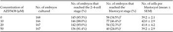
a,b,c Values with different superscripts in the same column are significantly different (P < 0.05).
Table 3 In vitro development of porcine embryos derived from parthenogenetic activation oocytes treated with 10 µM AZD5438 for different durations after exposure to electric pulse
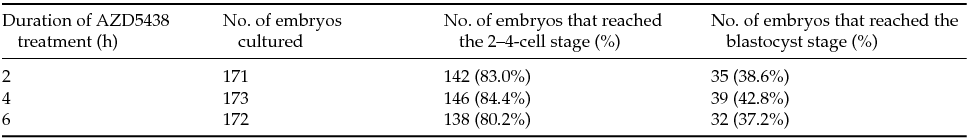

Figure 1 Karyotype analysis of a porcine blastocyst at day 7 derived from an AZD5438-treated parthenogenetic activation oocyte. A normal diploid karyotype of 38 chromosomes in metaphase is shown. Original magnification: ×1000.

Figure 2 (A) Blastocysts derived from AZD5438-treated parthenogenetic activation oocytes. (B) Blastocysts stained with Hoechst 33342. Scale bars = 100 μm.
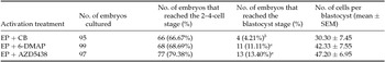
In vitro development of PA and SCNT embryos derived from oocytes activated with an EP and treated AZD5438 or 6-DMAP
After PA, there is no significant difference between the percentage (42.4% and 34.3%, P > 0.05; Table 4) of embryos that developed to the blastocyst stage in the AZD5438-treated and 6-DMAP-treated groups (Table 4). The cleavage rate (93.4% and 91.2%, respectively) and total number of cells per blastocyst (42.4 ± 13.1 and 42.8 ± 11.5, respectively) were similar in the AZD5438-treated and 6-DMAP-treated groups (Table 4).
Table 4 In vitro development of porcine parthenogenetic activation embryos activated with AZD5438 and 6-DMAP

6-DMAP, 6-Dimethylaminopurine; EP, electric pulse.
After SCNT, the percentage of embryos that developed to the blastocyst stage was significantly higher in the AZD5438-treated and 6-DMAP-treated groups than in the CB-treated group (13.4% and 11.1% vs. 4.2%, P < 0.05; Table 5). The cleavage rate (79.4%, 68.7%, and 66.7%, respectively) and total number of cells per blastocyst (47.2 ± 7.0, 42.3 ± 7.6, and 30.3 ± 7.5, respectively) were similar in each groups (Table 5).
Table 5 In vitro development of reconstructed porcine somatic cell nuclear transfer embryos derived from oocytes activated by EP and treated with AZD5438, 6-DMAP or CB
CB, Cytochalasin; 6-DMAP, 6-Dimethylaminopurine. EP, electric pulse;
a,b Values with different superscripts in the same column are significantly different (P < 0.05).
Levels of MPF and gene expression in porcine oocytes activated by an EP and AZD5438 treatment
The level of MPF was investigated in oocytes activated by an EP and treated with AZD5438 or CB. The level of MPF was significantly lower in the AZD5438-treated group than in the CB-treated group (125.9 pg/ml vs. 288.9 pg/ml, P < 0.05; Fig. 3).

Figure 3 MPF concentrations in oocytes activated by an electric pulse and treated with AZD5438 or cytochalasin B for 4 h. Bars with different superscripts are significantly different.
The mRNA expression level of genes related to apoptosis (Bax & Bcl-2) and pluripotency (Oct4, Nanog, and Sox2) were measured, together with RPL19 as an endogenous reference gene. The mRNA expression levels of these genes did not differ between the AZD5438- and CB-treated groups (Fig. 4).

Figure 4 Detection of porcine Bax, Bcl-2, Oct4, Sox2, Nanog, and RPL19 transcripts in parthenogenetic activation oocytes treated with AZD5438 and cytochalasin B by RT-PCR analysis (A) Expression of mRNA is shown. (B) Band intensities are shown. T = AZD5438 treatment, C = CB treatment.
Discussion
Present results clearly show that porcine oocytes can be efficiently activated by PA followed by AZD5438 treatment. This finding is in agreement with the previous findings that PA in combination with 6-DMAP (Liu et al., Reference Liu, Ju and Yang1998; Saikhun et al., Reference Saikhun, Kitiyanant, Songtaveesin, Pavasuthipaisit and Kitiyanant2004), cycloheximide (Presicce & Yang, Reference Presicce and Yang1994), and butyrolactone I (Dinnyés et al., Reference Dinnyés, Hirao and Nagai2000) treatment induces activation and the subsequent development of bovine oocytes more effectively than any single treatment.
This study aimed to improve the developmental competence of porcine embryos in PA and SCNT experiments using oocytes activated by an EP and treated with AZD5438 at different concentrations and for different durations. Treatment with 10 µM AZD5438 for 4 h significantly improved the developmental capacity of embryos following PA. Furthermore, the blastocyst formation rates in PA and SCNT experiments were similar between the group treated with 2 mM 6-DMAP for 4 h and the group treated with 10 µM AZD5438 for 4 h. 6-DMAP is a protein kinase inhibitor that has been widely exploited in nuclear transfer experiments and significantly enhances activation, accelerates pronuclear formation and parthenogenetic development, and inactivates cytostatic factor and MPF (Masui & Markert, Reference Masui and Markert1971; Masui, Reference Masui1991). However, 6-DMAP non-specifically affects several pathways in oocytes, induces a high incidence of chromosomal abnormality, and impairs further embryonic development (De La Fuente & King, Reference De La Fuente and King1998; Van De Velde et al., Reference Van De Velde, Liu, Bols, Ysebaert and Yang1999). In total, 66.67% of blastocysts derived from AZD5438-treated PA oocytes had a diploid karyotype. 6-DMAP treatment inhibits extrusion of the second polar body due to inhibition of phosphorylation necessary for the spindle apparatus (De La Fuente & King, Reference De La Fuente and King1998; Van De Velde et al., Reference Van De Velde, Liu, Bols, Ysebaert and Yang1999). The mechanism by which it inhibits extrusion of the second polar body is not known, although it may be related to inhibition of CDK substrate phosphorylation and blockade of cell cycling at the G2-M, S, and G1 phases (Dinnyés et al., Reference Dinnyés, Hirao and Nagai2000). The level of MPF was significantly decreased in PA oocytes treated with AZD5438. This result is consistent with previous research, and this supports the theory that a reduced level of MPF allows porcine oocytes to progress into interphase (Masui & Markert, Reference Masui and Markert1971; Masui, Reference Masui1991; Lee et al., Reference Lee, Kim, Kim, Lee, Ko and Lee2016). Moreover, previous studies have proved that treatment with CB, following parthenogenetic stimulation, enhanced developmental competence in cattle (Fukui et al., Reference Fukui, Sawai, Furudate, Sato, Iwazumi and Ohsaki1992; Presicce & Yang, Reference Presicce and Yang1994) and pig (Cha et al., Reference Cha, Kim, Lee, Baik, Lee and Chung1997). CB can prevent the release of the second polar body after activation of the oocytes, which results in diploid development (Fukui et al.,Reference Fukui, Sawai, Furudate, Sato, Iwazumi and Ohsaki1992). Therefore, CB-treated groups were used in these comparisons as a control.
To evaluate the effect of AZD5438 on the expression of genes related to apoptosis and pluripotency, RT-PCR was conducted. A previous study demonstrated that MPF plays important roles in oocyte cytoplasmic maturation by regulating gene expression (Zhang et al., Reference Zhang, Park, Sun, Xu, Li, Cui and Kim2011).
Bax and Bcl-2 play vital roles in apoptosis (Yang & Rajamahendran, Reference Yang and Rajamahendran2002). Oct4, Nanog, and Sox2 are considered to be important transcription factors in the regulation of early embryonic development (Sommer et al., Reference Sommer, Stadtfeld, Murphy, Hochedlinger, Kotton and Mostoslavsky2009). The mRNA expression levels of these genes did not differ between the AZD5438- and CB-treated groups.
In conclusion, our results indicate that treatment with 10 µM AZD5438 for 4 h can improve the developmental competence of porcine embryos in PA and SCNT experiments. This situation may be because AZD5438 treatment decreases MPF activity, reduces the incidence of abnormal karyotypes, and decreases the extrusion rate of the second polar body. AZD5438 improves the efficiency of porcine oocyte activation, and further studies are required to clarify its effects on early embryonic development.
Acknowledgements
This work was supported by the Institute for Basic Science (Grant No. IBS-R021-D1–2015-a02) and the Youth Scientific Research Foundation of Department of Science and Technology of Jilin Province of China (Grant No. 20160520057JH).
Conflict of interest
None of the authors has any conflict of interest to declare.


