


Introduction
With the rapid development of embryo engineering technology, oocytes have become one of the most important experimental materials used for animal cloning, nuclear reprogramming and epigenetic modification research. The efficient and practical way to obtain porcine oocytes was from ovaries that were collected at a slaughterhouse and through in vitro maturation (IVM). However oocyte nuclear modifications and cytoplasmic maturation undergo great alterations during IVM (around 42 h) that affect subsequent embryo development (Lonergan et al., Reference Lonergan, Rizos, Gutierrez-Adan, Fair and Boland2003). Therefore, an optimal culture system in vitro is needed to obtain large numbers of oocytes with good nuclear and cytoplasmic maturation.
Among the many factors that influence porcine oocyte IVM, hormonal requirements and antioxidant components play an important role in stimulating oocytes for nuclear and cytoplasmic maturation. Previous studies have shown that, at different IVM developmental stages, different kinds of hormones may be required for both nuclear and cytoplasmic maturation of porcine oocytes (Funahashi et al., Reference Funahashi, Cantley and Day1994; Shimada et al., Reference Shimada, Nishibori, Isobe, Kawano and Terada2003; Kawashima et al., Reference Kawashima, Okazaki, Noma, Nishibori, Yamashita and Shimada2008). Studies also recognized that reactive oxygen species (ROS) greatly affected porcine oocyte IVM because they cause damage to oocyte organelles (Favetta et al., Reference Favetta, St John, King and Betts2007). ITS (insulin–transferrin–selenium), known to promote glucose, amino acid and mineral uptake by cells and to detoxify oxygen radicals, has been used as an additive to culture medium to partially substitute for serum. Insulin may improve the cleavage rate and developmental potential of porcine oocytes and embryos during IVM and IVC (in vitro culture) (Lee et al., Reference Lee, Kang, Lee and Hwang2005). Selenium and transferrin may participate in the oocyte antioxidant defense system, which is essential for glutathione peroxidase catalytic activity (Cerri et al., Reference Cerri, Rutigliano, Lima, Araujo and Santos2009). ITS has been used, therefore, as a supplement for IVM in mouse (De La Fuente et al., Reference De La Fuente, O'Brien and Eppig1999), goat (Herrick et al., Reference Herrick, Behboodi, Memili, Blash, Echelard and Krisher2004) and pig (Jeong et al., Reference Jeong, Hossein, Bhandari, Kim, Kim, Park, Lee, Park, Jeong, Lee, Kim and Hwang2008). However, the ultimate culture condition and co-effects of ITS and hormones on porcine oocyte nuclear and cytoplasmic maturation have not been elucidated well. In this study, we tested variables, including hormone supplementation time with or without ITS, that influence the efficiency of porcine oocytes maturation in vitro, and set up a reproducible system to produce >80% IVM oocytes that subsequently could be used for somatic cloning and transgenic research.
Materials and methods
Chemicals and culture media
All chemicals used in the study were purchased from Sigma Chemical Company unless otherwise specified. Tissue Culture Medium (TCM)-199 was bought from Invitrogen. Insulin–transferrin–selenium (ITS) was obtained from Gibco and pregnant mare serum gonadotrophin (PMSG), human chorionic gonadotrophin (hCG) and follicle stimulating hormone (FSH) from Ni Bo Sangsheng Pharmaceutical Co., Ltd. The basic IVM medium was modified TCM-199 supplemented with 0.1% polyvinyl alcohol, 0.57 mmol/l cysteine, 3.05 mmol/l glucose and 0.91 mmol/l pyruvic acid Na-salt (mM199). The medium used for collecting and washing cumulus–oocyte complexes was composed of 10% newborn bovine serum (NBS) in Dulbecco's phosphate-buffered saline (PBS) (DPBS; Gibco).
Ovaries collection
Ovaries were collected at a local pig slaughterhouse from peripubertal gilts with no information on age and breeding. Ovaries stored in 0.9% (w/v) NaCl containing 100 mg/l penicillin and streptomycin at 30–37°C were transported to the laboratory within 2 h of slaughter.
Preparation and culture of cumulus–oocyte complexes
The follicular contents, including cumulus–oocyte complexes (COCs), were collected by aspirating the visible small antral follicles (around 2–6 mm diameter) with a 10 ml syringe equipped with an 8-gauge needle. Only COCs with a uniform ooplasm and a compact cumulus cell mass were collected and washed three times with Ca2+-/Mg2+-free PBS plus 10% NBS. Oocytes were classified into three types: type A, oocyte has more than five layers of cumulus cells symmetrically surrounding its surface and has the dim cytoplast; type B, oocyte has one to four layers of cumulus cells; and type C, abnormal oocytes have asymmetrical cytoplast or a few cumulus cells and naked oocytes. In this research, oocytes from types A and B were washed separately three times and cultured at 38.5 °C in 5% CO2 and a saturated humid air. After 42 h IVM, cumulus cells were removed by pipetting gently with a fine-bore pipette in saline supplemented with 0.3% hyaluronidase (Gibco Brl) for 3–5 min to investigate the matured oocytes. The signs of maturation oocytes are a protruding first polar body in combination with surrounding modality of oocytes.
Treatment by hormones and ITS
Four treatments were used to detect the effect of hormones during porcine oocyte IVM. Medium based on mM199 (TCM-199 medium plus 0.1% polyvinyl alcohol, 0.57 mmol/l cysteine, 3.05 mmol/l glucose and 0.91 mmol/l pyruvic acid Na-salt) was used. (i) Treatment A, oocytes were cultured in hormone-free medium A (mM199 plus 10 ng/ml EGF) for 42 h (control group). (ii) Treatment B, oocytes were cultured for 42 h in the hormone-supplemented medium B (mM199 supplemented with 10 ng/ml EGF, 10 IU/ml PMSG, 10 IU/ml hCG and 2.5 IU/ml FSH). (iii) Treatment C, in the first 21 h culture period oocytes were grown in medium B, and then cultured for another 21 h in medium A. (iv) Treatment D, in the first 21 h culture period oocytes were grown in medium A, and then cultured in medium B for 21 h.
To investigate if ITS was involved in porcine oocyte IVM, oocytes were cultured in medium B with or without 1% ITS for 42 h. The distribution of cortical granules (CGs) was categorized into three groups as described previously (Hosoe & Shioya, Reference Hosoe and Shioya1997). Nuclear maturation, when oocytes protrude the first polar body, and cytoplasmic maturation, when oocytes show type II CG distribution, were examined in groups treated with or without ITS after IVM.
Immunocytochemistry staining of cortical granules
After 42 h IVM, cumulus cell-free oocytes were washed twice in PBS, and fixed with 3.7% paraformaldehyde for 30 min, and then washed three times in PBS. Oocytes were permeabilized in PBS with 0.1% Triton X-100 for 5 min, and then rinsed four times in PBS. Oocytes were incubated in PBS with 100 g/ml fluorescent Lens culinaris agglutinin–fluorescein complex (LCA–FITC) for 30 min at 37 °C. Finally, oocytes were rinsed and mounted on histological slides and the localization of CGs was evaluated with a Nikon fluorescence microscope (wavelength: 488 nM). As before, the distribution of CGs in porcine oocyte IVM was evaluated based on three categories (types I–III) (Hosoe & Shioya, Reference Hosoe and Shioya1997).
Statistical analysis
Statistical analysis of the effects of different hormone supplementation was conducted using analysis of variance (ANOVA), and t-test using SPSS 10.0 (Statistical Package for Social Science) software, a p-value < 0.05 was considered significantly.
Results
Effect of hormone treatment on porcine oocytes IVM
Of the three types of oocytes described (A, B, C), oocytes from only types A and B were selected for these experiments (Fig. 1). In regular culture conditions with no hormone added (treatment A), the rate of porcine oocyte maturation was only 11.7% (Fig. 2A). Three different treatments were conducted to investigate oocyte maturation by reproductive hormones. In treatment A, the rate of IVM oocytes incubated in medium B for 42 h reached 47.8% (Fig. 2B). We then reduced the time of hormone treatment to 21 h in two separate experiments: (a) oocytes were treated with hormones for 21 h and then with non-hormone treatment for another 21 h; or (b) oocytes were treated without hormones for 21 h, and then with hormones for another 21 h. In these two experiments, the rates of IVM oocytes were in 45.4% and 44.9%, respectively, slightly lower than in treatment B (Fig. 2C,D). In summary, when hormones were added to the culture medium (treatments B–D), the rate of oocyte IVM increased to 44–48%, indicating that reproductive hormones could significantly improve the maturation of porcine oocytes. We observed that the rate of porcine oocyte maturation in hormone-treated groups was significantly higher than in the control group, but there was no significant difference among three groups with the different intervals of hormone treatments.

Figure 1 Category of cumulus–oocyte complexes. Oocytes were categorized into three types according to the quantity of cumulus cells and the degree of cytoplast refraction: type A, over five layers of cumulus cells surrounding oocytes symmetrically and a dim cytoplast; type B, contained 1–4 layers of cumulus cells; type C had an asymmetrical cytoplast or abnormal oocytes with a few cumulus cells, naked oocytes. (A) Oocyte with over five layers of cumulus cells. (B) Oocyte with 1–4 layers of cumulus cells. (C) Oocyte with a few cumulus cells. ×100 magnification.

Figure 2 In vitro maturation (IVM) of porcine oocytes with different hormone treatments. The IVM of porcine oocytes grown with or without hormone supplements for different time intervals. (A) Oocytes were grown without hormone supplements for 42 h (as control group). (B) Oocytes were grown with hormone supplements for 42 h. (C) Oocytes were firstly grown with hormone supplements for 21 h, and then without hormone supplements for 21 h. (D) Oocytes were firstly grown without hormone supplements for 21 h, and then with hormone supplements for 21 h. a,bValues with different superscripts indicate significant difference (p < 0.05). (See online for a colour version of this figure.)
ITS improves porcine oocyte nuclear maturation
The major sign of oocytes nuclear maturation is the first polar body protruding. To improve porcine oocyte nuclear maturation, 1% ITS was added in medium B that contained hormones. After 42 h incubation with or without ITS, the numbers of oocytes that protruded the first polar body were counted. Figure 3 shows that the ITS-treated group (Fig. 3A) had more mature oocytes than did the ITS-untreated group (Fig. 3B). The rate of nuclear maturation in the ITS-treated group was significantly higher than in the ITS-untreated group (78.5% vs. 54.35%, respectively, p < 0.05). This observation indicated that ITS could clearly improve porcine oocyte nuclear maturation as for hormone supplements (Table 1).

Figure 3 Maturation of porcine oocytes cultured with ITS. After 42 h incubation with or without ITS in vitro, the numbers of oocytes that protruded the first polar body were counted. (A) Oocytes grown with ITS. (B) Oocytes grown without ITS. The arrows indicate the polar bodies. ×50 magnification.
Table 1 Influence of 1% ITS on IVM of porcine oocytes
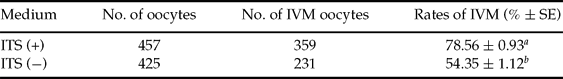
a,b Values with different superscripts indicate significant difference (p < 0.05), n = 4.
ITS stimulates oocyte cytoplasmic maturation
Three types of CG distribution were observed during the maturation of porcine oocytes. In type I oocytes, CGs were distributed in the cytoplasm, but not on the plasma membrane (Fig. 4A). In type II oocytes, CGs were distributed in the cortex beneath the zona pellucida and formed fluorescent halo on the plasma membrane (Fig. 4B). In type III oocytes, CGs were distributed partially on the plasma membrane and partially in the cytoplasm (Fig. 4C). When oocytes maturated in medium B without adding ITS, the oocyte ratios for different types of CG distribution were 17.9% for type I, 62.7% for type II and 19.4% for type III (Table 2). Following ITS treatment, the ratios of oocytes with type I or type III CG distribution were reduced significantly, and the per cent of oocytes with type II CG distribution significantly increased (85.3%, Table 2). These results indicated that ITS could enhance porcine oocyte cytoplasmic maturation.

Figure 4 Distribution of cortical granules (CGs) in porcine oocytes. Three types of CG distribution were identified during the maturation of porcine oocytes. (A) Type I: CGs distributed in cytoplasm, but not on plasma membrane. (B) Type II: CGs distributed in the cortex and forming a fluorescent halo around the plasma membrane. (C) Type III: CGs distributed partially on the plasma membrane and partially in the cytoplasm. ×200 magnification. (See online for a colour version of this figure.)
Table 2 Influence of 1% ITS on CG distribution in porcine oocytes

a,b Values with different superscripts indicate significant difference (p < 0.05), n = 3.
Discussion
Hormone supplement in culture medium was crucial for porcine oocytes nuclear and ooplasmic maturation. Previous studies have shown that medium supplemented with gonadotrophin or steroid, or with both, was necessarily to maintain the IVM of porcine oocytes. Funahashi and Day found that the use of both hCG and equine chorionic gonadotropin (eCG) greatly promoted porcine oocyte meiosis and cytoplasmic maturation (Funahashi & Day, Reference Funahashi and Day1993). Porcine oocyte maturation rates were enhanced and oocyte meiosis and cytoplasmic maturation were also facilitated when using PMSG, hCG and estradiol (E2) in culture medium for 20 h, and then withdrawing hormones from the medium, thus allowing oocytes to grow for another 20 h (Funahashi et al., Reference Funahashi, Cantley and Day1994). Mariana et al. showed that a 48 h treatment with eCG and hCG could reduce the maturation rate; while a better result was obtained using a treatment period for 24 h with hormones and a following period of 24 h without hormones (Viana et al., Reference Viana, Caldas-Bussiere, Matta, Faes, de Carvalho and Quirino2007). In our experiments, we noticed that the combination of PMSG, hCG and FSH supplemented in the medium could significantly increase the maturation rate of porcine oocytes during the 42 h period of cultivation. We discovered that the maturation rate was able to reach to 45% in both treatments C and D, although that rate was slightly lower than treatment B but not different statistically, therefore suggesting that the shorter period of hormone treatment (21 h) could also enhance IVM of porcine oocyte.
Glutathione (γ-glutamylcysteinylglycine, GSH), the major non-protein sulphydryl compound in mammalian cells, reduced oxidative damage and controlled the level of reactive oxygen species (ROS) in oocytes (Rodrigues & Rodrigues, Reference Rodrigues and Rodrigues2003). Tatemoto et al. found that antioxidants, such as GSH, transferrin and selenium, greatly enhanced maturation of oocytes through mitochondrial protection, decreasing polyspermy and increasing pronuclear formation (Tatemoto et al., Reference Tatemoto, Muto, Sunagawa, Shinjo and Nakada2004, Cerri et al., Reference Cerri, Rutigliano, Lima, Araujo and Santos2009). ITS supplementation could significantly increase glutathione content in oocytes and had an insulin-like function to raise the developmental potential of oocytes (Kim et al., Reference Kim, Lee, Lee, Kim, Jeong, Kim, Kang, Lee and Hwang2005; Lee et al., Reference Lee, Kang, Lee and Hwang2005, Jeong et al., Reference Jeong, Hossein, Bhandari, Kim, Kim, Park, Lee, Park, Jeong, Lee, Kim and Hwang2008). Although medium with hormone supplements could increase the rate of porcine oocyte maturation to over 50% (Table 1), we decided to add 1% ITS in the hormone-supplemented medium to illustrate if ITS was able to additionally raise the rate of oocyte maturation. Our results showed that after ITS treatment the maturation rate of the experimental group was significantly higher than that of the control group without ITS (p < 0.05, Table 1). Based on both previous reports and our observations, we concluded that ITS supplementation could help achieve the better maturation rate of porcine oocytes by accelerating oxygen elimination and reducing ROS in porcine oocytes.
Cytoplasmic quality in IVM oocytes played a major role in the reconstruction of embryonic development. An important indication of cytoplasmic maturation was the migration and redistribution of CGs that migrated to the oocyte cortical region of during IVM (Wang et al., Reference Wang, Sun, Hosoe, Shioya and Day1997b; Liu et al., Reference Liu, Mal, Miao, Liu, Bao and Tan2005; Cao et al., Reference Cao, Zhou, Luo, Zhao and Shi2009). Table 2 shows that after ITS treatment the percentage of types I and III oocytes decreased compared with the control (14.7% vs. 37.3%), while the percentage of type II oocytes was significantly increased (85.3% vs. 62.7%, p < 0.05). Our observation (Fig. 4 and Table 2) was consistent with previous reports that showed that oocytes with CG redistribution to the cortical region had the ability to fertilize and reprogramme other nuclear materials (Wang et al., Reference Wang, Hosoe, Li and Shioya1997a; Ferreira et al., Reference Ferreira, Vireque, Adona, Meirelles, Ferriani and Navarro2009), and highlighted the fact that ITS could enhance porcine oocyte cytoplasmic maturation.
In summary, ITS supplementation not only improved the maturation rate of porcine oocytes, but also augmented synchronization rates of nuclear and ooplasmic maturation to 67% (78.56% × 85.33%). Based on our results, the culture medium described (mM199, 10 ng/ml EGF, 10 IU/ml PMSG, 10 IU/ml hCG, 2.5 IU/ml FSH, and 1% ITS) was a suitable and stable medium for porcine oocytes IVM.
Acknowledgements
This work was supported by the Chinese National Natural Science Foundation (#30871786), the Chinese National Programs for High Technology Research and Development ‘863 program’ (#2008AA101005), and the Key Project of Chinese National Programs for Fundamental Research and Development ‘973 program’ (#2009CB941002).


