Introduction
In Bufo arenarum and other anuran species, the oviduct plays an essential role in the preparation of the female gamete for fertilization (Miceli & Cabada, Reference Miceli and Cabada1998). In vitro, sperm cannot penetrate the envelope surrounding plasma membrane of ovulated oocytes (known as coelomic envelope) (Miceli et al., Reference Miceli, Fernández, Raisman and Barbieri1978; Hedrick & Hardy, Reference Hedrick and Hardy1991). Conversion to a penetrable state occurs during passage through the pars recta (PR) portion of the oviduct, where a protease, oviductin, is secreted, which hydrolyzes the envelope glycoprotein gp43 to gp41 in Xenopus laevis (Hardy & Hedrick, Reference Hardy and Hedrick1992). Hydrolysis of gp40–52 by oviductin in Bufo japonicus results in the concomitant increase in gp39 and appearance of gp36 (Takamune & Katagiri, Reference Takamune and Katagiri1987). PR protease activity in B. arenarum is characterized by the disappearance of two glycoproteins, gp84 and gp55, and by the relative increase in concentration of gp39 and gp42 (Llanos et al., Reference Llanos, Barrera, Valz-Gianinet and Miceli2006).
Oviductin was originally characterized as a trypsin-like serine protease in the oviductal fluid of B. arenarum (Miceli & Fernández, Reference Miceli and Fernández1982) and later it was cloned and molecularly characterized in X. laevis and B. japonicus (Lindsay et al., Reference Lindsay, Matthew and Hedrick1999; Hiyoshi et al., Reference Hiyoshi, Takamune, Mita, Kubo, Sugimoto and Katagiri2002).
In all the species studied, proteolytic activity of oviductin is thought to produce rearrangement of certain envelope components (Mariano et al., Reference Mariano, de Martin and Pisano1984; Larabell et al., Reference Larabell, Hardy and Hedrick1989; Hedrick & Nishihara, Reference Hedrick and Nishihara1991) as well as uncover sperm binding domains on the egg envelope surface (Miceli et al., Reference Miceli, Fernández and Morero1980; Gerton & Hedrick, Reference Gerton and Hedrick1986; Takamune et al., Reference Takamune, Yoshizaki and Katagiri1986; Bakos et al., Reference Bakos, Kurosky and Hedrick1990). Sperm–envelope binding assays showed that coelomic envelopes treated with purified oviductin exhibited a dramatic increase in the level of sperm binding (Hardy & Hedrick, Reference Hardy and Hedrick1992; Llanos et al., Reference Llanos, Barrera, Valz-Gianinet and Miceli2006).
Bearing in mind the taxonomic proximity between both species, it is likely that gp39 from B. arenarum is similar to the 39 kDa glycoprotein from B. japonicus (Llanos et al., Reference Llanos, Barrera, Valz-Gianinet and Miceli2006), which has been reported to be involved in sperm adhesion (Omata & Katagiri, Reference Omata and Katagiri1996). Proteolysis of gp43 in X. laevis seems to be the only factor responsible for the increase in sperm binding. The Xenopus sperm binding site has been identified as gp69/64 (Tian et al., Reference Tian, Gong, Thomsen and Lennarz1997) and it seems that this site is exposed following the conformational change triggered by gp43 hydrolysis. However, Vo & Hedrick (Reference Vo and Hedrick2000) showed that gp41 is the primary ligand for sperm binding to the egg envelope.
Trypsin was able to mimic the action of oviductin by specifically cleaving the same glycoproteins without affecting other envelope glycoproteins (Lindsay & Hedrick, Reference Lindsay and Hedrick1998; Llanos et al., Reference Llanos, Barrera, Valz-Gianinet and Miceli2006). It is important to emphasize that although treatment of coelomic eggs in X. laevis and B. japonicus with trypsin or purified oviductin remarkably increased sperm binding to their envelopes, a concomitant increase in fertilization rates was not observed (Omata & Katagiri, Reference Omata and Katagiri1996; Lindsay et al., Reference Lindsay, Matthew and Hedrick1999). Assaying species-specific proteolytic oviductin activity showed that oviductin from B. japonicus exclusively hydrolyzed gp43 to gp41 in X. laevis and oviductin from X. laevis specifically hydrolyzed gp40–52 to gp39–36 in B. japonicus. However, treatment of coelomic eggs from B. japonicus with PR extracts from X. laevis did not result in eggs that were fertilizable with B. japonicus sperm (Takamune et al., Reference Takamune, Lindsay, Hedrick and Katagiri1987). Therefore, it is necessary to differentiate between the physiological effect of PR secretion and hydrolysis of coelomic envelope components caused by oviductin treatment. In B. arenarum, treatment of coelomic eggs with PR secretion allowed fertilization and generated embryos able to complete their development. However, eggs treated with purified oviductal protease were fertilizable, but their development stopped after the early gastrula stage (Miceli & Fernández, Reference Miceli and Fernández1982). This suggests the possibility that B. arenarum oviductin may play another role, complementary to remodeling the envelope: activation of other bioactive molecules of the oviductal fluid, some of which may be activated after proteolysis. Therefore, molecular information concerning functional domains in the oviductal molecules is essential to elucidate the oviductal mechanisms that convert an unfertilizable egg into a fertilizable one. In the current study oviductin cDNA from B. arenarum has been entirely sequenced and its protein domains have been predicted.
Taking into account that ovochymase 2 was identified as a mammalian orthologous sequence of the amphibian oviductin in mouse, rat and human degradome (Puente et al., Reference Puente, Sanchez, Overall and López-Otín2003), we studied its expression in the mouse reproductive tract as an approach in order to distinguish its role in the mammalian reproduction.
Materials and methods
Animals
Sexually mature female Bufo arenarum toads were collected in Tucumán, Argentina, between May and October and housed at 25 °C until used. Ovulation was induced in five specimens by injection with homologous hypophysis homogenate, at a ratio of 1 hypophysis/ml of Ringer–Tris solution (110 mM NaCl, 1.4 mM CaCl2, 2 mM KCl, 10 mM Tris–HCl, pH 7.6) per animal. After ovulation (7–12 h post-stimulation) the three main portions of the oviduct, PR (the uppermost, uncoiled region), pars preconvoluta (PPC) and pars convoluta (PC), as well as small pieces of ovary, liver and skeletal muscle were removed.
Adult female mice (Mus musculus, BALB/c) were used for assaying ovochymase 2 expression. Five animals were kept under controlled light (7 a.m. to 9 a.m.) and temperature (22 °C) conditions with free access to pelleted food and tap water. The animals were sacrificed and tissue samples of ovary, oviduct, uterus, intestine, kidney, liver and skeletal muscle were collected and immediately processed for total RNA extraction.
RNA isolation
Tissue samples of 50 mg each were used for isolation of total RNA using SV total RNA Isolation System Kit (Promega, Madison, WI, USA) according to the manufacturer's instructions. Contaminating genomic DNA was removed from total RNA preparations by digestion with one unit of RNase-free DNase I per microgram of RNA during the purification process. The integrity of the RNA samples was checked with agarose gel electrophoresis stained with ethidium bromide. RNA concentration was determined by absorbance at 260 nm.
Cloning and sequencing of the oviductin cDNA
A set of primers was designed according to cDNA sequences of oviductin from X. laevis (GenBank accession no. U81291) and B. japonicus (GenBank accession no. AB070367). The sequences were compared using the BLAST 2 alignment program of the National Center for Biotechnology Information (NCBI, http://www.ncbi.nlm.nih.gov/) and regions of high similarity were selected. The oligonucleotide primers used in these studies are given in Table 1.
Table 1 Primer set used to amplify the cDNA of B. arenarum oviductin.

Total RNA (2 μg) from PR was reversely transcribed using moloney murine leukemia virus (M-MLV) enzyme (Promega, Madison, WI, USA) and oligo(dT)15 primer. The 25 μl reaction mixture contained 50 mM Tris–HCl (pH 8.3), 75 mM KCl, 3 mM MgCl2, 0.5 mM of each dNTP, 25 pmol oligo(dT) and 200 units of reverse transcriptase, and was incubated at 42 °C for 90 min followed by a reverse transcriptase inactivation at 94 °C for 5 min.
Primers A and B were used in a first PCR reaction to amplify an internal fragment of the B. arenarum oviductin cDNA. The PCR reaction was performed in a final volume of 20 μl containing 2.5 μl cDNA, 10 mM Tris–HCl (pH 9.0), 50 mM KCl, 2.5 mM MgCl2, 200 μM of each dNTP, 1 μM of each primer and 2.0 units of Taq DNA polymerase (Invitrogen). PCR conditions consisted of an initial denaturing step at 94 °C for 1 min, 30 cycles at 94 °C for 15 s, 57 °C for 30 s, 72 °C for 30 s and a final extension step at 72 °C for 7 min.
For amplification of the cDNA downstream region, 3′ rapid amplification of cDNA ends (3′ RACE) was carried out with a modified oligo(dT) primer (an adapter primer) termed 3dtRACE: 5′-TAATACGACTCACTATAGGGT(17)V-3′ (V = A, C, G). An aliquot of isolated RNA was reversely transcribed with the 3dtRACE primer, and the first strand of cDNA was used as a template. PCR amplification was carried out with a specific reverse primer, designed from the cDNA sequence with high similarity (C primer) and a primer that anneals to the 3dt RACE adapter region (T7 primer). Amplification consisted of an initial denaturing step at 94 °C for 1 min, 30 cycles at 94 °C for 15 s, 55 °C for 30 s, 72 °C for 1 min 40 s and a final extension at 72 °C for 7 min.
The upstream 5′ region was amplified using a specific primer designed from the internal fragment of B. arenarum oviductin cDNA (E primer) and a primer designed from the conserved cDNA nucleotide sequences at the 5′ extreme of B. japonicus oviductin (D primer). PCR amplification consisted of an initial denaturing step at 94 °C for 1 min, 30 cycles at 94 °C for 15 s, 60 °C for 30 s, 72 °C for 50 s and a final extension step at 72 °C for 7 min.
PCR amplifications were performed in a Perkin Elmer GeneAmp® PCR system 2400 thermal cycler. PCR products were analyzed with 1.5% agarose gel electrophoresis and visualized by ethidium bromide staining, purified with a Prep-A-Gene Purification System, Master Kit (Bio-Rad), cloned in the pCR 2.1-TOPO plasmid vector (Invitrogen, CA, USA) and sequenced in both directions.
Nucleotide and amino acid sequence analysis
The open reading frame (ORF) in cDNA sequence was identified using the ORF finder tool (http://www.ncbi.nlm.nih.gov/projects/gorf/). Comparison of nucleotide and amino acid sequences was carried out by using BLAST in the DDBJ/EMBL/GenBank database. The amino acid sequence of B. arenarum oviductin was analyzed using the ProfileScan (http://hits.isb-sib.ch/cgi-bin/PFSCAN) and ScanProsite programs (http://ca.expasy.org/tools/scanprosite/). The PROSITE profile database and NCBI's Conserved Domain Database (CDD) were used for domain screening. Identification of the signal peptide and prediction of its cleavage site were performed using SignalP 3.0 server (http://www.cbs.dtu.dk/services/SignalP/). The molecular weight of B. arenarum oviductin was predicted using the ‘ExPASy ProtParam’ Molecular Weight Predictor (http://ca.expasy.org/tools/protparam.html). Multiple alignments were performed with ClustalW2 program (http://www.ebi.ac.uk/Tools/clustalw2/index.html) and Boxshade (http://www.ch.embnet.org/software/BOX_form.html) was used for editing. The phylogenetic tree of protease domains was constructed using the Basic GeneBee ClustalW 1.83 program with a neighbor-joining method.
Tissue expression analysis of oviductin
Detection of B. arenarum oviductin mRNA was carried out with RT-PCR by using total RNA (2 μg each) of PR, PPC, PC, ovary, liver and skeletal muscle samples. RNA extraction was performed as described above. A and B primers were used for amplification of oviductin cDNA. β-actin specific oligonucleotide primers (forward 5′-GTGGGTCGCCCAAGACATCA-3′ and reverse 5′-TTAGCTTTAGGATTCAGGGGTG-3′) were used as an internal control for RNA loading. PCR programming consisted of an initial denaturing step at 94 °C for 1 min, 30 cycles at 94 °C for 15 s, 57 °C for 30 s, 72 °C for 30 s and a final extension step at 72 °C for 7 min. RT-PCR amplifications were repeated at least three times for each sample with the same results.
Tissue expression analysis of ovochymase 2
Total RNA (2 μg of each tissue) of pooled ovaries, oviducts, uterus, skeletal muscles, livers, kidneys and intestines from five mice was reversely transcribed as described above. Primers with a span of 498 bp were designed, based on exon 3 and 6 of the ovochymase 2 protease domain identified in the mouse genome (GenBank accession no. NM_172908): forward primer 5′-CTGTGGAGGGACCATCATCT-3′ (exon 3) and reverse primer 5′-CCTCCTGAATCTCCCTGACA-3′ (exon 6). PCR amplifications were performed as described earlier and products were detected using electrophoresis in agarose gel with ethidium bromide. β-Actin amplification was used as an internal RNA loading control. RT-PCR amplifications were repeated at least three times for each sample with the same results. Nucleotide sequencing of the amplified fragment in both directions confirmed specificity of ovochymase 2.
Results
Nucleotide sequence of Bufo arenarum oviductin
In order to analyze the molecular identity of B. arenarum oviductin, amplification of its mRNA was carried out by means of primers designed according to conserved cDNA sequences of X. laevis and B. japonicus oviductin. Three fragments corresponding to the 5′ region (868 bp), an internal fragment (577 bp) and the 3′ region (1955 bp) were amplified, cloned and sequenced. Overlapping of these sequences showed that B. arenarum oviductin cDNA consisted of 3203 nucleotides. An ORF of 2943 bp, encoding a protein of 980 amino acids with an estimated molecular weight of 108.56 kDa, was distinctive. B. arenarum oviductin had a predicted ATG start codon in Kozak context at nucleotide 62 and a TAA stop codon at nucleotide 3,004. cDNA had an untranslated 3′ region of 199 bp without a typical (AATAAA) poly(A) signal sequence and a short, untranslated 5′ region of 61 bp. Comparison of the nucleotide sequence between oviductin cDNA from B. arenarum and B. japonicus indicated 93% identity.
The complete B. arenarum oviductin cDNA nucleotide sequence was submitted to the GenBank database under accession number AY704215.
Amino acid sequence of Bufo arenarum oviductin
The deduced amino acid sequence contained a potential secretion signal peptide (positions 1–21) with a predicted cleavage site after Gly21. Identification of conserved domains confirmed that B. arenarum oviductin comprises two protease domains and three CUB domains. A protease domain (oviductin α) at the N-terminal region was followed by CUB 1 and CUB 2 domains; an additional protease domain (oviductin β) at the C-terminus was followed by a CUB 3 domain (Fig. 1).

Figure 1 Predicted amino acid sequence of B. arenarum oviductin. (A) Amino acid sequence showing the secretion signal peptide (underlined) and the conserved domains identified (light grey: α and β protease domains; dark grey: CUB domains). Boxed amino acids form the catalytic triad. (B) Graphic representation of the B. arenarum oviductin structure showing the organization of its domains. The main molecular features are indicated. sp: signal peptide.
Application of the BLAST program for the complete amino acid sequence of B. arenarum oviductin revealed 90% identity with B. japonicus oviductin and 46% with X. laevis oviductin.
Protease domains
Alignment with other serine protease sequences (Fig. 2) showed that subsequent to the signal peptide, there is an Arg49 residue in a conserved activation motif (RIVGG) found in most members of the serine protease family. This amino acid is involved in the proteolytic cleavage and activation of the catalytic domain at the N-terminus (positions 50–299). Therefore, it is probable that the B. arenarum mature oviductin begins with an Ile50 residue. The catalytic triad required for proteolytic activity is included in this domain and comprises His90 (base), Asp140 (electrophile) and Ser238 (nucleophile) residues. Motif scanning of the amino acid sequence in B. arenarum oviductin confirmed that the first and last residue are included within the conserved motifs Thr–Ala–Ala–His–Cys (positions 87–91) and Gly–Asp–Ser–Gly–Gly (positions 236–240) present in most serine proteases.

Figure 2 Alignment of protease domains. Multiple alignment of Bufo arenarum (Ba) α and β domains with protease domains of Bufo japonicus (Bj) oviductin, Xenopus laevis (Xl) oviductin, Xenopus laevis (Xl) polyprotease (ovotryptase 1, ovotryptase 2 and ovochymase), Xenopus laevis (Xl) trypsin, Mus musculus (Mm) ovochymase 2; Homo sapiens (Hs) ovochymase 2 and Homo sapiens (Hs) plasmatic kallikrein. Conserved and similar amino acids are shaded in black and grey, respectively. The arginine amino acid of the motif (RIVGG) is marked with an arrow. Amino acids of the catalytic triad are indicated with asterisks and the conserved motifs are underlined in the consensus file. The predicted cystein residues are indicated with arrows. Amino acids involved in calcium binding are indicated with circles. The aspartate amino acids that contribute to substrate specificity are shown by a box.
The TCT codon, encoding for Ser238 of the active site, which is regarded a molecular marker of serine proteases, was also detected. The domain revealed eight cysteine residues that probably form intramolecular disulfide bridges in the following positions: 75–91, 174–244, 205–223 and 234–263. Comparison with trypsin-like proteases revealed the presence of Glu114, Glu117 and Val112 residues, predicted to be involved in Ca2+ binding, in the oviductin α domain. Asp232 at the bottom of the pocket with Gly260 and Gly282 at its upper side give substrate specificity. Potential N-glycosylation sites at positions 39, 766, 858 and 932 were also present. Because of the features described above, B. arenarum oviductin could be classified within the serine proteases belonging to the Clan PA family S1.
An additional protease domain at the C-terminal region of the protein (positions 593–822) was identified as oviductin β. Unlike oviductin α, this second domain is preceded by a NIIKA sequence and not linked to the activation motif characteristic of serine proteases, RIVGG. The catalytic pocket of the oviductin β domain showed important changes when compared with oviductin α. Sequence alignment of oviductin β with oviductin α revealed that the oviductin β domain displays the characteristic Asp682 and Ser773 residues of the active site, but lacks the His residue, which is substituted by Asn in position 633 (Fig. 2). Furthermore, it is remarkable that the catalytic Ser residue in oviductin β is surrounded by other amino acids than in oviductin α: the highly conserved Gly–Asp–Ser–Gly–Gly sequence, characteristic of active serine protease domains, is replaced by Ala–Gln–Ser–Gly–Ala. Cysteine residues (at positions 618–634, 716–779, 744–757, 769–798) exist both in the oviductin β and α domains and participate in the formation of disulfide bridges. However, the loops had different lengths when matching up with oviductin α domain.
A phylogenetic tree was obtained from multiple alignment of B. arenarum, B. japonicus and X. laevis oviductin, X. laevis ovotryptase 1, ovotryptase 2 and ovochymase, X. laevis trypsin, Mus musculus ovochymase 2, Homo sapiens ovochymase 2 and Homo sapiens plasmatic kallikrein. It may be inferred that B. arenarum oviductin α and β domains followed different evolutionary ways (Fig. 3). This proposition could explain the low sequence identity between oviductin α and β domains: 28%.

Figure 3 Phylogenetic tree of the different protease domains related to B. arenarum oviductin. Bufo arenarum oviductin α and β domains are indicated. Ba: Bufo arenarum, Bj: Bufo japonicus, Xl: Xenopus laevis, Hs: Homo sapiens, Mm: Mus musculus.
It is interesting that amino acid sequences of the oviductin α domain showed high identity with its orthologous protease, ovochymase 2, identified in the degradome of human (42%) and mouse (46%) genome.
CUB domains
Analysis of the amino acid sequence of B. arenarum oviductin indicated the occurrence of three CUB domains at positions 312–425 (CUB 1), 435–547 (CUB 2) and 870–997 (CUB 3). Alignment of the three CUB domains, which span about 110 residues, revealed the presence of four conserved cysteine residues in CUB 1 (Cys312, Cys342, Cys369, Cys388) and CUB 2 (Cys435, Cys462, Cys489, Cys510) domains, while in CUB 3 domain only two cysteine residues (Cys870, Cys897) were observed (Fig. 4). Secondary structure prediction analysis using PSIPRED software (http://bioinf.cs.ucl.ac.uk/psipred/) confirmed that B. arenarum oviductin CUB 1 and CUB 2 domains had the typical antiparallel β-barrel similar to immunoglobulins (results not shown).

Figure 4 Alignment of CUB domains. Multiple alignment between CUB domains of B. arenarum, B. japonicus and X. laevis oviductin (Ba, Bj, Xl ovi); X. laevis Bone Morphogenetic Protein 1 (Xl BMP-1); S. scrofa AWN zona pellucida-binding protein (Ss AWN); H. sapiens Bone Morphogenetic Protein 1 (Hs BMP-1); H. sapiens Tolloid-like protein 2 precursor (Hs Tll2); M. musculus and H. sapiens ovochymase 2 (Mm Ovch2, Hs OVCH2). Conserved and similar amino acids are shaded in black and grey, respectively. The four conserved cysteine residues are indicated with asterisks.
Expression analysis of oviductin mRNA
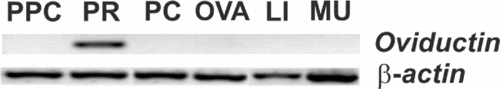
Expression analysis of oviductin in oviductal segments and other tissues was performed using RT-PCR. Our results showed that the oviductin gene was differentially expressed in the oviductal PR region, whereas the PPC and PC segments showed no expression. Ovary, liver and skeletal muscle showed no expression either (Fig. 5).

Figure 5 Expression analysis of oviductin in B. arenarum tissues. Total RNA from pars preconvoluta (PPC), pars recta (PR), pars convoluta (PC), ovary (OVA), liver (LI) and skeletal muscle (MU) of hormone stimulated female specimens was isolated and analyzed for oviductin mRNA expression by RT-PCR. The expected band of 577 bp was exclusively visualized in PR. Amplification of β-actin was performed as control.
Ovochymase 2 gene expression in the reproductive tract of female mouse
RT-PCR analysis was used for examining presence of ovochymase 2 mRNA transcripts in mouse RNA samples of ovary, oviduct and uterus. The experiments demonstrated that ovochymase 2 was only expressed in uterus tissue (Fig. 6A). To confirm that this expression was exclusive to the reproductive tract, as it was seen for oviductin gene in amphibians (Lindsay et al., Reference Lindsay, Matthew and Hedrick1999; Hiyoshi et al., Reference Hiyoshi, Takamune, Mita, Kubo, Sugimoto and Katagiri2002), other female mouse tissues like intestine, kidney, liver and skeletal muscle were also analyzed for presence of ovochymase 2 mRNA, but none of these tissues showed expression (Fig. 6B). The amplified fragment of uterine ovochymase 2 cDNA was purified, cloned and sequenced. The nucleotide sequence was 100% identical to ovochymase 2 mRNA of the mouse genome (GenBank accession no NM_172908).

Figure 6 Expression analysis of ovochymase 2 in mouse female reproductive tract. (A) Total RNA from ovary (OVA), oviduct (OVI) and uterus (UTE) was isolated and analyzed for ovochymase 2 (Ovch2) mRNA expression using RT-PCR. The specific product of 498 bp was only detected in uterus cDNA samples. (B) RT-PCR analysis of Ovch2 expression in female mouse non-reproductive tissues: intestine (INT), kidney (KI), liver (LI) and skeletal muscle (MU). No expression of Ovch2 was detected. β-actin was amplified as internal control.
Discussion
The oviduct and oviductal secretions in B. arenarum and other anuran amphibians perform a crucial function in the preparation of the female gamete for fertilization. Hence, changes in the coelomic envelope during passage of the egg through the oviduct are essential for successful fertilization. These changes are produced by a protease, oviductin, which is synthesized and secreted under hormonal regulation by the oviductal PR epithelium (Miceli et al., Reference Miceli, Fernández, Raisman and Barbieri1978; Miceli & Fernández, Reference Miceli and Fernández1982; Katagiri et al., Reference Katagiri, Yoshizaki, Kotani and Kubo1999; Llanos et al., Reference Llanos, Barrera, Valz-Gianinet and Miceli2006; Hedrick, Reference Hedrick2008). The current study describes for the first time the molecular features of B. arenarum oviductin and analyzes gene expression of its putative mammalian orthologous. This latter enzyme, ovochymase 2, shows a structural and evolutionary relationship with amphibian oviductin, identified in different species.
cDNA sequences of X. laevis and B. japonicus oviductin (Linsday et al., 1999; Hiyoshi et al., Reference Hiyoshi, Takamune, Mita, Kubo, Sugimoto and Katagiri2002) were used to design primers that allow us to amplify B. arenarum oviductin mRNA. Sequential RT-PCR experiments, using oviductal PR cDNA as a template, amplified full-length cDNA (3203 bp) coding for B. arenarum oviductin. This oviductin cDNA showed a complete ORF, a stop codon and 5′- and 3′-untranslated regions. Deduced amino acid sequence indicated that B. arenarum oviductin has 980 amino acids with a predicted molecular weight (MW) of 108.56 kDa.
The predicted MW of B. japonicus and X. laevis oviductin is 107.5 and 110.6 kDa, respectively. Post-translational processing at both the N- and C-terminal ends is thought to be the mechanism that produces the 66 kDa active oviductin in both species (Takamune & Katagiri, Reference Takamune and Katagiri1987; Hardy & Hedrick, Reference Hardy and Hedrick1992; Lindsay et al., Reference Lindsay, Matthew and Hedrick1999; Hiyoshi et al., Reference Hiyoshi, Takamune, Mita, Kubo, Sugimoto and Katagiri2002). The main protein synthesized and secreted in the PR lumen in B. arenarum also weighs 66 kDa (Whitacre & Miceli, Reference Whitacre and Miceli1996). In order to know whether B. arenarum oviductin has a post-translational processing similar to the one proposed for X. laevis and B. japonicus, we checked presence of potential cleavage sites in its amino acid sequence by using Peptide Cutter software (http://ca.expasy.org/tools/peptidecutter/) (data not shown). Putative cleavage sites were found at Arg49, Arg587 and Lys615 residues.
Bioinformatics analysis of B. arenarum oviductin showed a multi-domain protein with a protease domain at the N-terminal region (oviductin α) followed by two CUB domains and toward the C-terminal region another protease domain (oviductin β) and one CUB domain. Similar data have been reported for X. laevis and B. japonicus oviductin (Lindsay et al., Reference Lindsay, Matthew and Hedrick1999; Hiyoshi et al., Reference Hiyoshi, Takamune, Mita, Kubo, Sugimoto and Katagiri2002). Although the α and β domains could have a common ancestor, they may have followed different evolutionary ways, bearing in mind that both domains only have 28% identity.
The oviductin α domain had a conserved Asp232 residue in the substrate-binding pocket that is known to interact with basic amino acids. This molecular feature suggests that B. arenarum oviductin has substrate specificity similar to trypsin, which is in agreement with biochemical characteristics that indicated specific activity toward synthetic trypsin substrates but no activity toward chymotrypsin substrates (Miceli & Fernández, Reference Miceli and Fernández1982).
The oviductin α domain, as X. laevis and B. japonicus oviductin (Swiss-Prot acc. no. P79953 and Q90WD8), covered an area of 250 amino acids. Within this domain, there were strictly conserved sequence motifs including the His90, Asp140 and Ser238 amino acids that constitute the catalytic triad of the active site (Hedstrom, Reference Hedstrom2002). In addition, this domain was preceded by a typical conserved sequence (RIVGG) of serine protease zymogens for proteolytic processing (Kitamoto et al., Reference Kitamoto, Yuan, Wu, McCourt and Sadler1994), features that were absent in the oviductin β domain. Instead, the IIKAE and not RIVGG sequence preceded the oviductin β domain. An important fact is that only two of the three amino acids in the catalytic triad were conserved: His was replaced by Asn at position 633. In addition, the amino acids that surround Ser733 were different from those characteristic of the domain with catalytic activity. Regarding oviductin β, it seems likely that this domain may present a three-dimensional folding that would allow it to join its substrate, but without proteolytic activity toward the substrate. There exist other cases of proteases with one domain able to bind and cleave the substrate and an additional domain devoid of catalytic activity that preserves the binding ability. Recently, serine proteases with three (polyserase-1 and -2) and two tandem protease domains (polyserase-3) have been identified and characterized in the human genome (Cal et al., Reference Cal, Moncada-Pazos and López-Otín2007). The functional benefits of complex structural features with different protease domains in the same polypeptide chain are still unknown.
The CUB domain was first found in the complement subcomponents C1s and C1r and later in the embryonic sea urchin protein or fibropellin (Uegf) and bone morphogenetic protein 1 (BMP-1). It is present in some extracellular proteases and many proteins involved in developmental functions. There is strong evidence that the CUB domain is involved in adhesion mechanisms and/or recognition of substrates and it is thought to mediate protein–protein interactions (Bork & Beckmann, Reference Bork and Beckmann1993). To date, the role of the oviductin CUB domains is unknown. The CUB 1 and CUB 2 domains of B. arenarum oviductin have all the four conserved cysteine residues, whereas CUB 3 lacks two of them. The cysteine residues most likely form disulfide bridges involved in the secondary structure. CUB 1 and CUB 2 domains also have four highly conserved aromatic residues and 15 out of 17 invariant hydrophobic residues, similar to spermadhesin (Varela et al., Reference Varela, Romero, Sanz, Romão, Töpfer-Petersen and Calvete1997). The CUB 1, CUB 2 and oviductin α domains would be part of the mature oviductin, while oviductin β and the CUB 3 domain are apparently detached from the mature protein. Taking into account that the presumed cleavage sites at Arg49, Arg587 and Lys615 residues were detected in the B. arenarum oviductin sequence, it is likely that proteolysis could occur. Proteolytic cleavage at Arg587 and Lys615 residues, which are localized just before the oviductin β domain, would give a protein of around 66 kDa. However, further studies for examining the posttranslational process of the oviductin are necessary to understand the mechanism that produces a mature and active 66 kDa protein from oviductal secretion fluid. It is possible that a proteolytic process occurs prior to its secretion into the lumen.
Based on previous observations (Tao et al., Reference Tao, Peng, Nolasco, Cal, López-Otín, Li, Moake, López and Dong2005; Blanc et al., Reference Blanc, Font, Eichenberger, Moreau, Ricard-Blum and Moali2007), it seems possible that CUB domains anchor the oviductin to its substrates in the coelomic envelope of eggs while they pass through the oviductal PR. This idea is supported by the fact that the oviductin CUB domain may bind to glycans of envelope glycoproteins (Hedrick, Reference Hedrick2008).
It would be very interesting to establish phylogenetic relationships between analogous molecules and mechanisms of action on the fertilization process in different species. The amphibian oviductin has its orthologous gene in mammals: ovochymase 2. Mouse genome analysis has revealed the presence of this enzyme in the degradome (Puente et al., Reference Puente, Sanchez, Overall and López-Otín2003). When analyzing the distribution of ovochymase 2 in mouse tissues by RT-PCR we determined that the expression pattern of ovochymase 2 was constrained to uterine tissues. It has been well documented that genes encoding reproductive proteins are among the most highly diverged ones (Makalowski & Boguski, Reference Makalowski and Boguski1998; Swanson & Vacquier, Reference Swanson and Vacquier2002) and there is evidence that protease gene families involved in reproduction and host defense played a role during the evolution of some mammalian species (Puente et al., Reference Puente, Sánchez, Gutiérrez-Fernández, Velasco and López-Otín2005). Thus, the absence of ovochymase 2 RNA transcripts in oviduct could be one of the signs of the transition from a primitive fertilization mode (external fertilization) to a more complex form (internal fertilization). Our hypothesis is that ovochymase 2 could produce proteolytic changes in the zona pellucida similar to the action of amphibian oviductin, which would play a role in embryo attachment to the uterine wall during implantation.
Acknowledgements
This work was partially supported by research grants from Consejo Nacional de Investigaciones Científicas y Técnicas (CONICET), Agencia Nacional de Promoción Científica y Tecnológica (ANPCYT) and Universidad Nacional de Tucumán, Argentina. D.B. is a research fellowship recipient from CONICET.