


Introduction
During fertilization the membrane of the spermatozoon fuses with the oolemma (the oocyte's plasma membrane). The problem of when the oolemma acquires the ability to fuse with spermatozoa has been studied in the hamster (Zuccotti et al., Reference Zuccotti, Yanagimachi and Yanagimachi1991) and in the mouse (Zuccotti et al., Reference Zuccotti, Piccinelli, Marziliano, Mascheretti and Redi1994). These studies demonstrated that in both species small primary oocytes were unable to fuse with spermatozoa. Hamster and mouse oocytes acquired the ability to fuse with spermatozoa during oogenesis, and first became fusion-competent when they reached about 20 μm in diameter (Zuccotti et al., Reference Zuccotti, Yanagimachi and Yanagimachi1991, Reference Zuccotti, Piccinelli, Marziliano, Mascheretti and Redi1994).
More recent findings have demonstrated that a crucial role in gamete fusion is played by the integral oolemma tetraspanin CD9 (Chen et al., Reference Chen, Tung, Coonrod, Takahashi, Bigler, Chang, Yamashita, Kincade, Herr and White1999; Miyado et al., Reference Miyado, Yamada, Yamada, Hasuwa, Nakamura, Ryu, Suzuki, Kosai, Inoue, Ogura, Okabe and Mekada2000). Female mice lacking CD9 are infertile due to the failure of sperm–egg fusion (Kaji et al., Reference Kaji, Oda, Shikano, Ohnuki, Uematsu, Sakagami, Tada, Miyazaki and Kudo2000; Le Naour et al., Reference Le Naour, Rubinstein, Jasmin, Prenant and Boucheix2000; Miyado et al., Reference Miyado, Yamada, Yamada, Hasuwa, Nakamura, Ryu, Suzuki, Kosai, Inoue, Ogura, Okabe and Mekada2000). The aim of our study was to verify the hypothesis that the acquisition of fusibility with spermatozoa by growing oocytes is correlated with the appearance of CD9 protein on their surfaces. To achieve this goal we used indirect immunofluorescence to detect the CD9 protein on oolemma of growing mouse oocytes.
Materials and methods
Reagents
All chemicals, unless otherwise stated, were obtained from Sigma-Aldrich (Poznan, Poland).
Animals
F1 (C57Bl/10 × CBA/H and C57Bl/6 × CBA/H) female mice between 1 and 15 days after birth and adult F1 female mice (3–6 months old) were used for the experiments.
Collection of growing oocytes
To induce superovulation adult females were injected with 10 IU of pregnant mare serum gonadotrophin (PMSG, Folligon, Intervet, The Netherlands) followed 48 h later by injection of 10 IU of human chorionic gonadotrophin (hCG, Chorulon, Intervet, The Netherlands). Ovaries were isolated into M2 (M-16 medium buffered with HEPES; Fulton & Whittingham Reference Fulton and Whittingham1978). Oocytes were release from ovaries of females which were 1–3 days old by puncturing ovaries with a fine needle. Ovaries isolated from older mice (6–15 days old) were fragmented and exposed to mixture of enzymes (collagenase 0.5 mg/ml, hyaluronidase 0.5 mg/ml, lysozyme 0.5 mg/ml phosphate-buffered saline (PBS); Mangia & Epstein, Reference Mangia and Epstein1975) to release oocytes from growing follicles. Subsequently follicular cells were removed from oocytes by pipetting with a fine capillary. The diameter of oocytes was measured before subsequent fixation and staining.
Collection of ovulated oocytes
Ovulated secondary oocytes were collected from the oviducts 15–18 h after hCG injection, and were freed from cumulus cells by treatment with 0.1% bovine testicular hyaluronidase (200 IU/mg) in PBS for a few minutes. The oocytes were subsequently rinsed thoroughly in M2.
Removal of the zona pellucida
Growing oocytes obtained from females 6–15 days old and ovulated secondary oocytes were freed of zona pellucida by brief exposure to acidic Tyrode solution (pH 2.5, Nicolson et al., Reference Nicolson, Yanagimachi and Yanagimachi1975). Oocytes isolated from younger females (1–3 days old) did not have a visible zona pellucida and were not exposed to acidic Tyrode solution.
Indirect immunofluorescence
Oocytes were fixed with 4% paraformaldehyde (20 min at room temperature), then washed twice in PBS and once in PBS with 10 mM glycine for 10 min. To block non-specific antibody binding sites, oocytes were incubated for 1 h in 2% bovine serum albumin (BSA) solution in PBS at room temperature. Oocytes were then incubated with monoclonal rat antibody raised against mouse CD9 (Clone KMC8, PharMingen, 50 μg/ml in 1% BSA solution in PBS) for 1 h at room temperature or overnight at 4 °C, and washed three times (15 min per wash) in PBS with 1% BSA. Then eggs were exposed to secondary antibody (fluorescein isothiocyanate (FITC)-conjugated mouse anti-rat IgG, Clone MRK-1, PharMingen, 1.25 μg/ml in 1% BSA solution in PBS) during a 1 h incubation at room temperature. Oocytes were washed three times in PBS with 1% BSA (15 min per wash) and incubated for 20 min with propidium iodide (50 μg/ml in PBS) to stain chromatin. Then the oocytes were washed for 20 min in 1% BSA solution in PBS and mounted on glass slides in a drop of antifading mounting medium (Citifluor AF1, Citifluor Ltd). To check the specificity of staining, control oocytes were exposed to the secondary antibody without previous treatment with the primary antibody. Oocytes were examined with confocal laser microscopy (LSM 510 Zeiss).
Results
We isolated primary oocytes from female mice between 1 and 15 days of age to collect growing oocytes of different sizes. At birth the mouse ovary is populated with non-growing oocytes arrested in meiosis. It is known that during the first 2 weeks following birth more oocytes begin to grow (∼5%) than at any other period in the lifetime of the mouse (Wassarman & Albertini, Reference Wassarman, Albertini, Knobil and Neill1994). In our experiments we used growing oocytes which belonged to this first ‘wave’ of oocytes growth.
We found that in the ovaries of 1- to 3-day-old females only oocytes at the very early stages of growth are present (diameter between 10 and 24 μm). Larger oocytes (25–74 μm) appeared in the ovaries of females which were 6–9 days old, and the largest oocytes (75–80 μm) dominated among the growing oocytes isolated from ovaries of 15-day-old females. The relation between the age of females and the diameter of growing oocytes is shown in Table 1.
Table 1 The relationship between the age of newborn mice and the diameter of growing oocytes isolated from their ovaries

We next used immunocytochemistry to examine whether CD9 is present in growing oocytes. We observed expression of CD9 only in the oolemma and not in the cytoplasm of the eggs. Subsequently the amount of CD9 in the oolemma of growing oocytes, reflected by the intensity of immunostaining, was analysed. In each experiment ovulated eggs were collected and stained together with growing oocytes. The intensity of staining of oocytes isolated from the ovary was subjectively compared with the fluorescence of the antibody-stained, ovulated secondary oocytes (Fig. 1). We found that the intensity of staining increased during the earliest stages of the growth of oocytes, when they enlarged approximately from 10 to 20 μm in diameter. All oocytes larger than 20–22 μm were stained with anti-CD9 antibody with the same intensity as ovulated secondary oocytes (Fig. 1). Three classes of growing oocytes were distinguished according to the intensity of staining in relation to the diameter of the oocyte:

Figure 1 Comparison of the intensity of staining of CD9 protein between a growing oocyte of diameter 17–22 μm (left) and an ovulated oocyte (right). Both oocytes were stained during the same experiment. Pictures are single slices taken at the level of the centre of the oocytes. CD9 labelling is visualized in green, the nucleus of the growing oocyte is shown in red. The metaphase chromosomes of the ovulated oocyte are not visible because they were located at a different level. Scale bar represents 20 μm.
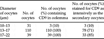
1. 10–13 μm: The majority of oocytes of this size (90%, 28/31) were not stained for CD9, or stained very weakly (Fig. 2A). Only a few oocytes in this group (3/31) were stained to a similar degree as secondary oocytes.
2. 13–17 μm: All oocytes of this size were stained for CD9 but in this group of oocytes the greatest variation of the intensity of staining was observed. Some oocytes (29.1%, 32/110) were stained for CD9 in their oolemma, but staining was much weaker than that observed for ovulated eggs, and CD9 was not uniformly distributed on the egg surface (Fig. 2B). However, 70.9% (78/110) of oocytes in this group were stained as intensively as the secondary oocytes (Fig. 2C).
3. 17–22 μm: All oocytes of this group were stained for CD9 on the cell membrane. The majority of them (85%, 33/39) were stained with the same intensity as ovulated mature eggs (Fig. 2D).

Figure 2 Detection of CD9 protein on the surface of growing oocytes. (A) An oocyte in the diameter range 10–13 μm. (B), (C) Oocytes between 13 and 17 μm in diameter. (D) Oocytes in the diameter range 17–22 μm. Pictures are single slices taken at the level of the centre of the oocytes. CD9 labelling is visualized in green, the nuclei of growing oocytes are shown in red. Scale bar represents 10 μm.
The relation between the diameter of growing oocytes and the presence of CD9 in the oolemma is shown in Table 2.
Table 2 The relation between the diameter of growing oocytes and the presence of CD9 in the oolemma
Control oocytes, for which the incubation with the primary anti-CD9 antibody was omitted from the staining procedure, did not stain with secondary antibody (not shown). These data show that CD9 appears on the oolemma during the early stages of growth of the oocyte.
Discussion
In the present study we investigated the time during mouse oogenesis at which CD9 protein appears on the surface of the oocytes. We found that CD9 emerges on the oolemma during the early stages of growth of the oocyte, when it measures 13–22 μm in diameter. It was shown previously that at this period of growth of the mouse oocyte, the oolemma acquires the ability to fuse with spermatozoa (Zuccotti et al., Reference Zuccotti, Piccinelli, Marziliano, Mascheretti and Redi1994). Thus we hypothesize that the development of the fusibility of the oocyte with spermatozoa may be related to the threshold amount of CD9 present in the oolemma. We observed that enrichment of the oolemma with CD9 is accomplished when the oocyte reaches a diameter of approximately 17–22 μm. At this stage of growth the density of CD9 in the oolemma, evaluated by the examination of the intensity of staining in indirect immunofluorescence, is similar to the density of this protein in the cell membrane of the fully grown secondary oocyte. During subsequent growth of the oocyte CD9 has to be continuously synthesized and incorporated into the expanding oolemma.
The presence of CD9 in immature oocytes ranging from 30 to 100 μm in diameter has been demonstrated previously by Chen et al. (Reference Chen, Tung, Coonrod, Takahashi, Bigler, Chang, Yamashita, Kincade, Herr and White1999), but these authors did not study the time during oogenesis at which CD9 begins to appear on the oolemma. In the present study we show that CD9 starts to accumulate in the oolemma soon after the beginning of growth of the oocyte, and soon reaches a density sufficient to enable the oocyte to fuse with spermatozoa (Zuccotti et al., Reference Zuccotti, Piccinelli, Marziliano, Mascheretti and Redi1994). However, the presence of CD9 on the egg membrane apparently does not itself suffice to achieve its fusibility with sperm. It was demonstrated for the mouse (Zuccotti et al., Reference Zuccotti, Piccinelli, Marziliano, Mascheretti and Redi1994), as well as for the hamster (Zuccotti et al., Reference Zuccotti, Yanagimachi and Yanagimachi1991), that the fusibility of growing oocytes with spermatozoa increases with increasing oocyte diameter, even though, according to our study, the concentration of CD9 seems to be the same in all oocytes larger than approximately 20 μm. Since fusibility is measured by the number of spermatozoa fusing with a single oocyte, it could be expected that fusibility increases as the oocyte grows since the surface area of the oocyte increases with growth. However, it cannot be excluded that synthesis and incorporation, or modification, of elements other than CD9 in the oolemma or cortex of the oocyte is required for the development of oolemma's ability to fuse with spermatozoa. For instance, integrins of the oolemma (Chen et al., Reference Chen, Tung, Coonrod, Takahashi, Bigler, Chang, Yamashita, Kincade, Herr and White1999) or cortical actin filaments (McAvey et al., Reference McAvey, Wortzman, Williams and Evans2002) could also be involved in the fusibility of oolemma with spermatozoa.
The tetraspanin CD9 is the only protein of the mouse oolemma currently known to function in the fusion of gametes (Chen et al., Reference Chen, Tung, Coonrod, Takahashi, Bigler, Chang, Yamashita, Kincade, Herr and White1999; Kaji et al., Reference Kaji, Oda, Shikano, Ohnuki, Uematsu, Sakagami, Tada, Miyazaki and Kudo2000; Le Naour et al., Reference Le Naour, Rubinstein, Jasmin, Prenant and Boucheix2000; Miyado et al., Reference Miyado, Yamada, Yamada, Hasuwa, Nakamura, Ryu, Suzuki, Kosai, Inoue, Ogura, Okabe and Mekada2000). It is known that tetraspanins mediate protein interactions in the cell membrane to create unique cell-surface domains (Hemler, Reference Hemler2003; Stipp et al., Reference Stipp, Kolesnikova and Hemler2003). In our previous paper (Komorowski et al., Reference Komorowski, Szczepanska and Maleszewski2003) we demonstrated that the concentration of CD9 in the oolemma of mouse zygotes which cannot be penetrated by additional spermatozoa due to the oolemma block to polyspermy is similar to the concentration of CD9 in the ovulated oocytes which are able to fuse with spermatozoa. When CD9 is removed from the oocytes by proteolytic enzymes, they lose fusibility with sperm (Komorowski et al., Reference Komorowski, Szczepanska and Maleszewski2003). We observed that after 17 h of culture in vitro CD9 is restored in the oolemma, but this does not restore the ability of pronase-treated oocytes to fuse with sperm (Bialecka, Komorowski and Maleszewski, unpublished data). Thus the presence of CD9 in the oolemma is not sufficient to make the oocyte able to fuse with spermatozoa.
Acknowledgements
We thank Professor A.K. Tarkowski and Drs K. Swann, M.A. Ciemerych and E. Borsuk for critical reading of the manuscript.


