


Introduction
Post-traumatic stress disorder (PTSD) is a serious mental health condition that may occur following exposure to traumatic events, such as combat, serious accidents, abuse, or violent crime. Characteristic symptoms of PTSD include increased distress and physiological reactivity to reminders of the traumatic event (American Psychiatric Association [APA], 2013). The biological correlates of these symptoms can be studied using the script-driven imagery (SDI) paradigm, a standard symptom provocation task (Pitman et al. Reference Pitman, Orr, Forgue, de Jong and Claiborn1987). During SDI, participants recall and imagine personal life events while psychophysiological and brain responses are measured. An important advantage of SDI over other symptom provocation tasks is that it employs autobiographical stimuli, which allows it to more accurately represent each individual's unique experiences (Pitman et al. Reference Pitman, Orr, Forgue, de Jong and Claiborn1987).
Neuroimaging studies have consistently reported decreased activation of the rostral regions of medial prefrontal cortex (mPFC), including the anterior cingulate cortex (ACC) and medial frontal gyrus (MFG), during traumatic v. neutral SDI in individuals with PTSD, compared with control participants (Bremner et al. Reference Bremner, Narayan, Staib, Southwick, McGlashan and Charney1999; Shin et al. Reference Shin, McNally, Kosslyn, Thompson, Rauch, Alpert, Metzger, Lasko, Orr and Pitman1999, Reference Shin, Orr, Carson, Rauch, Macklin, Lasko, Peters, Metzger, Dougherty, Cannistraro, Alpert, Fischman and Pitman2004; Lanius et al. Reference Lanius, Williamson, Densmore, Boksman, Gupta, Neufeld, Gati and Menon2001, Reference Lanius, Williamson, Hopper, Densmore, Boksman, Gupta, Neufeld, Gati and Menon2003; Liberzon et al. Reference Liberzon, Britton and Phan2003; Lindauer et al. Reference Lindauer, Booij, Habreken, Uylings, Olff, Carlier, den Heeten, van Eck-Smit and Gersons2004; Britton et al. Reference Britton, Phan, Taylor, Fig and Liberzon2005; reviewed in Hayes et al. Reference Hayes, Hayes and Mikedis2012). Furthermore, PTSD symptom severity has been found to be inversely correlated with mPFC activation during SDI in some (Osuch et al. Reference Osuch, Benson, Geraci, Podell, Herscovitch, McCann and Post2001; Shin et al. Reference Shin, Orr, Carson, Rauch, Macklin, Lasko, Peters, Metzger, Dougherty, Cannistraro, Alpert, Fischman and Pitman2004), but not all (Lanius et al. Reference Lanius, Williamson, Boksman, Densmore, Gupta, Neufeld, Gati and Menon2002; Gold et al. Reference Gold, Shin, Orr, Carson, Rauch, Macklin, Lasko, Metzger, Dougherty, Alpert, Fischman and Pitman2011) studies.
Individuals with PTSD also exhibit increased psychophysiological [e.g. heart rate, skin conductance (SC), and facial electromyographic] responses to trauma-related SDI, compared with trauma-exposed participants without PTSD (Pitman et al. Reference Pitman, Orr, Forgue, de Jong and Claiborn1987, Reference Pitman, Orr, Forgue, Altman, de Jong and Herz1990; Orr et al. Reference Orr, Pitman, Lasko and Herz1993, Reference Orr, Lasko, Metzger, Berry, Ahern and Pitman1998; Shalev et al. Reference Shalev, Orr and Pitman1993; Shin et al. Reference Shin, Orr, Carson, Rauch, Macklin, Lasko, Peters, Metzger, Dougherty, Cannistraro, Alpert, Fischman and Pitman2004; reviewed in Orr et al. Reference Orr, McNally, Rosen, Shalev and Rosen2004). Elevated psychophysiological responses in PTSD are associated with increased PTSD symptom severity (reviewed in Orr & Roth, Reference Orr and Roth2000).
Recent studies have shown that abnormal brain activation and psychophysiological responses to imagery of traumatic events in PTSD may extend to imagery of trauma-unrelated stressful events. The use of trauma-unrelated stressful events as stimuli allows for the inclusion of trauma-unexposed comparison groups; scripts describing trauma-unrelated stressful events (e.g. divorce, job loss) can be used for all participants, regardless of whether they have been exposed to traumatic events severe enough to meet the criterion A requirements for PTSD diagnosis. For example, Britton et al. (Reference Britton, Phan, Taylor, Fig and Liberzon2005) measured regional cerebral blood flow during Stressful v. Neutral Imagery in combat veterans with and without PTSD and in combat-unexposed control participants. They found that the PTSD group had significantly greater deactivation in the rostral ACC (rACC) than the two comparison groups. Similarly, Gold et al. (Reference Gold, Shin, Orr, Carson, Rauch, Macklin, Lasko, Metzger, Dougherty, Alpert, Fischman and Pitman2011) found relatively diminished rACC responses and elevated SC responses to Stressful v. Neutral Imagery in PTSD compared to trauma-exposed control participants without PTSD.
Although previous research has provided evidence for diminished mPFC activation and increased psychophysiological responses during the recollection and imagery of stressful life events in PTSD, the origin of these abnormalities remains unclear. They may reflect familial vulnerability factors that increase the risk of PTSD after trauma exposure, or result from trauma exposure, or be acquired characteristics of PTSD. Determining the origin of these abnormalities could have important clinical implications. For example, an abnormality that reflects a familial vulnerability for PTSD could be identified before potential exposure to traumatic events and hence guide primary or secondary prevention efforts. An abnormality that is an acquired characteristic could potentially assist in the diagnosis of PTSD or in the assessment of treatment response.
The origin of these abnormalities can be clarified with twin studies (Gilbertson et al. Reference Gilbertson, Shenton, Ciszewski, Kasai, Lasko, Orr and Pitman2002; Shin et al. Reference Shin, Lasko, Macklin, Karpf, Milad, Orr, Goetz, Fischman, Rauch and Pitman2009, Reference Shin, Bush, Milad, Lasko, Brohawn, Hughes, Macklin, Gold, Karpf, Orr, Rauch and Pitman2011). The present study examined identical twin pairs discordant for combat exposure; within each pair, one twin was exposed (Ex) and the other was unexposed (Ux) to combat. The Ux co-twin served as a proxy of what the Ex twin would be like if combat had not been experienced. Two types of twin pairs were included in the current design: PTSD (P+) twin pairs, in which the Ex twin had a current diagnosis of PTSD, and non-PTSD (P−) twin pairs, in which the Ex twin did not have a history of PTSD (Supplementary Fig. S1). Thus, our design included four distinct participant groups: combat-exposed participants with PTSD (ExP+) and their combat-unexposed identical co-twins without PTSD (UxP+), and combat-exposed participants without PTSD (ExP−) and their combat-unexposed identical co-twins without PTSD (UxP−). In this design, any abnormalities demonstrated in the P+ twin pairs (both ExP+ and UxP+ participants) would indicate a familial vulnerability for PTSD; abnormalities demonstrated in the Ex participants (both ExP+ and ExP− participants) would reflect combat exposure; and abnormalities demonstrated in only the ExP+ participants (the only participants diagnosed with PTSD) would indicate an acquired characteristic of PTSD (for review of the twin study design see Pitman et al. Reference Pitman, Gilbertson, Gurvits, May, Lasko, Metzger, Shenton, Yehuda and Orr2006).
In an attempt to resolve the origin of mPFC and psychophysiological abnormalities in PTSD, we examined functional magnetic resonance imaging (fMRI) and SC responses (SCRs) during SDI in these twins. Based on previous findings, we hypothesized that during (trauma-unrelated) stressful v. neutral SDI, combat-exposed individuals with PTSD (ExP+) would show diminished mPFC activation and exaggerated SCRs relative to combat-exposed individuals without PTSD (ExP−). Due to a lack of prior research, we had no basis for predicting whether these abnormalities would represent familial vulnerabilities (observed in both twins of the P+ pairs) or acquired characteristics of PTSD (observed in only the ExP+ participants). In the event that familial vulnerability factors were identified, we planned to examine the relationship between Ex participants’ PTSD symptom severity and their Ux co-twins’ mPFC activation and SCRs; significant inverse correlations would provide further evidence of a familial vulnerability for PTSD. In the event that acquired characteristics were identified, we planned to examine the relationship between the ExP+ participants’ PTSD symptom severity and their own mPFC activation and SCRs.
Methods and materials
Participants
Participants were male identical twins recruited from the Vietnam Era Twin (VET) Registry (Henderson et al. Reference Henderson, Eisen, Goldberg, True, Barnes and Vitek1990), the University of Washington Twin Registry (Strachan et al. Reference Strachan, Hunt, Afari, Duncan, Noonan, Schur, Watson, Goldberg and Buchwald2013), letters sent through the Veterans Benefits Administration (Washington, DC; Orr et al. Reference Orr, Metzger, Lasko, Macklin, Hu, Shalev and Pitman2003), or by advertisements on electronic media. There were four distinct participant groups: ExP+ (n = 12), UxP+ (n = 12), ExP− (n = 14), and UxP− (n = 14). ExP+ participants were exposed to combat during the Vietnam War (n = 11) or a serious accident (n = 1). (Analyses were completed with and without the latter P+ twin pair; because these analyses yielded similar results, we included this twin pair.) ExP− participants were exposed to combat during the Vietnam War (n = 13) or Operation Desert Storm/Shield (n = 1). No participant reported neurological disorders or major head trauma involving loss of consciousness for more than 10 min. A complete description of the study was provided to the participants, and written informed consent was obtained. This research was approved by the Institutional Review Boards of the Partners Healthcare System at Massachusetts General Hospital and the VET Registry.
Demographic and clinical characteristics
A trained clinician (N.B.L.) administered the Clinician-Administered PTSD Scale (CAPS; Blake et al. Reference Blake, Weathers, Nagy, Kaloupek, Gusman, Charney and Keane1995) and the Structured Clinical Interview for the DSM-IV (SCID; First et al. Reference First, Spitzer, Gibbon and Williams2002) to all participants in order to determine PTSD diagnostic status/symptom severity and comorbidity, respectively. Four of the ExP+ participants reported partial remission of PTSD symptoms; however, all of these individuals reported at least mild-to-moderate current PTSD symptoms (as defined by Weathers et al. Reference Weathers, Keane and Davidson2001) and were therefore included in the analyses. According to the SCID, ExP+ participants met criteria for the following current comorbid diagnoses: dysthymia (n = 3), major depression (n = 2), specific phobia (n = 2), substance abuse/dependence (n = 2), alcohol abuse/dependence (n = 1), social phobia (n = 1), generalized anxiety disorder (n = 1), panic disorder (n = 1), and eating disorders (n = 1). UxP+ participants met criteria for the following current diagnoses: alcohol abuse/dependence (n = 3), PTSD (n = 1), dysthymia (n = 1), specific phobia (n = 1), and panic disorders (n = 1). Among ExP− participants, current diagnoses included: alcohol abuse/dependence (n = 2), substance abuse/dependence (n = 1), and paranoid/delusion disorders (n = 1). One UxP− participant met criteria for current alcohol abuse/dependence (n = 1). Analyses were run with and without the pair in which the UxP+ twin had PTSD; the results were the same, so we retained this pair in the final analyses.
Participants also completed the Childhood Trauma Questionnaire (CTQ; Bernstein et al. Reference Bernstein, Fink, Handelsman, Foote, Lovejoy, Wenzel, Sapareto and Ruggiero1994), Beck Depression Inventory (BDI; Beck & Steer, Reference Beck and Steer1987), Beck Anxiety Inventory (BAI; Beck & Steer, Reference Beck and Steer1993), the Michigan Alcoholism Screening Test (MAST; Selzer, Reference Selzer1971), and a combat exposure severity index for Vietnam era veterans (combat-exposed participants only; Janes et al. Reference Janes, Goldberg, Eisen and True1991).
Script-driven imagery task procedures
One day before the fMRI session, participants completed an interview in which they provided detailed descriptions of two neutral and two trauma-unrelated stressful personal events. Additionally, the Ex participants (ExP+ and ExP−) provided descriptions of two combat-related personal events (results not reported here). After describing each event, participants examined a list of bodily responses (e.g. ‘heart races’, ‘labored breathing’) and circled those that they recalled having experienced during the event. Immediately after this interview, the investigators wrote scripts (i.e. brief narratives describing each event) in the second person, present tense, including an average of about four (but no more than five) of the bodily response cues that each participant selected. The scripts were audio-recorded in an emotionally neutral, male voice for playback during fMRI scanning the next day.
Each participant was scanned during two Neutral, two Stressful, and two Combat script blocks across two functional runs. (Because the Ux participants did not experience combat, they heard standardized Combat scripts. Given the differing personal relevance of the Combat scripts across Ex and Ux participants, the combat condition was not included in the analyses.) Before each scan, participants were instructed to close their eyes, listen carefully to each script, and imagine the described event as vividly as possible, as if they were actually back in the situation. fMRI and SC data were collected at five different epochs for each script: (1) Baseline (30 s), when participants focused on a fixation point; (2) Read (~50 s), when they listened to the recorded scripts; (3) Imagery (30 s), when they recalled and imagined the event as if reliving the experience; (4) Recovery (30 s), when they opened their eyes, stopped imagining the event, and relaxed; and (5) Rating (60 s), when they used a button box to rate each script on valence, arousal, and imagery vividness.
To be consistent with previous SDI studies, although all of the above-described epochs were included in the statistical model, our contrasts included only the Imagery and Baseline epochs. (Contrasts involving the Imagery and Read epochs yielded similar findings.)
MRI parameters
All MRI scans were completed using a Siemens Trio Tim 3 Tesla MRI with a 12-channel head coil at the Massachusetts General Hospital (MGH) Martinos Center for Biomedical Imaging (Charlestown, Massachusetts, USA). High-resolution, three-dimensional structural MRI scanning was completed for each participant using a multi-echo magnetization-prepared rapid gradient echo (MEMPRAGE) sequence in 176 sagittal slices [repetition time (TR) = 2530 ms, echo time (TE)1 = 1.64 ms, TE2 = 3.5 ms, TE3 = 5.36 ms, TE4 = 7.22 ms, flip angle = 12.50°, thickness = 1.00 mm]. fMRI blood-oxygen-level-dependent images were acquired using a gradient echo T2-weighted sequence (TR = 2500 ms, TE = 30 ms, flip angle = 90°) in 46 slices (thickness = 2.5 mm, 20% distance factor, 0.5 mm skip). Total scan time was approximately 10 min per run, 20 min total.
SC parameters
Participants’ SC levels (SCLs; microsiemens, μS) were measured at a sampling rate of 10 Hz by an isolated SC coupler (Coulbourn Instruments LLC, Whitehall, Pennsylvania, USA) during fMRI, according to established procedures (Pitman et al. Reference Pitman, Orr, Forgue, de Jong and Claiborn1987, Reference Pitman, Orr, Forgue, Altman, de Jong and Herz1990; Orr et al. Reference Orr, Lasko, Metzger, Berry, Ahern and Pitman1998; Shin et al. Reference Shin, McNally, Kosslyn, Thompson, Rauch, Alpert, Metzger, Lasko, Orr and Pitman1999, Reference Shin, Orr, Carson, Rauch, Macklin, Lasko, Peters, Metzger, Dougherty, Cannistraro, Alpert, Fischman and Pitman2004). In vivo Metric (Healdsburg, California, USA) Ag/AgCl electrodes filled with an isotonic conductive paste were placed on the hypothenar surface of participants’ non-dominant hand, in accordance with published guidelines (Fowles et al. Reference Fowles, Christie, Edelberg, Grings, Lykken and Venables1981).
SCLs were calculated by averaging the SC data within each individual Baseline and Imagery period for each script. The mean SCL during the Baseline period was subtracted from the mean SCL during the Imagery period for each script condition to yield a SCR score. Additionally, a script condition difference score (Stressful SCR–Neutral SCR) was calculated to assess the difference between script conditions. Given that two scripts were presented for each condition, SC data from both scripts were averaged and used in the final analyses. SC data were not usable from three of the P+ twin pairs (adjusted n = 9) and two of the P− twin pairs (adjusted n = 12).
Data analysis
For the SC data, we performed 2 (PTSD diagnosis: P+, P−) by 2 (exposure: Ex, Ux) mixed-model analyses of variance (ANOVAs) using the Statistical Package for the Social Sciences (SPSS) program, version 22 (IBM, Armonk, New York, USA). A significant between-subjects main effect of PTSD diagnosis (P+ v. P− twin pairs) would indicate a familial vulnerability to the development of PTSD. A significant within-subjects main effect of exposure (Ex v. Ux) would indicate a consequence of combat exposure, independent of PTSD. A significant PTSD diagnosis by exposure interaction in which the ExP+ participants differed from all the other groups would indicate an acquired characteristic of PTSD. Pearson correlations were used to assess the relationships among PTSD symptom severity, fMRI, and SC data.
For the fMRI data, whole-brain voxelwise comparisons were performed using the statistical parametric mapping (SPM8) software package (http://www.fil.ion.ucl.ac.uk/spm/software/spm8). Each participant's functional images were co-registered to his MEMPRAGE image, spatially normalized in standard stereotactic space (Montreal Neurological Institute, MNI), and smoothed (8 mm). We used an approach that consisted of hierarchical levels of analysis in which each level's random-effects analysis absorbs the random effects from the level beneath it. The first level required contrasting two conditions (e.g. Stressful Imagery v. Neutral Imagery) to generate a contrast map per participant. Movement >5 mm translation or 3° of rotation was exclusionary for this study. However, no participant exceeded these movement thresholds; most participants’ movement was 1–2 mm with <1.5° rotation. Additionally, ANOVAs on movement (translation and rotation) data revealed no significant main effects or interactions.
In the first set of analyses, which were an attempt to replicate findings from previous cross-sectional studies of PTSD (Bremner et al. Reference Bremner, Narayan, Staib, Southwick, McGlashan and Charney1999; Shin et al. Reference Shin, McNally, Kosslyn, Thompson, Rauch, Alpert, Metzger, Lasko, Orr and Pitman1999, Reference Shin, Orr, Carson, Rauch, Macklin, Lasko, Peters, Metzger, Dougherty, Cannistraro, Alpert, Fischman and Pitman2004; Lanius et al. Reference Lanius, Williamson, Densmore, Boksman, Gupta, Neufeld, Gati and Menon2001, Reference Lanius, Williamson, Hopper, Densmore, Boksman, Gupta, Neufeld, Gati and Menon2003; Liberzon et al. Reference Liberzon, Britton and Phan2003; Lindauer et al. Reference Lindauer, Booij, Habreken, Uylings, Olff, Carlier, den Heeten, van Eck-Smit and Gersons2004), the contrast images of the ExP+ and ExP− groups were compared by independent-samples t test. The second set of analyses utilized the twin design. To assess the main effect of exposure, the Stressful Imagery v. Neutral Imagery contrast images of the Ex twins were compared with their Ux co-twins using a paired t test. To assess the main effect of PTSD diagnosis (P+ pairs v. P− pairs), contrast images of the Ex and Ux co-twins within each diagnostic group were averaged, and the P+ and P− groups were compared by independent-samples t test. To assess the PTSD diagnosis by exposure interaction, Ex v. Ux contrast images were generated, and the P+ and P− groups were then compared by independent-samples t test. To verify results, we repeated the fMRI analyses using a 2 (PTSD diagnosis: P+ v. P−) by 2 (Exposure: Ex v. Ux) ANOVA.
For the replication analyses comparing ExP+ v. ExP−, given our directional a priori hypotheses, we applied a significance threshold of p < 0.001, one-tailed, uncorrected (z ⩾ 3.09) to activations in the mPFC, including the ACC and MFG. This is the same threshold used in the previous studies we are attempting to replicate (Shin et al. Reference Shin, Orr, Carson, Rauch, Macklin, Lasko, Peters, Metzger, Dougherty, Cannistraro, Alpert, Fischman and Pitman2004, Reference Shin, Wright, Cannistraro, Wedig, McMullin, Martis, Macklin, Lasko, Cavanagh, Krangel, Orr, Pitman, Whalen and Rauch2005; Brohawn et al. Reference Brohawn, Offringa, Pfaff, Hughes and Shin2010). We did not have directional hypotheses for the twin-design analyses; therefore, we applied a significance threshold of p < 0.0005, two-tailed, uncorrected (z ⩾ 3.29) for activations in the mPFC. For all other regions about which we had no a priori predictions, we applied a more conservative significance threshold of p < 0.00002, two-tailed, uncorrected (z ⩾ 4.27) in accordance with previous studies (Shin et al. Reference Shin, Wright, Cannistraro, Wedig, McMullin, Martis, Macklin, Lasko, Cavanagh, Krangel, Orr, Pitman, Whalen and Rauch2005; Brohawn et al. Reference Brohawn, Offringa, Pfaff, Hughes and Shin2010). Activations in the mPFC were identified by their highest local maximum, and their location was verified using the Mai et al. (Reference Mai, Majtanik and Paxinos2015) and Talairach & Tournoux (Reference Talairach and Tournoux1988) brain atlases, as well as the SPM Anatomy Toolbox. Following the whole-brain voxelwise analyses, we extracted parameter estimates for individual participants from identified functional regions of interest (ROI; spherical, 4 mm radius) using the MarsBaR SPM toolbox (Brett et al. Reference Brett, Anton, Valabregue and Poline2002) and further analyzed these data using SPSS.
Results
The demographic and clinical characteristics of the study participants are summarized in Table 1. All groups were similar in terms of age, years of education, current depressed mood (BDI), magnitude of childhood trauma (CTQ), and alcohol use (MAST). Consistent with their PTSD diagnosis, ExP+ participants reported significantly greater PTSD symptom severity (CAPS) compared with ExP− participants. Additionally, the ExP+ participants reported higher levels of combat severity than the ExP− participants. A PTSD diagnosis by exposure interaction was observed for anxiety ratings (measured by the BAI) and reflected relatively high anxiety scores in the ExP+ group.
Table 1. Demographic and clinical characteristics for combat-exposed participants with (P+) and without (P−) PTSD and their combat-unexposed identical co-twins
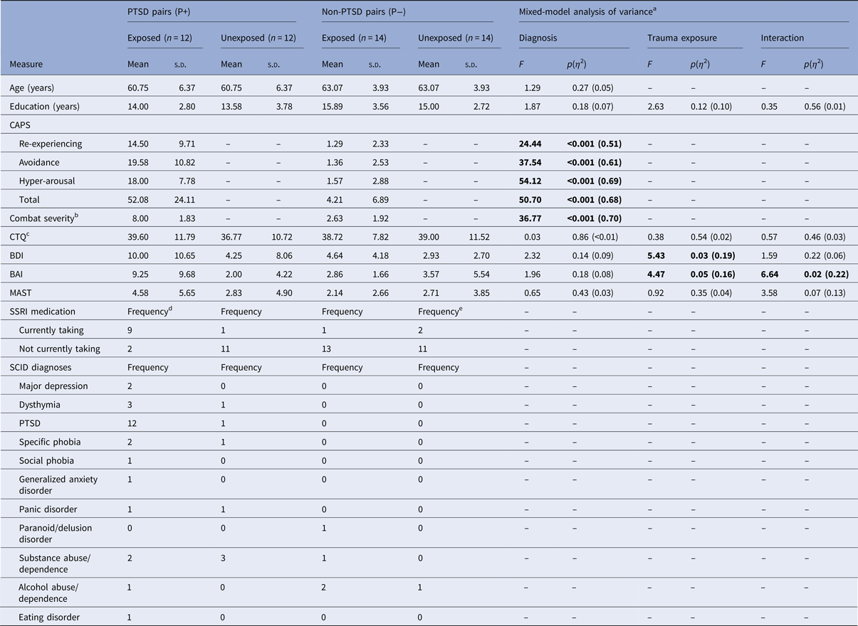
Significant effects in bold.
a df = 1,24 unless noted otherwise.
b df = 1,16.
c df = 1,21.
d One participant did not report current medications.
e Two participants did not report current medications.
BAI, Beck Anxiety Inventory; BDI, Beck Depression Inventory; CAPS, Clinician-Administered PTSD Scale; CTQ, Childhood Trauma Questionnaire; MAST; Michigan Alcoholism Screening Test; PTSD, post-traumatic stress disorder; SCID, Structured Clinical Interview for DSM; SSRI, selective serotonin reuptake inhibitor.
fMRI results
Replication analyses: ExP+ v. ExP−
The ExP+ group, relative to the ExP− group, showed less activation in the rACC (MNI 14, 38, 24) in the Stressful v. Neutral Imagery contrast, replicating previous findings (Table 2). Inspection of the means (Fig. 1a) revealed that the ExP+ group showed rACC deactivation, whereas the ExP− group showed rACC activation.

Fig. 1. Significant PTSD diagnosis by exposure interactions during Stressful v. Neutral Imagery in (a) the rACC (MNI 14, 38, 34; z = 3.18), (b) the ACC (MNI 10, 26, 44; z = 3.71), and (c) the MFG (MNI −8, 40, 46; z = 3.58). Error bars represent standard error of the mean. ROI masked by cluster for display purposes only. Abbreviations: ACC, anterior cingulate cortex; BOLD, blood-oxygen-level-dependent; fMRI, functional magnetic resonance imaging; MFG, medial frontal gyrus; MNI, Montreal Neurological Institute; PTSD, post-traumatic stress disorder; rACC, rostral anterior cingulate cortex; ROI, region of interest. Group abbreviations: Ex, combat-exposed; Ux, combat-unexposed; P+, PTSD twin pair; P−, control twin pair.
Table 2. ExP+ v. ExP−: voxelwise analyses of Stressful v. Neutral Imagery in combat-exposed participants only
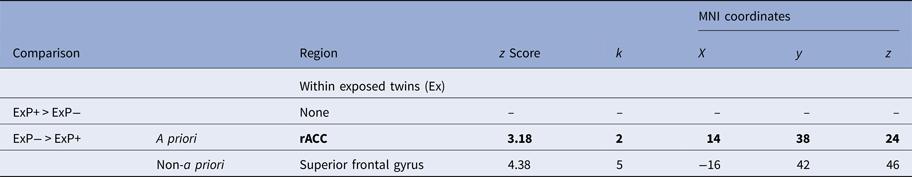
Participant groups: ExP+, participants exposed to combat with PTSD; ExP−, participants exposed to combat without PTSD.
MNI, Montreal Neurological Institute; k, cluster size at the significance threshold (see below); rACC, rostral anterior cingulate cortex.
For a priori regions, a significance threshold was applied at z ⩾ 3.09, p ⩽ 0.001, one-tailed.
For non-a priori regions, the applied significance threshold was z ⩾ 4.27, p ⩽ 0.00002, two-tailed. Significant a priori regions are listed in bold print.
Twin-design analyses
First, using the rACC (MNI 14, 38, 24) functional ROI that was identified in the preceding ExP+ v. ExP− analysis, we extracted data from all four subject groups. An ANOVA demonstrated a significant PTSD diagnosis by exposure interaction; ExP+ participants showed lower rACC activation during Stressful v. Neutral Imagery, relative to all other groups, F(1,24) = 5.447, p = .028, η 2 = .185 (Fig. 1a). The main effects of PTSD diagnosis and exposure were both non-significant (p ⩾ 0.09).
Second, we conducted whole-brain voxelwise twin-design analyses of the Stressful v. Neutral Imagery contrast images. These also yielded significant PTSD diagnosis by exposure interactions in the mPFC, which included a region located on the boundary of the dorsal ACC and rACC (ACC; MNI 10, 26, 44) and a region in the MFG (MNI −8, 40, 46). There were no significant main effects of exposure (Table 3). A main effect of PTSD diagnosis, in which the P+ pairs showed significantly less activation compared with P− pairs, was found in one non-a priori region (postcentral gyrus). The 2 (PTSD diagnosis: P+ v. P−) by 2 (Exposure: Ex v. Ux) ANOVA yielded nearly identical findings, although the z-scores were lower in the ANOVA (z = 3.09 for the ACC; z = 3.14 for the MFG; and z = 3.76 for the postcentral gyrus).
Table 3. Twin analyses: voxelwise analyses of Stressful v. Neutral Imagery in all participants

Participant groups: P+ twin pairs in which the exposed twin has PTSD; P− twin pairs in which the exposed twin does not have PTSD; Ex, combat-exposed twins; Ux, combat-unexposed twins.
MNI, Montreal Neurological Institute; k, cluster size at the significance threshold (see below); ACC, anterior cingulate cortex; MFG, medial frontal gyrus.
For a priori regions, a significance threshold was applied at z ⩾ 3.29, p ⩽ 0.0005, two-tailed.
For non-a priori regions, the applied significance threshold was z ⩾ 4.27, p ⩽ 0.00002, two-tailed. Significant a priori regions are listed in bold print.
In order to fully examine the PTSD diagnosis by exposure interactions, values for each participant were extracted from each functional ROI using the MarsBaR SPM toolbox. Analyses of the data extracted from the ACC (MNI 10, 26, 44) confirmed a significant PTSD diagnosis by exposure interaction; ExP+ participants showed less ACC activation during Stressful v. Neutral Imagery relative to all other groups, F(1,24) = 12.690, p = 0.002, η 2 = 0.346 (Fig. 1b). Similarly, analyses of the extracted data from the MFG (MNI −8, 40, 36) confirmed a significant PTSD diagnosis by exposure interaction; ExP+ participants showed less MFG activation during Stressful v. Neutral Imagery, relative to all other groups, F(1,24) = 12.395, p = 0.002, η 2 = .341 (Fig. 1c). An examination of the extracted fMRI data further broken down by Stressful Imagery v. Baseline and Neutral Imagery v. Baseline confirmed that the results from the Stressful v. Neutral Imagery contrast were due to mPFC deactivation in the ExP+ group during Stressful Imagery (Supplementary Fig. S2).
Correlations with mPFC activation
The ExP+ participants’ Stressful v. Neutral SDI activation in the rACC (MNI 14, 38, 24), ACC (MNI 10, 26, 44), and MFG (MNI −8, 40, 46) did not significantly correlate with their own total CAPS scores (ps ⩾ 0.074, one-tailed).
SC results
Results for the SC data are presented in Table 4. No significant effects were found for average SCL during the Imagery and Baseline periods of the Stressful and Neutral script conditions. Additionally, no significant effects were found for average SCR (Imagery SCL–Baseline SCL) during the Stressful or Neutral script conditions. When the Stressful SCR–Neutral SCR difference score was assessed, a significant PTSD diagnosis by exposure interaction emerged, F(1,19) = 4.531, p = 0.047, η 2 = 0.193. ExP+ individuals had significantly smaller average Stressful SCR–Neutral SCR difference scores relative to all other groups, which appears to be due to higher average SCLs (although not significantly so) in ExP+ participants throughout the SDI paradigm, particularly during both the Baseline and Imagery periods of the Stressful condition (Supplementary Fig. S3). Stressful v. Neutral SCR difference scores were not significantly correlated with PTSD symptom severity [r(19) < 0.065, p > 0.355].
Table 4. Skin conductance (μS) during the Stressful and Neutral Imagery conditions
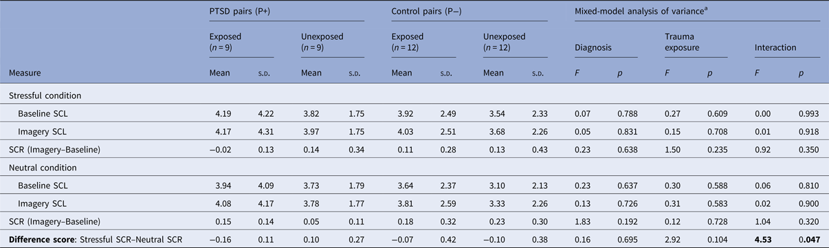
Significant effects in bold.
a df = 1,19.
PTSD, post-traumatic stress disorder; SCL, skin conductance level; SCR, skin conductance response.
Covariate analyses
Assessment of potential confounders
Using a method we have previously employed (Gilbertson et al. Reference Gilbertson, Shenton, Ciszewski, Kasai, Lasko, Orr and Pitman2002; Kasai et al. Reference Kasai, Yamasue, Gilbertson, Shenton, Rauch and Pitman2008; Shin et al. Reference Shin, Lasko, Macklin, Karpf, Milad, Orr, Goetz, Fischman, Rauch and Pitman2009, Reference Shin, Bush, Milad, Lasko, Brohawn, Hughes, Macklin, Gold, Karpf, Orr, Rauch and Pitman2011), the following variables were tested as potential confounders: age, years of education, left-handedness, birth weight, BDI, BAI, MAST, CTQ, and selective serotonin reuptake inhibitor (SSRI) use. Specifically, we examined their associations with the fMRI- and SC-dependent measures using correlation analyses with a screening threshold of p ⩽ 0.20. Any variable that met this criterion was subsequently assessed using analysis of covariance (ANCOVA).
rACC (MNI 14, 38, 24)
fMRI data extracted from this rACC ROI identified in the ExP+ v. ExP− contrast did not correlate with any of the potential confounders below the p ⩽ 0.20 screening threshold.
ACC (MNI 10, 26, 44)
fMRI data extracted from this ROI in the PTSD diagnosis by exposure interaction correlated with BDI and SSRI use below the p ⩽ 0.20 screening threshold. Separate ANCOVAs controlling for Ex participants’ BDI and SSRI use showed that the previously significant PTSD diagnosis by exposure interactions remained significant [F(1,23) = 11.283, p = 0.003, η 2 = 0.329 and F(1,22) = 7.614, p = 0.011, η 2 = 0.257, respectively].
MFG (MNI −8, 40, 46)
fMRI data extracted from this ROI in the PTSD diagnosis by exposure interaction correlated with age and SSRI use below the p ⩽ 0.20 screening threshold. A separate ANCOVA controlling for Ex participants’ age showed that the previously significant PTSD diagnosis by exposure interaction remained significant [F(1,23) = 10.536, p = 0.004, η 2 = 0.314]. A separate ANCOVA controlling for Ex participants’ SSRI use reduced the previously significant PTSD diagnosis by exposure interaction to a trend level [F(1,22) = 14.003, p = 0.058, η 2 = 0.154].
Stressful v. Neutral SCR difference scores
These difference scores were correlated with age below the p ⩽ 0.20 threshold. An ANCOVA controlling for age showed that the previously significant PTSD diagnosis by exposure interaction was reduced to a trend level [F(1,18) = 3.513, p = 0.077, η 2 = 0.163].
Discussion
Our analyses confirmed previous findings of relatively diminished mPFC activation during Stressful v. Neutral Imagery in individuals with PTSD compared with trauma-exposed individuals without PTSD (Bremner et al. Reference Bremner, Narayan, Staib, Southwick, McGlashan and Charney1999; Shin et al. Reference Shin, McNally, Kosslyn, Thompson, Rauch, Alpert, Metzger, Lasko, Orr and Pitman1999, Reference Shin, Orr, Carson, Rauch, Macklin, Lasko, Peters, Metzger, Dougherty, Cannistraro, Alpert, Fischman and Pitman2004; Lanius et al. Reference Lanius, Williamson, Densmore, Boksman, Gupta, Neufeld, Gati and Menon2001, Reference Lanius, Williamson, Hopper, Densmore, Boksman, Gupta, Neufeld, Gati and Menon2003; Liberzon et al. Reference Liberzon, Britton and Phan2003; Lindauer et al. Reference Lindauer, Booij, Habreken, Uylings, Olff, Carlier, den Heeten, van Eck-Smit and Gersons2004; Britton et al. Reference Britton, Phan, Taylor, Fig and Liberzon2005; Gold et al. Reference Gold, Shin, Orr, Carson, Rauch, Macklin, Lasko, Metzger, Dougherty, Alpert, Fischman and Pitman2011). Furthermore, we found reduced mPFC activation in ExP+ relative to other groups, providing evidence that this abnormality is an acquired characteristic of PTSD. Contrary to our predictions, mPFC activation was not inversely correlated with PTSD symptom severity in this study. However, previous evidence supporting this relationship has been inconsistent (Lanius et al. Reference Lanius, Williamson, Boksman, Densmore, Gupta, Neufeld, Gati and Menon2002; Gold et al. Reference Gold, Shin, Orr, Carson, Rauch, Macklin, Lasko, Metzger, Dougherty, Alpert, Fischman and Pitman2011).
Controlling for the use of SSRI medications did not affect the PTSD diagnosis by exposure interaction observed for the rACC and ACC, but reduced it to a trend (p = 0.058) for the MFG (MNI −8, 40, 36). Thus, SSRI use may affect MFG activation during SDI, but have little or no impact in other mPFC regions. No other potential confounders changed the significance of the PTSD diagnosis by exposure interactions for the mPFC.
There is significant evidence that the mPFC plays a critical role in fear extinction learning and extinction recall; mPFC malfunction may be related to the development of PTSD and maintenance of symptoms by impairing the extinction of fear (reviewed in Yehuda & LeDoux, Reference Yehuda and LeDoux2007; VanElzakker et al. Reference VanElzakker, Dahlgren, Davis, Dubois and Shin2014). Our imaging finding of reduced mPFC activation in trauma-exposed individuals with PTSD is consistent with previous studies, particularly Kasai et al. (Reference Kasai, Yamasue, Gilbertson, Shenton, Rauch and Pitman2008), who reported that diminished gray matter density in the rACC is an acquired characteristic of PTSD. However, we acknowledge that the current twin study finding could alternatively reflect an acquired vulnerability factor (e.g. due to early non-shared life experience or stochastic variation during neurodevelopment) rather than an acquired characteristic of PTSD itself. Our study design cannot eliminate this possibility.
Previous findings have typically demonstrated increased psychophysiological reactivity to trauma-related scripts in PTSD (Pitman et al. Reference Pitman, Orr, Forgue, de Jong and Claiborn1987, Reference Pitman, Orr, Forgue, Altman, de Jong and Herz1990; Orr et al. Reference Orr, Pitman, Lasko and Herz1993, Reference Orr, Lasko, Metzger, Berry, Ahern and Pitman1998; Shalev et al. Reference Shalev, Orr and Pitman1993; Shin et al. Reference Shin, Orr, Carson, Rauch, Macklin, Lasko, Peters, Metzger, Dougherty, Cannistraro, Alpert, Fischman and Pitman2004; Gold et al. Reference Gold, Shin, Orr, Carson, Rauch, Macklin, Lasko, Metzger, Dougherty, Alpert, Fischman and Pitman2011; reviewed in Orr et al. Reference Orr, McNally, Rosen, Shalev and Rosen2004). In contrast, our results indicate that ExP+ individuals show smaller average Stressful SCR–Neutral SCR difference scores, which may be due to higher average SCLs throughout the SDI paradigm, particularly during both the Baseline and Imagery periods of the Stressful condition. ExP+ participants’ SCL may have already been high during the Baseline period leading to ceiling effects and lack of upwards modulation during the Imagery period, which could be interpreted as higher arousal as an acquired characteristic of PTSD. However, it is important to note that only the SCR difference scores showed a significant interaction, and that comparisons of the SCL levels, which were non-significant, did not provide a clear interpretation of the data; therefore, the SCR results should be interpreted with caution. Additionally, the average SCL during Stressful Imagery in the ExP+ participants (M = 4.17 µS) did not represent a large magnitude SCL (for review see Orr et al. Reference Orr, McNally, Rosen, Shalev and Rosen2004). In fact, all groups showed relatively low magnitude SCLs, which could be due to a variety of factors including older age, time since the events, etc. Our assessment of potential confounding variables suggests that age may affect SCL. Indeed, when age was controlled for, the previously significant PTSD diagnosis by exposure interaction of the Stressful SCR–Neutral SCR difference score was no longer significant.
If the current findings are replicated in future twin or longitudinal studies, these acquired, objectively measured biological characteristics could potentially assist in the diagnosis of PTSD or in the assessment of treatment response. In fact, recent research has shown that SCR during SDI has good convergent validity with PTSD symptom severity as measured by the CAPS total score (Bauer et al. Reference Bauer, Reuf, Pineles, Japuntich, Macklin, Lasko and Orr2013). Additionally, decreases in psychophysiological reactivity during trauma-related imagery have been observed in successful treatment of PTSD (Boudewyns & Hyer, Reference Boudewyns and Hyer1990; Shalev et al. Reference Shalev, Orr and Pitman1992) and increased mPFC activation has been shown following successful SSRI treatment of PTSD (Fani et al. Reference Fani, Ashraf, Afzal, Jawed, Kitayama, Reed and Bremner2011) providing further evidence of their potential clinical utility. More recently, a pilot study indicated that deep transcranial magnetic stimulation of the mPFC can successfully reduce PTSD symptoms (Isserles et al. Reference Isserles, Shalev, Roth, Peri, Kutz, Zlotnick and Zangen2013).
Limitations and future directions
The small sample size of the present study provided limited statistical power. Data lost due to unmeasurable SCR further reduced our sample size. The repeated-measures analyses techniques employed for this twin study design helped maximize statistical power, but the sample size may have contributed to type II errors. The nature of our sample, with its unique entry criteria, made it challenging to acquire, but also made the sample uniquely suited to address the question of whether a given PTSD characteristic is acquired or a familial vulnerability factor.
This study is also limited by the characteristics of the study sample; for example, participants were all men. However, similar findings in the mPFC have been observed in all-male (Britton et al. Reference Britton, Phan, Taylor, Fig and Liberzon2005), all-female (Bremner et al. Reference Bremner, Narayan, Staib, Southwick, McGlashan and Charney1999; Shin et al. Reference Shin, McNally, Kosslyn, Thompson, Rauch, Alpert, Metzger, Lasko, Orr and Pitman1999), as well as mixed-sex (Lanius et al. Reference Lanius, Williamson, Densmore, Boksman, Gupta, Neufeld, Gati and Menon2001, Reference Lanius, Williamson, Hopper, Densmore, Boksman, Gupta, Neufeld, Gati and Menon2003; Osuch et al. Reference Osuch, Benson, Geraci, Podell, Herscovitch, McCann and Post2001; Lindauer et al. Reference Lindauer, Booij, Habreken, Uylings, Olff, Carlier, den Heeten, van Eck-Smit and Gersons2004; Shin et al. Reference Shin, Orr, Carson, Rauch, Macklin, Lasko, Peters, Metzger, Dougherty, Cannistraro, Alpert, Fischman and Pitman2004; Gold et al. Reference Gold, Shin, Orr, Carson, Rauch, Macklin, Lasko, Metzger, Dougherty, Alpert, Fischman and Pitman2011) studies. In addition, almost all Ex participants reported exposure from combat that had occurred decades earlier; consequently the results may not be generalizable to individuals with more recent or other types of traumatic experiences.
This study required participants to travel to Boston for 2 full days of testing, necessitating that participants be relatively high functioning. This may have unintentionally excluded more severely symptomatic participants. Orr & Roth (Reference Orr and Roth2000) suggested that less severe PTSD symptoms reduce the size of psychophysiological effects, increasing the likelihood of type II errors. However, our sample of ExP+ individuals all presented with at least moderate PTSD symptom severity (as defined by Weathers et al. Reference Weathers, Keane and Davidson2001), which lessens the impact of this limitation.
Conclusions
This study provides further evidence of reduced mPFC activation in PTSD and provides evidence that this abnormality is an acquired characteristic. These findings have important clinical implications because acquired characteristics of PTSD could potentially assist in diagnosis or the assessment of treatment response.
Supplementary material
The supplementary material for this article can be found at https://doi.org/10.1017/S003329171700263X.
Acknowledgements
This research was supported by National Institute of Mental Health (NIMH) grant R01MH054636. The Massachusetts General Hospital (MGH) Clinical Research Center was supported by Harvard Catalyst grants 1UL1RR025758, 8UL1TR000170, 1UL1TR001102. The MGH Athinoula A. Martinos Imaging Center was supported by the Center for Functional Neuroimaging Technologies grant P41RR14075. M.B.V. was supported by a National Defense Science & Engineering Graduate fellowship from the US Department of Defense. L.M.S. received additional support from the Faculty Research Awards Committee at Tufts University. The Cooperative Studies Program (CSP) of the Office of Research & Development of the United States Department of Veterans Affairs (VA) has provided financial support for the development and maintenance of the Vietnam Era Twin (VET) Registry.
All statements, opinions, or views are solely of the authors and do not necessarily reflect the position or policy of the NIMH, VA, or United States Government. M.K.D. previously presented preliminary analyses of these data in her Master's Thesis.
The authors would like to thank Mary Foley and Lawrence White for neuroimaging technical assistance, and the current and past members of the Tufts University Posttraumatic Stress Disorder Neuroimaging Lab, especially Navneet Kaur, Rachel Korus, Lisa Sangermano, Julia Russell, Kelsey Keyser, Phil Panic, Belal Hakim, Tyler Chang, Eliza White, Nikki Chen, Tiffany Tu, and Nicole Carter. Most importantly, the authors gratefully acknowledge the continued cooperation and participation of the members of the VET and University of Washington Twin Registries and their families. Without their contribution this research would not have been possible.
R.O. is employed at Glooko (Mountain View, California, USA), and reports having received teaching honoraria from Metis Data Science Bootcamp and Springboard Data Science Intensive. He has also received data science consulting fees from 1010 Data, Columbia University, the Massachusetts Institute of Technology, Irrational Games, and the Fletcher School.
All other authors do not have any financial conflicts of interest to disclose.
Ethical standards
The authors assert that all procedures contributing to this work comply with the ethical standards of the relevant national and institutional committees on human experimentation and with the Helsinki Declaration of 1975, as revised in 2008.


