


I. INTRODUCTION
Until the 1930s opals were considered, based on their physical and optical properties, as typical examples of amorphous matter, similarly with glass and other hydrogel colloidal materials (Garavelli, Reference Garavelli1964). This assumption was also supported by the first X-ray powder diffraction (XRPD) investigations on samples of hydrophane, hyalite, and milk opals that showed the presence of just two diffraction peaks, i.e., typical of synthetic glass or liquid materials (Rinne, Reference Rinne1924). Similarly, other authors carried out XRPD investigations on opals and did not identify any lines on the films or, at best, identified only some weak interferences (Kerr, Reference Kerr1924). Even Baier Reference Baier(1932), into his long article about the optical properties of opals, underlined that it was not possible to observe any crystalline phases through XRPD analyses of opals.
However, following investigations showed that some diffraction peaks could be observed in several types of opals, which were more intense than the weak broad diffraction peaks typical of glasses. In an XRPD study of opals, silica glass, and silica gel, Levin and Ott Reference Levin and Ott(1933) reported for the first time that these anomalies were attributed to the presence of cristobalite. This high-temperature phase of silica was effectively identified, together with small amounts of quartz, in different samples of common opal, fire opal, and wood opal. Levin and Ott Reference Levin and Ott(1932, Reference Levin and Ott1933) also highlighted the existence of completely amorphous opals such as hyalite and geyserites that showed in their XRPD patterns a broad peak at ∼4 Å, typical of silica glass or artificial silica gel. Levin and Ott Reference Levin and Ott(1933) and Dwyer and Mellor Reference Dwyer and Mellor(1934) were the first to make a classification of the opals on the basis of their crystallinity. For this reason these authors have always been considered as pioneers in the investigation of opals through X-ray diffractometry (Taliaferro, Reference Taliaferro1935; Cirilli and Giannone, Reference Cirilli and Giannone1941; Fenoglio and Sanero, Reference Fenoglio and Sanero1943; Raman and Jayaraman, Reference Raman and Jayaraman1953; Flörke, Reference Flörke1955; Frondel, Reference Frondel1962; Garavelli, Reference Garavelli1964; Jones and Segnit, Reference Jones and Segnit1971; Mitchell and Tufts, Reference Mitchell and Tufts1973; Wilson et al. Reference Wilson, Russell and Tait1974; Graetsch, Reference Graetsch1994; Smith, Reference Smith1998).
With the development of more sophisticated technologies, several other authors continued their investigations of opals, substantially confirming the results of previous studies but also producing new results and theories (Wilm et al. Reference Wilm, Hofmann and Endell1934; Taliaferro, Reference Taliaferro1935; Laves, Reference Laves1939). For instance, some authors have reported the occurrence of the β phase of cristobalite in opals (Greig, Reference Greig1932; Sosman, Reference Sosman1932; Dwyer and Mellor Reference Dwyer and Mellor1932, Reference Dwyer and Mellor1934). Consequently, it was suggested that opals enclosed crystallites of high-temperature cristobalite.
Due to the increasing interest of the scientific community about the opal crystallinity, several investigations were carried out on this topic. The presence of α - and β-cristobalite was therefore hypothesised in several samples of opals from different areas (Cirilli and Giannone, Reference Cirilli and Giannone1941). However, the interpretation of the origin of cristobalite in opals was very controversial, especially because the mechanism of evolution of the silica gel into a crystalline state, at natural conditions, was still unknown.
Fenoglio and Sanero Reference Fenoglio and Sanero(1943) carried out X-ray investigations on their rich collection of opals from magnesite ores in the Piedmont Alpine foothills and observed that the enclosed β-cristobalite was characterised by a relatively low degree of crystallinity.
Further researches highlighted that two different phases of silica, characterised by d basal spacings of 4.04 and 4.11 Å, occurred in several types of opals. These two different phases implied a high and a low degrees of order and were interpreted as α- and β-cristobalite, respectively, because the former showed a lower symmetry with respect to the latter, which has cubic symmetry (Raman and Jayaraman, Reference Raman and Jayaraman1953). However, it was still unexplained how, in the same microcrystalline sample of opal, both the high- and low-temperature phases of cristobalite might be present.
The publication of the work by Flörke, “Zur frage des hoch-cristobalit in opalen, bentoniten und gläsern,” in the 1955s was particularly significant for the modern interpretation of the XRPD results on opals. The analyses reported in this work were carried out using a high-resolution instrument, which allow for the identification of diffraction peaks of tridymite. It was therefore assessed that, in natural opals, tridymite could be present together with low-temperature cristobalite (α(-cristobalite). Tridymite and cristobalite in opals do not occur as individual crystallites: areas with stackings corresponding to cristobalite occur together with others corresponding to tridymite. This structure is typical of a strong disorder (Flörke, Reference Flörke1955).
The reasons preventing a recognition of the presence of α-cristobalite, exchanging it often for β or that led to the assumption that both α-cristobalite and β-cristobalite could be present in the same sample of opal, were probably because of the fact that many of the diffraction peaks of tridymite overlap with those of α-cristobalite.
As confirmed by many his contemporaries (Wahl et al., Reference Wahl, Grim and Graf1961; Frondel, Reference Frondel1962; Jones et al., Reference Jones, Segnit and Nickson1963, Reference Jones, Sanders and Segnit1964; Jones and Segnit, Reference Jones and Segnit1971; Wilson et al., Reference Wilson, Russell and Tait1974; Mizutani, Reference Mizutani1977) and by modern authors, the contribution of Flörke for an understanding of the complex α-cristobalite/α-tridymite mixture, connected to the structural order/disorder in opals, was of fundamental importance. Performing photometric curves on the Guinier data, Flörke also observed a strong diffraction peak at d=4.3 Å, which he correctly attributed it to the (200) reflection of tridymite. We have to point out that this interference had been previously observed also by other authors (Levin and Ott, Reference Levin and Ott1933; Wilm et al., Reference Wilm, Hofmann and Endell1934), although they did not interpret it correctly probably because it was related, through a dense darkening, to the (111) peak of cristobalite. In particular, the interference observed in the Debye-Scherrer data was wrongly interpreted as a split of the (111) interference due either to the modalities of sample preparation or to an overlap of the diffuse band of the amorphous component.
In conclusion, Flörke observed that, with an increase in disorder, the X-ray patterns becoming more poorly defined, there was the emergence of a strong band characteristic of α-tridymite, while several interferences typical of cristobalite tended to disappear. Flörke Reference Flörke(1955) also explained the shift of the main peak of cristobalite towards that of tridymite because of an increase in disorder.
The important work of Garavelli Reference Garavelli(1964)Ordine e disordine negli opali (“Order and disorder in opals”), concerning a large number of analyses on several types of opals, represented the “Italian” answer to the studies of Flörke, which were considered as milestones in this field of research. Garavelli confirmed the subdivision of opals in two types based on their degrees of structural order. In particular, he included into the disordered type the opaline silica of biogenic origin, the noble opals, the limpid hyalites, and the geyserites. In the ordered type Garavelli included the common opals, the xiloid opals, and the various types of “resinite” opals. He also observed that a small number of opals showed intermediate characteristics between the ordered and the disordered groups.
A true classification of opals was elaborated only in 1971 when Jones and Segnit in their work The nature of opal-nomenclature and constituent phases presented a classification of opals that today, almost 40 years after, is still being used. Until then, the preceding classifications substantially divided opals in two groups, ordered and disordered (as described above); even the existence of intermediate characteristics was recognised (Levin and Ott, Reference Levin and Ott1933; Dwyer and Mellor, Reference Dwyer and Mellor1934; Flörke, Reference Flörke1955; Garavelli, Reference Garavelli1964).
Conversely, based on the results of their previous works (Jones et al., Reference Jones, Segnit and Nickson1963, Reference Jones, Sanders and Segnit1964) Jones and Segnit identified three well-defined typologies or structural groups of opals. Even if this classification appeared to be very simple, it turned out to be correct and was accepted by the scientific community. As a matter of fact, this classification was very suitable, according to what was reported by the same authors, because every opal analysed through X-ray diffractometry could be included into one of these three groups. However, we must point out that Jones and Segnit Reference Jones and Segnit(1971) did not include into the term of “opal” the materials of biogenic origin or the concretions (such as diatomites and geyserites) but assigned to these phases the term of “opaline silica.”
According to Jones and Segnit Reference Jones and Segnit(1971), the opals were classified as follows:
— opal C: opal with a very ordered structure; the diffraction peaks in the X-ray pattern of α-cristobalite can be clearly identified even if accompanied by some weak peaks typical of tridymite and, in some cases, quartz.
— opal CT: opal with a disordered structure; some diffraction peaks typical of α-cristobalite and α-tridymite in the X-ray pattern can be identified, the latter becoming more evident with an increase in disorder.
— opal A: opal with a high disordered, almost amorphous, structure; only a diffuse band at about d=4.3 Å in the X-ray pattern can be identified.
The classification of Jones and Segnit Reference Jones and Segnit(1971) subsequently underwent some small modifications. Some authors studied in more detail the sequence and disposition of the cristobalite/tridymite layers and the influence they could exert on the main diffraction peaks (Wilson et al., Reference Wilson, Russell and Tait1974). Other authors subdivided opal A into two groups, namely, opal AN, amorphous opals with a pseudostructure similar to a glass (for instance, hyalite), and opal AG, amorphous opals with a pseudostructure similar to that of silica gel like, for instance, the noble opal. This subdivision was supported by analytical evidences and by the different origins of the two kinds of material. It is known that opal AN forms through deposition process, by the quenching of fluid phases at high temperatures on cold substrates, while opal AG forms through precipitation from aqueous solutions (Langer and Flörke, Reference Langer and Flörke1974).
In this paper we propose a new classification of opals through XRPD. This work was made possible because we had the possibility of accessing and analysing several samples collected from different worldwide areas. The analyses were carried out with two main purposes:
1. Identification of the crystallinity degree of opals, i.e., the degree of structural order/disorder of the minerals.
2. Identification of the eventual presence of accessory minerals in the samples.
Even XRPD is not commonly used in the field of gemology because it is a destructive technique, this review should be useful also to gemologists for the identification of common and noble opals.

Figure 1. (Color online) XRPD patterns of opals analysed in this work: opal A (sample 1—light noble opal from Australia), disordered opal CT (sample 2—red fire opal from Australia), more ordered opal CT (sample 3—green opal bGW from Peru), opal C (sample 4—pink opal bVD from Peru), and quartz (sample 5—white chalcedony from Germany).
II. EXPERIMENTAL
A. Opal samples
About 75 samples of opal were analysed by XRPD: most come from Peru and others came from Tanzania, Brazil, Australia, Mexico, Madagascar, Slovakia, Italy, and Argentina. They mostly belong to the collection “Caucia-Ghisoli” (Ghisoli, Reference Ghisoli2008), while few other samples belonged to the collection of the museum “Don Bosco” of Torino (Italy). Further analyses were carried out on opals showing different colours and on some matrices. On the whole we carried out a total of 93 analyses. In particular, several differently coloured samples from Peru came from few centimeters thick veins in a copper ore in the area of Acari near Nazca, in the Arequipa Department, 600 km southeast of Lima (Ghisoli, Reference Ghisoli2008). Samples of crisopal came from the Hanety Hill, North of Dodoma, in the central part of Tanzania. Opals from Brasil came from the areas of Castelo do Piauí and of Varzea Grande in the state of Piauí. Other fire opals analysed in this work came from Western Australia, from the State of Queretaro in Mexico, and from the area around Bemia in the southeast of Madagascar. European white opals came from Slovakia and the Italian Alps and Italian hyalite came from Sardinia. Play colour opals came from the area of Lightning Ridge (Australia). Brown opal came from Patagonia (Argentina).
B. Specimen preparation
Opal samples were each manually grinded into an agate mortar and sieved to obtain a powder with a grain diameter <33 μm. Then, the powders have been pressed and mounted on the slab for the analyses.
C. Data collection
The XRPD analyses of samples of opal were carried out using a PW1800/10 Philips X-ray diffractometer, with a divergent slit and a graphite diffracted-beam monochromator, operated at 40 kV and 40 mA with Cu Kα radiation (Kα1=1.540 60 Å(and Kα2=1.544 39 Å). The speed and the angular range of the analyses were 0.02°2θ/sand between 2° and 65°2θ, respectively.
The experimental XRPD data were analysed using the software X’PERT HIGH SCORE, V.2.0a (PANalytical). The phase identification of the XRPD results was done using the 1986 version of PDF database.
III. RESULTS AND DISCUSSION
A. Opal XRD patterns: Previous works
It is well known that the XRD patterns of opals do not have sharp diffraction peaks but show a broad and diffuse reflection at around ∼21.5°2θ (see Figures 1 and 2). This broad peak was named, by some authors, with the term of “glass-peak” (Dódony and Takàcs, Reference Dódony and Takàcs1980) because of its resemblance with the diffuse peak produced by glass and merged with the background without interruptions. The corresponding value of d was frequently reported in literature (e.g., Giuseppetti and Veniale, Reference Giuseppetti and Veniale1969; Jones and Segnit, Reference Jones and Segnit1971; Sanders, Reference Sanders1975; Dódony and Takàcs, Reference Dódony and Takàcs1980; Morse and Casey, Reference Morse and Casey1988; Herdianita et al., Reference Herdianita, Browne, Rodgers and Campbell2000a; Herdianita et al., Reference Herdianita, Rodgers and Browne2000b) even if some authors reported d values between 4.01 and 3.88 Å (Garavelli, Reference Garavelli1964) or between 4.1 and 3.8 Å (Kastner et al., Reference Kastner, Keene and Gieskes1977). Also the “Gilson” opal is characterised by a broad peak centered at about ∼21.4°2θ corresponding to a d value of ∼4.1 Å, although this opal also shows accessory peaks related to well-crystallized cubic tetragonal zircon (Simonton et al., Reference Simonton, Rustum, Komarneni and Breval1986). Some authors also highlighted the presence of weak peaks corresponding to d values of about 2.0, 1.5, and 1.2 Å (Jones and Segnit, Reference Jones and Segnit1971; Rondeau et al., Reference Rondeau, Fritsch, Guiraud and Renac2004).
With regard to the opals showing a well-defined sequence of peaks (opal C), we considered at first the peaks typical of cristobalite and tridymite because these minerals mainly represent the crystalline component of opals. Successively, also the typical peak sequence of the sole well-crystallized α-cristobalite was considered.

Figure 2. (Color online) Diagnostic peaks of opals (cristobalite/tridymite mixture): A, B , C , D , E and of cristobalite: F and G. The pattern of sample 1 is related to an orange fire opal from Mexico, while the pattern is of sample 2 to blue opal bVD from Peru.
The diffraction peaks have been indicated with a capital letter. In particular, the peaks related to cristobalite/tridymite were named with the letters from A to E while those related to only cristobalite with the letters F and G (Figure 2).
As shown in Figures 1 and 2, the diffraction peaks indicating the presence of cristobalite/tridymite in opals can be described as follows:
Peak A: a “shoulder” at ∼20.65°2θ, corresponding to d4.30 Å. This is a weak peak diagnostic of tridymite, which can be presented along with the main peaks (q.v. B and C ) (Flörke, Reference Flörke1955; Reference Flörke1967; Wilson et al., Reference Wilson, Russell and Tait1974; Kastner et al., Reference Kastner, Keene and Gieskes1977) and is probably related to the (040) plane of α-tridymite. This peak can be observed only in the opals with a quite ordered structure because it is masked by the background as the crystallographic disorder increases. Therefore, the intensity of this peak was reported to vary with the variation of the structural disorder (Flörke et al., Reference Flörke, Graetsch, Martin, Röller and Wirth1991). It also corresponds to the most intense peak of tridymite (Graetsch et al., Reference Graetsch, Gies and Topalovic1994), but it can also be diagnostic of α-cristobalite as is not present in the β phase (White et al., Reference White, Hutchinson and Keith1988). Although this reflection is very evident in opal C, some authors considered it as the characteristic of opal CT (Jones and Segnit, Reference Jones and Segnit1971; Smith, Reference Smith1997; Herdianita et al., Reference Herdianita, Browne, Rodgers and Campbell2000a; Herdianita et al., Reference Herdianita, Rodgers and Browne2000b). While it generally features a d value of 4.3 Å (Flörke, Reference Flörke1967; Wilson et al., Reference Wilson, Russell and Tait1974; Flörke et al., Reference Flörke, Graetsch, Martin, Röller and Wirth1991; Graetsch et al., Reference Graetsch, Gies and Topalovic1994), it can also show in opal-CT d values of 4.32 Å (Banergee and Wenzel, Reference Banergee and Wenzel1999), 4.318 to 4.300 Å (Bustillo et al., Reference Bustillo, García and García Pérez1999), 4.33 to 4.28 Å (Esenli et al., Reference Esenli, Kumbasar, Esenli and Kırıkoğlu2003), 4.28 Å (Herdianita et al., Reference Herdianita, Browne, Rodgers and Campbell2000a; Herdianita et al., Reference Herdianita, Rodgers and Browne2000b), and, lastly, 4.23 Å for the mixtures of the opal CT/A (Lynne and Campbell, Reference Lynne and Campbell2004).
Peak B: a main peak at ∼21.80°2θ, with a d value of ∼4.07 Å. It is the most important peak and corresponds to the combination of the reflections hkl (101) of α-cristobalite and α(−404) of α-tridymite (Flörke, Reference Flörke1955) or the hkl (002) of the hexagonal polytype H of tridymite (Graetsch, Reference Graetsch1994). It is frequently accompanied by the previously described shoulder A and can also show a secondary peak that can be very weak or also absent in opal C (Smith, Reference Smith1998). The d values vary with the distance between the levels of tetrahedrons (Flörke et al., Reference Flörke, Graetsch, Martin, Röller and Wirth1991), and their shift allows us to define the degree of structural order in the opals, highlighting the gradual transition from opals CT to C (Kano, Reference Kano1983; Graetsch et al., Reference Graetsch, Gies and Topalovic1994).
The d values of α-tridymite and α-cristobalite forming this peak are 4.107 and 4.04 Å, respectively; in opals the observed d values of this peak range depending from the authors: from 4.13 to 4.07 Å (De Jong et al., Reference De Jong, Van Hoek, Veeman and Manson1987), from 4.12 to 4.07 Å (Herdianita et al., Reference Herdianita, Browne, Rodgers and Campbell2000a; Herdianita et al., Reference Herdianita, Rodgers and Browne2000b), from 4.12 to 4.04 Å (Murata and Nakata, Reference Murata and Nakata1974; Graetsch, Reference Graetsch1994; Graetsch et al., Reference Graetsch, Gies and Topalovic1994), from 4.11 to 4.04 Å (Jones and Segnit, Reference Jones and Segnit1971), from 4.11 to 4.03 Å (Elzea and Rice, Reference Elzea and Rice1996; Viti and Gemmi, Reference Viti and Gemmi2006), and from 4.10 to 4.05 Å (Mizutani, Reference Mizutani1977).
With regard to opal CT, the following d values for this peak were reported to range from 4.14 to 4.09 Å by Esenli et al. Reference Esenli, Kumbasar, Eren and Uz(2001, Reference Esenli, Kumbasar, Esenli and Kırıkoğlu2003), from 4.12 to 4.07 Å by Bustillo et al. Reference Bustillo, García and García Pérez(1999), from 4.11 to 4.06 Å by Cady et al. Reference Cady, Wenk and Downing(1996), from 4.11 to 4.07 Å by Flörke et al. Reference Flörke, Graetsch, Martin, Röller and Wirth(1991), and from 4.10 to 4.07 Å by Elzea et al. Reference Elzea, Odom and Miles(1994). The values of d for opal C were reported to range from 4.062 to 4.056 Å by Jones and Segnit Reference Jones and Segnit(1971), from 4.06 to 4.05 Å by Flörke et al. Reference Flörke, Graetsch, Martin, Röller and Wirth(1991), from 4.055 to 4.036 Å by Esenli et al. Reference Esenli, Kumbasar, Eren and Uz(2001), and 4.05 Å by Bareille et al. Reference Bareille, Labracherie, Maillet and Latouche(1990). After heating, the d value can decrease down to 4.025 Å (Jones and Segnit, Reference Jones and Segnit1971). This peak has also been reported as a variable single diffraction peak at around ∼4.1 Å (Wilson et al., Reference Wilson, Russell and Tait1974; Banergee and Wenzel, Reference Banergee and Wenzel1999) or as a peak characteristic of opal CT (Kastner et al., Reference Kastner, Keene and Gieskes1977).
Peak C : a secondary peak at ∼36.05°2θ corresponding to a d value of ∼2.50 Å. Like the preceding one, this peak is also formed by a juxtaposition of two reflections, in particular the hkl (200) of α-cristobalite (Wilson et al., Reference Wilson, Russell and Tait1974) and the (341) of α-tridymite. It therefore derives from the sequence of stackings of the two phases (Flörke et al., Reference Flörke, Graetsch, Martin, Röller and Wirth1991).
This peak is the second with relevance in paracrystalline opals and is usually associated to the B peak and, sometimes, also to the A peak. This peak is also an indicator of the abundance of well-ordered cristobalite (Jones and Segnit, Reference Jones and Segnit1971) and is important for the identification of the degree of structural order in opals (Graetsch, Reference Graetsch1994; Esenli et al., Reference Esenli, Kumbasar, Eren and Uz2001). According to some authors, this peak can be related to the presence of tridymite (Banergee and Wenzel, Reference Banergee and Wenzel1999) and is also reported as a “low-tridymite peak” in opal CT (Kastner et al., Reference Kastner, Keene and Gieskes1977). Moreover, the variations of this peak were found to relate to the dimensions of the particles in opals (Guthrie et al., Reference Guthrie, Bish and Reynolds1995). It has a d value of 2.5 Å (Wilson et al., Reference Wilson, Russell and Tait1974; Kastner et al., Reference Kastner, Keene and Gieskes1977; Flörke et al., Reference Flörke, Graetsch, Martin, Röller and Wirth1991) but also of 2.51 Å (Banergee and Wenzel, Reference Banergee and Wenzel1999), of 2.485 Å (Viti and Gemmi, Reference Viti and Gemmi2006), of 2.50 to 2.52 Å for the case of opal CT and 2.49 Å for opal C (Esenli et al., Reference Esenli, Kumbasar, Eren and Uz2001; Reference Esenli, Kumbasar, Esenli and Kırıkoğlu2003).
Peak D : a secondary peak at ∼44.50°2θ corresponding to a d value of ∼2.03 Å. It is a rather weak peak and is frequently ignored by many authors but sometimes can provide significant information on the structure of the opals (Guthrie et al., Reference Guthrie, Bish and Reynolds1995). It results from the juxtaposition of the values hkl (202) of α-cristobalite and of the (−432) of α-tridymite.
Peak E : a secondary peak at ∼56.85°2θcorresponding to a d value of ∼1.62 Å. This reflection is also rather weak and is usually ignored, but it can characterise the opals from some specific ores, such as the fire-coloured opals from Turkey (Esenli et al., Reference Esenli, Kumbasar, Eren and Uz2001). This peak corresponds to the juxtaposition of hkl (301) of α-cristobalite and (−513) of α-tridymite.
Peak F : a peak of a medium intensity at ∼28.50°2θcorresponding to a d value of ∼3.13 Å. It is consistent with the hkl (111) value of α-cristobalite. It was reported together with peak G by some authors (e.g., Jones and Segnit, Reference Jones and Segnit1971).
Peak G : a peak of medium intensity at ∼31.40°2θ corresponding to a d value of ∼2.84 Å. It matches the hkl value (102) of α-cristobalite (q.v. F).
Besides the previous peaks, some authors highlighted the occurrence of some other weak diffraction peaks, having d values of 4.48 Å (Jones and Segnit, Reference Jones and Segnit1971) and/or of 3.8 Å (Garavelli, Reference Garavelli1964), which would indicate the presence of tridymite.
B. Opal XRD patterns: This work
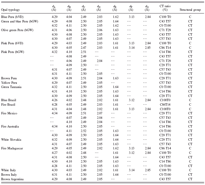
The values relative to peaks A to E and F and G observed in the opals examined in this work are discussed in this section. Examples of the XRD patterns of the examined opals are reported in Figures 1 and 2, while the weighted means of the d values (〈d〉) for each peak in the different typologies of opals are reported in Table I. Some diffraction peaks have not been detected because they were very weak or because they juxtaposed with those of other minerals (for instance, phyllosilicates). The abbreviation bVD and bGW are used to indicate materials with vitreous-dull and greasy-waxy lusters for the opals from Peru, respectively. Most results are in agreement with those in literature even if some are uncommon or also unpublished.
Shoulder A was observed in all the samples, with the exception of some pink opals from Peru containing palygorskite and most fire opals, especially from Mexico. In any case, when detected, this diffraction showed a d value ranging from 4.340 to 4.263 Å (〈d〉=4.301 Å).
Peak B was detected in all the analysed opals and shows d values ranging from 4.114 to 4.017 Å (〈d〉=4.073 Å).
Peak C was observed in all samples. The d values of this diffraction peak range from 2.520 to 2.480 Å (〈d〉=2.487 Å).
Peak D was observed in all the samples, with the exception of some pink opals from Peru and some fire opals from Madagascar, which showed poorly defined XRD patterns. When detected, this diffraction peaks showed d values ranging from 2.071 and 2.017 Å (〈d〉=2.041 Å).
Peak E was observed in all the samples, with the exception of some bGW pink opals from Peru containing palygorskite, some fire opals from Madagascar that showed poorly defined XRD peaks. In the bGW pink opals, this diffraction peak is partly masked by the juxtaposition of the diffraction at d=1.6104 Å due to enclosed palygorskite.
Diffraction peaks F and G were always observed together, forming a couple that is a characteristic of some specific categories, such as the bVD blue opals and bVD pink opals from Peru and blue opals from Brazil. These diffraction peaks were also observed in some samples of fire opals from Madagascar, from Brazil, and from Cuneo of Italy. As shown in Table I, peak F showed d values ranging from 3.140 to 3.121 Å (〈d〉=3.130 Å), while peak G had d values ranging from 2.851 to 2.832 Å (〈d〉=2.841 Å).
C. Accessory minerals present in opals of this work
The remaining diffraction peaks detected in our opals are generally related to some other accessory mineralogical phases. The most intense peaks are those of sepiolite, palygorskite, clay minerals, quartz, calcite, and gypsum. Regarding to palygorskite, this mineral can be recognised by its main reflection corresponding to d 100∼10.4 to 10.5 Å, which is partially embedded into the diffraction peak at 10 Å corresponding to illite. Other diagnostic diffraction peaks correspond to 6.44, 5.42, 4.49, 4.18, and 3.69 Å even if they are not always observable in the XRD patterns (Isphording, Reference Isphording1973; Chisholm, Reference Chisholm1992). The presence of palygorskite was determined into 14 pink bGW samples of opals from Peru and into some related matrices. We also analysed the differently coloured parts of some samples and found that the different colour intensities are related to a relatively large presence of palygorskite, especially with respect to sepiolite.
Sepiolite has a diagnostic reflection with a corresponding d value of ∼12 Å. Other reflections correspond to d values of 7.6, 5.0, 4.6, and 3.8 Å but can show variable intensities. Sepiolite was confidently detected only in four green and blue bGW samples from Peru. Some samples also feature the presence of clay minerals s.l., such as the bGW olive green opals from Peru, the green opals from Tanzania, the white opals from Italy, and the fire opals from Mexico and Madagascar.
The presence of α quartz was detected, in variable amounts, in 33 samples. In the bVD opals from Peru, which showed the presence of bands or coloured zones, the quartz is present in high amounts. Conversely, in the bGW olive green and blue opals from Peru, the quartz is relatively low. In the other typologies of samples, quartz generally occurs only as an accessory phase but is slightly higher in some opals from Patagonia.
TABLE I. CT ratios, structural groups, d values of the diagnostic peaks of the opals analysed in this work. bVD=vitreous-dull luster; bGW=greasy-waxy luster.
D. Discussion
The XRD analyses carried out in this work revealed that the structure of six of the samples of opal is completely amorphous, while α-cristobalite and α-tridymite (crystalline phases) are abundant in 63 samples. Several samples of opals are paracrystalline and characterised by a variably ordered structure due to the intercalation of cristobalite and tridymite layers (Graetsch, Reference Graetsch1994). Consequently, we were able to recognise all the different typologies of opals (A, CT, and C as shown in Figure 1) with their related “intermediate crystallization” (Jones and Segnit, Reference Jones and Segnit1971). In several samples, variable quantities of accessory phases such as palygorskite, sepiolite, clay s.l., α quartz, and, in few cases, calcite and gypsum were identified.
Concerning the amorphous opals, in three samples it was not possible to identify significant diffraction peaks, while other three samples showed the characteristic broad and diffuse band (at about 4.1 Å) typical of the amorphous opals (see Figures 1 and 2).
On the contrary, the paracrystalline opals showed intense diffraction peaks. The intensity is not probably related to the amount of cristobalite but to the degree of order of the opal. The variably amorphous structure of the opals becomes progressively more ordered during the diagenetic process (Juchem et al., Reference Juchem, Lubachesky, De Brum, Waichel, Pecchio, Dias de Andrade, D’Agostino, Kahn, Sant’Agostino and Lé Tassinari2004; Lynne and Campbell, Reference Lynne and Campbell2004). According to some authors, the tridymite in the opals also shows a similar behaviour, transforming into α-cristobalite (Graetsch, Reference Graetsch1994). With the increasing of the diagenetic process, the degree or order increases following the sequence opal-A→opal-CT→opal-C→chalcedony→quartz. In this sequence the transition from opal CT to opal C takes place gradually and slowly, as it mostly consists in a transformation of tridymite into cristobalite. However, it seems that in some siliceous volcanic deposits, the opals C and CT may form directly in situ without following the previous sequence (Graetsch et al., Reference Graetsch, Gies and Topalovic1994).
Discriminating the paracrystalline opals (C and CT opals) from the opals A through XRD is relatively easy, while it is more difficult to discriminate the opals C and CT. Probably, the opals C and CT represent a continuous series of intergrowths among the stacking sequences of the end members cristobalite and tridymite (Elzea and Rice, Reference Elzea and Rice1996). As a consequence, the local order increases with the decreasing of the disordered layers, so that the passage from opals CT to C can take place through intermediate steps (Graetsch et al., Reference Graetsch, Gies and Topalovic1994).
Cristobalite in natural environment shows two different structural forms of low and high temperatures: tetragonal α-cristobalite and cubic β-cristobalite, respectively. In the opals, as previously mentioned, cristobalite is nearly exclusively represented by the low-temperature phase. Differently, tridymite in natural environment presents low-temperature and a high-temperature forms, α-tridymite and β-tridymite, respectively. The low-temperature phase can be monocline and orthorhombic. The high-temperature phase is hexagonal and presents several polytypes, but the most common are the 10H and the 20H. A large part of the previous forms of tridymite can be present in the opals, but their identification and quantification through XRD are difficult. The investigations on the structure of the different forms of tridymite are still not conclusive even if interesting studies on this topic are present in literature (e.g., Graetsch and Flörke, Reference Graetsch and Flörke1991).
For the aims of our work it was necessary to define a parameter to evaluate the degree of order/disorder in the opals C and CT, which is related to the “content” of cristobalite/tridymite. The parameter is represented by the shift of the main peak B , which has already been used successfully by several authors. Peak B is the most important reflection and always detectable. It can indicate the different arrangements of tetrahedrons in the sequence of stackings of cristobalite and tridymite. According with the previously cited works, we considered the value d of 4.11 Å as diagnostic of tridymite and the value of 4.04 Å as diagnostic of cristobalite. The ratio between the amounts of tridymite and cristobalite in the crystalline fraction of the opals can be defined as

The calculated values of C/T for the analysed opals are reported in Table I.
The value of 4.11 Å is considered to be ideal for d Tryd, assuming that all the tridymite is present as the monoclinic low-temperature phase. Also in the case that the polytype 20H of the high-temperature phase was present, then the diffraction peak at 4.11 Å would combine with the 20H diffraction peak at 4.075 Å. This peak is due to the diffraction d 100 at 4.27 Å that causes a decreasing of the d values of shoulder A.
Once the values of C/T were determined for all the analysed samples, it was necessary to find a criterion that goes beyond a numerical value, which can be influenced by a different degree of disorder in a particular zone of a sample. Previous works in literature did not define a univocal way to separate opal-C values d 100 and opal CT. Therefore, on the basis of the collected results, we try to advance a new proposal. In particular, we establish that the d value of the B peak (d B) corresponding to 4.05 Å is the limit for opal C. Consequently, one deduces that the opals that show values d B≥4.06 Å must be classified as opal CT. This differentiation is strictly related to the presence or absence of the two F and G peaks at ∼30°2θ, typical for α-cristobalite. However, the limit at 4.06 Å for opal C can have some exceptions because of the degree of order/disorder. There are opals that show poorly defined diffraction patterns that make hard to identify the F and G peaks even if they show the d value of 4.06 Å (see Table I). On the contrary, the d value of 4.06 Å and a shaper XRD pattern for opals, these two peaks, diagnostic of α-cristobalite, are evident. Nevertheless, we must point out that most of samples showed XRD patterns that supported the previous consideration.
Once the general parameter is defined, some considerations concerning the behaviour of other diffraction peaks can be discussed in order to estimate the validity of the above assumed criterion.
The diffraction peaks A , B , and C are characterised by variable shifts. The d values of the different opal groups, related to the 2θ angular position, increase with increasing structural disorder (see Table I). Particularly, the diffraction peaks D and E , which show more shifts, evidence this behaviour as they separate more clearly the two types of opal (see Table I). The only data that yielded slightly different results are those relative to shoulder A. As previously reported, this peak is characteristic of the presence of tridymite. It is, therefore, likely that this value depends on the relative proportion of the different polytypes of tridymite. The group of fire opals of Madagascar (see Table I) presents a great heterogeneity: a group is formed nearly exclusively by α-cristobalite bearing opals, while another group features high contents of α-tridymite. The structural difference of these opals is probably related to the various unknown origins of these materials.
In addition of peaks F and G for opal C, other peaks are also present at the angular positions of ∼42.5°, 47°, 48°, 5°, 54°, 60°, and 62°2θ. However, these diffraction peaks were not considered, but for a confirmation, because their intensities are small and often not evident due to the general structural disorder. In addition, α-cristobalite has other additional 15 diffraction peaks, but they are not in the 2θ range used in this study. These diffraction peaks have d values ranging from 1.398 to 1.183 Å and are very weak. In opal CT we mostly observe a general and increasing trend to a broadening of peaks B and D , accompanied by a progressive fading of shoulder A and an increasing of the disorder of the entire diffraction pattern.
Some values of peak B showed values of d<4.04 Å; in particular, some pink bVD opals from Peru and one opal from Italy showed d values of ∼4.03 Å and blue opals from Brazil and fire opals from Madagascar showed d values of ∼4.02 Å. It is known that, under heating, opal may convert into well-ordered forms of cristobalite (Wahl et al. Reference Wahl, Grim and Graf1961). In addition, Jones and Segnit Reference Jones and Segnit(1971) heated some opal CT having d values of 4.114 Å up to temperatures of 1150 and 1300 °C and observed that the d values decreased to 4.038 and 4.025 Å, respectively. Therefore, we can hypothesise that our opals, which were extracted in volcanic settings, underwent a natural heating process that caused the decreasing of their d B values.
On the other hand, in our samples we have never found d values higher than 4.114 Å, such as d=4.127 Å reported for fire opals from Turkey by Esenli et al. Reference Esenli, Kumbasar, Eren and Uz(2001).
For a correct identification of opal, it would be important to identify a correct d value that allows the differentiation between opal CT and opal T. This limit could be the d value of 4.10 Å. However, because of the high disorder of the structure, other secondary peaks that can confirm the previous differentiation are frequently weak or not observable. For this reason, in according with other authors (e.g., Elzea and Rice, Reference Elzea and Rice1996), we will consider the opals with d values higher than 4.10 Å to be high disordered opal CT but not opal T.
The accessory minerals that can characterise the different typologies of opals turn out to be sepiolite and palygorskite. Palygorskite and especially sepiolite are also very rich in silica. Therefore, they can also form in the same silica rich environments where opals form.
Sepiolite and palygorskite have never been found together in the opals analysed in this work, which is different from what was reported by other authors on similar opals from Peru (Brajkovic et al., Reference Brajkovic, Rolandi, Vignola and Grizzetti2007). These minerals have been used as markers of particular typologies of opals. For instance, orthorhombic sepiolite was found only in blue and green bGW opals from Peru, although in modest amount and less frequently than palygorskite. Monocline palygorskite, on the contrary, was exclusively found in the pink bGW opals from Peru, in particular into the pink, white, and yellow parts. A large amount of palygorskite frequently occurs in opal; this is in agreement with reported palygorskite contents up to around 40% (Brajkovic et al., Reference Brajkovic, Rolandi, Vignola and Grizzetti2007; Fritsch et al., Reference Fritsch, Gaillou, Ostroumov, Rondeau, Devouard and Barreau2004). The amount of palygorskite present in opal appears proportional to the saturation of their colour, as can be deduced by the strong differences in intensities in various coloured parts of a same sample; this is due to the presence of organic colourings absorbed into palygorskite that could also be the cause of the pink colour. Similarly, in the crusts that cover coarse samples, palygorskite is sometimes associated with zeolites: stilbite and barrerite (Brajkovic et al., Reference Brajkovic, Rolandi, Vignola and Grizzetti2007). Different from sepiolite and palygorskite, clay minerals in the samples analysed in this work do not seem relevant for the determination of the colour, as observed, for instance, in the red opals from Macedonia (Bermanec et al., Reference Bermanec, Tomašić, Karfunkel, Scholz, Kniewald, Jaksch, Pecchio, Dias de Andrade, D’Agostino, Kahn, Sant’Agostino and Lé Tassinari2004).
In a few cases the presence of α quartz can provide some information for the characterisation of the opals. Conversely, calcite occurs only in trace and cannot be used for the previous purposes.
IV. CONCLUDING REMARKS
The purposes of this work are as follows:
1. Drawing up a detailed review of the application of XRD analysis on opals.
2. Describing the most important results of XRD analyses in literature and compare them with the new data of the analyses obtained from opals extracted from ores worldwide.
3. Proposing of a simple method to classify the opals based on their degrees of order/disorder and to discriminate opal C from opal CT.
Based on the XRD analyses, it is concluded that the original methods for the identification of different kinds of opals (namely, A, CT, and C) based on the XRPD technique previously reported in literature by Jones and Segnit Reference Jones and Segnit(1971) and Elzea and Rice Reference Elzea and Rice(1996) are still valid. These three types of opal can be easily identified based on their XRPD patterns. In particular, opal C is characterised by a peak corresponding to a d value between 4.02 and 4.05 Å and by the presence of two other peaks at 3.13 and 2.84 Å. Opal CT shows only a peak corresponding to a d value ranging from ∼4.06 to 4.11 Å. Also, it is not possible to distinguish an opal CT from a theoretical opal T (in this work, d=4.11 Å) ) as previously reported by Elzea and Rice Reference Elzea and Rice(1996). As a matter of fact, cristobalite crystallites are stacked in a tridymite superstructure. Hence, a single silica molecule can be considered as a part of a cristobalite crystal in one direction and a tridymite crystal in another direction.
Regarding to the accessory minerals, palygorskite and sepiolite are particularly important for the colour of opals. For instance, the opals from Acari Mine of Peru are characterised by the presence of palygorskite and sepiolite with a greasy-waxy luster. On the other hand, the opals with a vitreous-dull luster obtained from Lily Maine of Peru contain neither palygorskite nor sepiolite.


