


I. INTRODUCTION
The maximum entropy method (MEM) is an effective way to establish models from a finite number of observed physical quantities by maximizing information entropy (Izumi, Reference Izumi2004) By combining MEM (Sakata and Sato, Reference Sakata and Sato1990) with the Rietveld refinement (Rietveld, Reference Rietveld1969), one can determine three-dimensional (3D) charge and nuclear density distributions from X-ray and neutron diffraction data, respectively. This technique has extensively been applied to structural studies of manganites (Takata et al., Reference Takata, Nishibori, Kato, Sakata and Moritomo1999), ferroelectric perovskites, (Kuroiwa et al., Reference Kuroiwa, Aoyagi, Sawada, Harada, Nishibori, Takata and Sakata2001; Aoyagi et al., Reference Aoyagi, Kuroiwa, Sawada, Tanaka, Harada, Nishibori, Takata and Sakata2002) ionic conductors, (Nishimura et al., Reference Nishimura, Kobayashi, Ohoyama, Kanno, Yashima and Yamada2008; Yashima, Reference Yashima2009) and clathrates (Hoshikawa et al., Reference Hoshikawa, Igawa, Yamauchi and Ishii2005; Igawa et al., Reference Igawa, Taguchi, Hoshikawa, Fukazawa, Yamauchi, Utsumi and Ishii2010). For instance, MEM can reveal electronic orbital hybridization and orbital ordering from a limited amount of X-ray diffraction data (Takata et al., Reference Takata, Nishibori, Kato, Sakata and Moritomo1999; Kuroiwa et al., Reference Kuroiwa, Aoyagi, Sawada, Harada, Nishibori, Takata and Sakata2001; Aoyagi et al., Reference Aoyagi, Kuroiwa, Sawada, Tanaka, Harada, Nishibori, Takata and Sakata2002). MEM can also resolve possible ionic pathways in ionic or mixed conductors and gas molecular distributions in clathrates (Hoshikawa et al., Reference Hoshikawa, Igawa, Yamauchi and Ishii2005; Nishimura et al., Reference Nishimura, Kobayashi, Ohoyama, Kanno, Yashima and Yamada2008; Yashima, Reference Yashima2009; Igawa et al., Reference Igawa, Taguchi, Hoshikawa, Fukazawa, Yamauchi, Utsumi and Ishii2010). Therefore, the MEM/Rietveld analysis is a very powerful approach to reconstruct the atom distribution and interatomic charge densities associated with thermal motion and phase transition.
In the present study, we choose PbTiO3, a classic and extensively studied ferroelectric material (Shirane and Hoshino, Reference Shirane and Hoshino1951; Glazer and Mabud, Reference Glazer and Mabud1978; Fontana et al., Reference Fontana, Idrissi and Wojcik1990; Cohen, Reference Cohen1992), to corroborate the origin of tetragonal ferroelectric distortion, electron and nuclear distributions, and spontaneous polarization. All of these properties can be derived and calculated by the MEM/Rietveld analysis of neutron and X-ray diffractions. It is well known that at room temperature PbTiO3 possesses a tetragonal structure with an axial ratio of c/a = 1.06 (Shirane and Hoshino, Reference Shirane and Hoshino1951), distorted from the cubic, paraelectric phase below the Curie temperature of 490 °C. In ABO3 ferroelectric perovskite such as BaTiO3 and KNbO3, the anisotropic distortion of tetragonal phase from cubic phase arises from the covalent nature of B–O bonds. It has been pointed out that the hybridization between the Ti 3d states and O 2p states is essential to the ferroelectric instability in PbTiO3 (Cohen, Reference Cohen1992). In PbTiO3, the 6s 2 lone-pair effect from Pb2+ gives rise to additional distortion (Kuroiwa et al., Reference Kuroiwa, Aoyagi, Sawada, Harada, Nishibori, Takata and Sakata2001), which results in high spontaneous polarization in the tetragonal phase. There are two distinct atomic positions for oxygen in the crystal structure of tetragonal PbTiO3. The Pb–O2 bonds (parallel to the c-axis) are covalent, whereas the Pb–O1 bonds (normal to the c-axis) are ionic. Direct visualization of temperature-dependent nuclear and electron distributions will not only help understand the observed structural and electronic behaviors, but will also be essential for validating first-principles calculations from local bonding characteristics to 3D unit-cell models.
We performed in situ neutron and X-ray diffraction experiments to investigate the evolution of nuclear and charge densities of PbTiO3 as a function of temperature. The MEM/Rietveld analysis of neutron diffraction data provides an insight into the nuclear configuration, relative atom position distributions, and anisotropic thermal motion. From the X-ray diffraction data one can determine the charge density distributions around the atomic nuclei, bonding characteristics from interatomic charge densities, strength of covalent nature, as well as the effective ionic charges. Most importantly, spontaneous polarization can be deduced from the nuclear and charge distributions.
II. EXPERIMENTAL
Phase-pure tetragonal PbTiO3 powders were obtained from Alfa Aesar (99.9% metal based purity). The high-temperature neutron diffraction experiment was conducted with an ILL-type furnace (Vogel et al., Reference Vogel, Hartig, Lutterotti, Von Dreele, Wenk and Williams2004) at the flightpath of High-Pressure-Preferred Orientation (HIPPO) (Wenk et al., Reference Wenk, Lutterotti and Vogel2003; Vogel et al., Reference Vogel, Hartig, Lutterotti, Von Dreele, Wenk and Williams2004), Los Alamos Neutron Science Center (LANSCE). The sample was loaded into a vanadium can of 1 cm diameter, which has low attenuation for neutrons and can hold temperature up to 1200 °C. The time-of-flight neutron diffraction data were collected with three detector banks at Bragg angles of 2θ = 144.45°, 90.00°, and 39.30°, covering the d-spacing range of 0.58–4.8 Å. The high-temperature X-ray diffraction experiment was performed in a Philips X'pert diffractometer using CuKα 1 radiation. Diffraction data were collected at steps of 0.03°, and the counting time was 5 s for each step. The Rietveld analyses were performed using the General Structure Analysis System (GSAS) program software package (Larson and Von Dreele, Reference Larson and Von Dreele2004). Observed structure factors, F o, and standard uncertainties, σ(|F o|), which were estimated with Alchemy (Izumi and Kawamura, Reference Izumi and Kawamura2006) from relevant data in files output by GSAS, were analyzed by the MEM with Dysnomia (Izumi and Momma, Reference Izumi and Momma2011). In MEM analysis, we used all three banks of neutron data with carefully selected TOF regions and X-ray diffraction data in a scanning range of 2θ = 10–135°, corresponding to the d-spacing range of 0.77–4.44 Å. The unit cell was divided into 128 × 128 × 134 for the tetragonal PbTiO3. The detailed method for MEM analysis can be found in Ref. (Izumi and Momma, Reference Izumi and Momma2011).
III. RESULTS AND DISCUSSION
The Rietveld refinements of neutron and X-ray diffraction data for tetragonal PbTiO3 at room temperature are shown in Figures 1(a) and 1(b), respectively. The intensity residual values, R B, for all of the refinements are smaller than 5%, which indicates reasonable agreement between the experimental data and structural model for the title compound. The refinements with fairly low R B values also provide good starting points for the subsequent MEM analysis. For neutron diffraction, the use of data from multi-detector banks, as shown in the inset of Figure 1(a), can minimize the complication from background and region of overlapping reflection to obtain more accurate nuclear density distributions.

Figure 1. Rietveld refinements of neutron (a) and X-ray diffraction data (b); for tetragonal PbTiO3. Inset of (a) shows diffraction patterns from three different detector banks, which are combined for the MEM analysis.
Figure 2 shows nuclear density distributions at four different temperatures of tetragonal PbTiO3 in one unit cell with an isosurface level of 0.1 fm/Å3; the 3D visualization was conducted by VESTA (Momma and Izumi, Reference Momma and Izumi2011). The (blue) balls in the unit-cell center are Ti ions with a negative neutron scattering length. The Pb ions are located at the unit-cell corners. The O ions are on the six surface planes. For clearer discussion, the two O ions on the (0 0 1) planes are denoted as O1, and the rest of O ions on the (1 0 0) and (0 1 0) planes are denoted as O2. An inspection of Figure 2 indicates that the nuclear density distributions for all atoms expand with increasing temperature as a result of thermal vibrations. The expansion, however, is substantially more pronounced for Ti and O atoms than for Pb atoms. Note that O and Ti atoms also have a similar anisotropic temperature factor. The nuclear density distribution for O2 atoms stretches symmetrically along the c-axis and has an egg-shaped geometry. On the other hand, the O1 atoms gradually elongate along the b-axis with increasing temperature and eventually are pan-caked at 460 °C. This behavior indicates that the interaction between Ti and O2 is different from that between Ti and O1, which is further confirmed by the charge density analysis, as will be discussed below. The relatively smaller nuclear density distribution for Pb atoms indicates that the interaction between Ti and O is stronger than that between Pb and O. For all three atoms, the shape evolutions are consistent with the mean-square thermal amplitude variations reported for single-crystal PbTiO3 (Nelmes and Kuhs, Reference Nelmes and Kuhs1985).

Figure 2. 3D nuclear density distributions of tetragonal PbTiO3 at 30, 310, 410, and 460 °C, all at an isosurface level of 0.1 fm/Å3.
MEM analysis of X-ray diffraction data is a straightforward approach to derive the interatomic charge-density distributions and hence the interaction and bonding characteristics between adjacent atoms. Figure 3 shows the charge-density distributions for PbTiO3 at four different temperatures, all with an isosurface level of 0.8 e/Å3. Figures 4(a)–4(c) display the 2D charge density configurations in the range of 0–1.0 e/Å3 for (0 0 1), (1 0 0), and (0 2 0) lattice planes, respectively. It is evident from Figure 4(a) that there is no charge density overlap between neighboring Pb and O1, indicating an ionic nature for the Pb–O1 bonds. The Pb–O2 bond is covalent with ~0.17 e/Å3 charge density value in critical point, as revealed by the charge density distributions on the (1 0 0) lattice plane in Figure 4(b). The Ti–O bond is also covalent with ~0.75 e/Å3 charge density value in critical point, relatively stronger than the Pb–O2 bond as shown in Figure 4(c). In titanate perovskite such as PbTiO3 and BaTiO3, the Ti–O covalent bonding is primarily responsible for ferroelectric polarization, whereas the Pb–O2 covalent bonding presents a large energy barrier for the ferroelectric to paraelectric phase transition (Kuroiwa et al., Reference Kuroiwa, Aoyagi, Sawada, Harada, Nishibori, Takata and Sakata2001). Figure 4(d) shows the charge density distributions and atom positions of PbTiO3 at room temperature. The 3D vision of the charge density geometry of PbTiO3 unit cell is straightforward and clear to describe the bond characters.

Figure 3. 3D charge density distributions of tetragonal PbTiO3 at 30, 310, 410, and 460 °C, all with an isosurface level of 0.8 e/Å3.

Figure 4. 2D charge density configurations of PbTiO3 at 30 °C in the range of 0–1.0 e/Å3. The images of (a), (b), and (c) correspond to the (0 0 1), (1 0 0), and (0 2 0) lattice planes, respectively, as illustrated in (d). Figure 4(d) also shows charge density distributions and positions of the constituent atoms in 3D version.
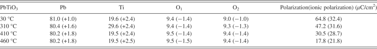
The bond lengths determined from our neutron diffraction data for Pb–O1, Pb–O2, Ti–O1, and Ti–O2 are 2.79(1) , 2.52(1), 1.76(3), and 1.97(2) Å, at room temperature and are 2.80(2), 2.82(1), 1.94(1), and 1.98(2) Å, respectively, at 460°. The relatively large increase in Ti–O1 and Pb–O2 bond lengths indicates that both tetragonal distortion and bonding strength decrease with increasing temperature. To further describe the bonding character at elevated temperature, the ionic states of constituent atoms were calculated by counting the numbers of charges around the atoms with a minimum charge density surface as a boundary condition. The spontaneous polarization of tetragonal PbTiO3 was then calculated from two distinct components: the ionic polarization calculated from the effective ionic states of constituent atoms and displacements of Pb and Ti sublattices relative to the oxygen octahedron (Kuroiwa et al., Reference Kuroiwa, Aoyagi, Sawada, Harada, Nishibori, Takata and Sakata2001); the electron polarization of all atoms, which was calculated from the relative shift of the electrons (X-ray refinements) to the nuclear position (neutron diffraction refinements). Briefly, the ionic spontaneous polarization is calculated by multiplying the effective charges and the distance between the negative and positive valence weighted mean center along the c-axis then dividing by the volume of the unit cell. The electron polarisation is the sum of the contribution of each constituent ion: the total electrons of each ion times the distance between the electrons center (negative charge) and nuclear center (positive charge) along c-axis and then divided by the volume of the unit cell. The effective ionic valence and spontaneous polarization calculated for tetragonal PbTiO3 at different temperatures are listed in Table I. With increasing temperature from 30 to 460 °C, the ionic state changes from +1 to +1.8 for Pb and from −1 to −1.4 for O2. The ionic states of Ti and O1 are essentially unchanged in this temperature range. The results indicate that the covalency of the Pb–O2 bond is suppressed with increasing temperature. Eventually, after phase transition above 490 °C, the Pb–O2 bond becomes ionic in the cubic phase (Kuroiwa et al., Reference Kuroiwa, Aoyagi, Sawada, Harada, Nishibori, Takata and Sakata2001). The calculated spontaneous polarization from the charge distributions only at room temperature is 32.4 μC/cm2, which is consistent with the conclusion in (Kuroiwa et al., Reference Kuroiwa, Aoyagi, Sawada, Harada, Nishibori, Takata and Sakata2001), but it is far less than total polarization obtained from the experimental measurements (Gavrilyachenko et al., Reference Gavrilyachenko, Spinko, Martynenko and Fesenko1970; Remeika and Glass, Reference Remeika and Glass1970). This indicates that the relative shifts of electrons from nuclei cannot be ignored. By taking the advantage of MEM analysis of neutron and X-ray diffraction data, we can calculate the contribution of such shifts to spontaneous polarization. The thus-calculated total spontaneous polarization for tetragonal PbTiO3 is 64.8 μC/cm2 at room temperature, which agrees with those obtained from dielectric hysteresis loop measurements, 75 μC/cm2 (Gavrilyachenko et al., Reference Gavrilyachenko, Spinko, Martynenko and Fesenko1970) and 57 μC/cm2 (Remeika and Glass, Reference Remeika and Glass1970). The calculated spontaneous polarization decreases rapidly with increasing temperature, indicating that the electrons are more symmetrically distributed around the nuclei at elevated temperature. This behavior dominates the contribution to spontaneous polarization even though the effective ionicity increases with increasing temperature.
Table I. Effective charges and ionic states (in parentheses) of constituent atoms and polarizations in tetragonal PbTiO3 as a function of temperature. The polarization is calculated from the ionic states, ionic displacements, and relative shifts between electron and nucleus positions. The ionic polarization values in the brackets are only from charge densities derived from X-ray diffractions.
IV. CONCLUSION
In summary, by a combination of MEM and Rietveld analyses of neutron and X-ray diffraction data, spontaneous polarization for ferroelectric PbTiO3 was accurately determined. This method would be particularly useful and effective for the study of multiferroics as an experimental measurement of polarization is greatly hindered and screened by the low electrical resistance and high-current leakage in bulk samples. In addition, MEM analysis of neutron and X-ray diffraction data can offer accurate structural and electronic parameters for verifying theoretical models and predictions.
ACKNOWLEDGEMENTS
This work was supported by the laboratory-directed research and development (LDRD) program of Los Alamos National Laboratory, which is operated by Los Alamos National Security LLC under DOE contract no. DE-AC52-06NA25396. The experimental work has benefited from the use of the Lujan Neutron Scattering Center at Los Alamos Neutron Science Center, which is funded by the US Department of Energy's Office of Basic Energy Sciences. The work at IOPCAS was supported by NSF & MOST through the research projects.


