I. INTRODUCTION
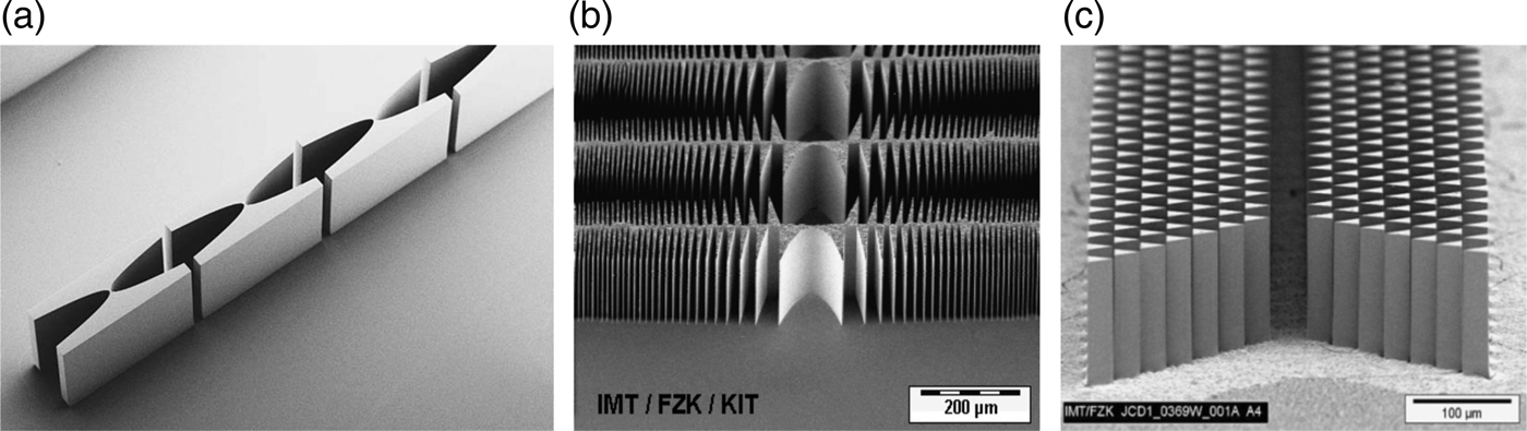
The Institute of Microstructure Technology (IMT) at Karlsruhe Institute of Technology (KIT) produces multiple types of refractive X-ray optics. They are fabricated using the LIGA (Lithographie, Galvanoformung, Abformung) process (Saile, Reference Saile2009), which yields high aspect ratios and high surface quality. Figure 1 shows multiple examples of such refracting structures [from Figures 1(a) to 1(c)]: a compound refractive lens, a kinoform lens, which allows for larger apertures and less absorption (Nazmov et al., Reference Nazmov, Shabel'nikov, Pantenburg, Mohr, Reznikova, Snigirev, Kouznetsov and DiMichiel2004), and a refractive prism lens (Simon et al., Reference Simon, Reznikova, Nazmov, Last, Jark, Khounsary, Morawe and Shunji2008).

Figure 1. Different types of refractive collecting lens types for X-rays: a compound refractive lens (a), a kinoform (b) and a prism lens (c).
Applications for these optics range from illumination to imaging. Whereas in X-ray microscopy application of both types of optics is required to produce high-resolution images, in material analysis applications, such as X-ray diffractometry, no imaging properties of the optics are required. For this type of application, the prism lens is the most useful refractive optics, since it offers the possibility for comparatively large apertures and low absorption. Nevertheless this lens type has three down-sides: first the aperture is limited by the height of the structures: the high columns become unstable when a certain aspect ratio is exceeded. Second, to create a two-dimensional (2D) refracting lens, two of these lens sets need to be aligned, tilted to 90° and third, the structures are fabricated using a high aspect ratio process, which is cost-intensive, and time-consuming.
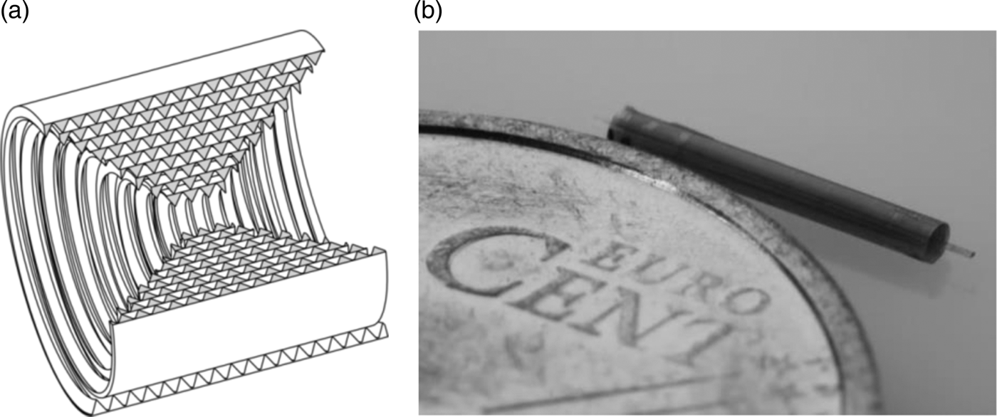
To improve this situation a new kind of refracting lens was developed at our institute: the so-called Rolled X-ray Prism Lenses (RXPL) [Simon, Reznikova et al., Reference Simon, Reznikova, Nazmov, Grund and Last2010]. Figure 2(a) shows a sketch with a cut-out of such a lens and Figure 2(b) shows a photograph of such a lens.
Figure 2. Principle sketch of an RXPL (a) and a photograph of a produced lens (b).
The lens consists of a polyimide foil with triangular-shaped corrugations on one side, which is cut and rolled around a glass fiber to form a refracting element for X-rays. The lens aperture is not limited to the stability of the single refracting elements, since they are not free-standing. The absorption is low because of the low Z-number material, the lens has point-focusing properties and for fabricating the lens, no high aspect ratio process is required.
With all these properties, the lens shows a promising potential to become a low-cost alternative for X-ray illumination applications.
II. FABRICATION
To fabricate an RXPL three major steps are necessary: first, the foil needs to be produced, second, layout calculation and foil cutting, and third, rolling process to form the actual lens.
A. Foil fabrication
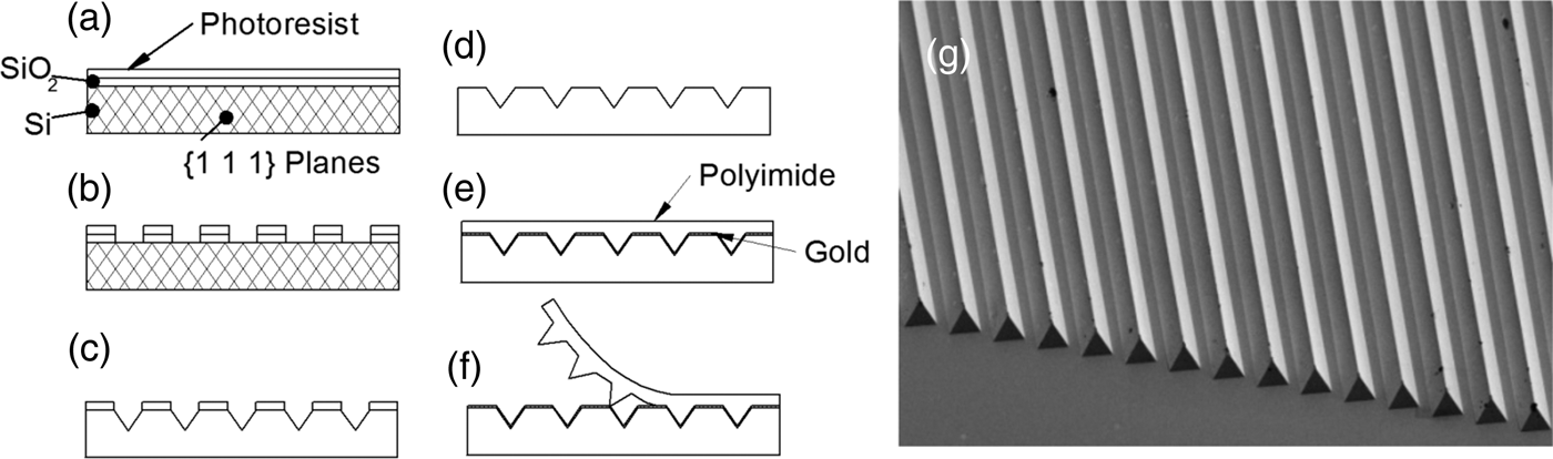
The foil is fabricated using a molding process. The mold itself is a structured silicon wafer. Figures 3(a)–3(f) show the principal steps to create the structure on the wafer.
Figure 3. Principal steps to create a mold wafer for RXPL-Foils (a–f) and SEM picture of the structures on the foil (g).
An oxidized wafer is coated with an ultraviolet (UV) photoresist by spin-coating [Figure 3(a)]. Then the photoresist is structured by UV-lithography using a chromium mask with a parallel line pattern, followed by an etching process that transfers this pattern into the silicon oxide [Figure 3(b)]. After removing the photoresist, V-shaped grooves are realized by anisotropic KOH etching of the (100)-silicon wafer to form the final mold [Figure 3(c)]. The wafer is oxidized again to prevent the gold layer, deposited in the next step, from amalgamating with the silicon. To produce the foil, a thin gold separation layer is deposited on the wafer and a liquid polyimide precursor (FUJIFILM™ Durimide® 32) is spin-coated on top. After backing, a thermal release tape (Nitto Denko™ Revalpha) is glued onto the foil to be able to peel the foil off the wafer without tearing it [Figure 3(f)]. To remove the foil from the thermal release tape the tape is heated to neutralize its adhesion properties and the foil, approximately 3 µm thick with 7.1 µm high triangular-shaped corrugations on one side, is released. Figure 3(g) right shows a scanning electron microscope (SEM) picture of the structures on one side of the foil.
B. Shape calculation and cutting
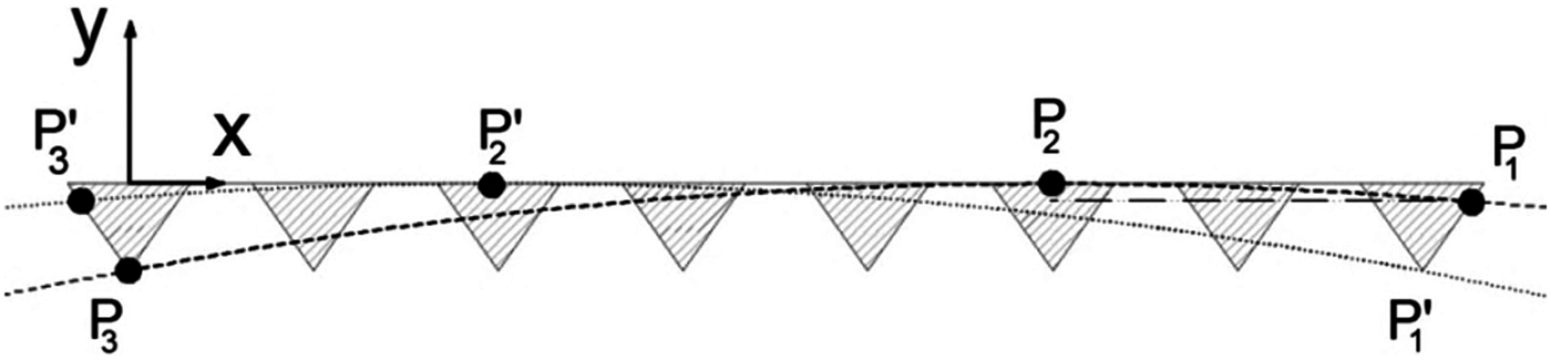
To derive the actual lens shape, the number of refracting elements in a certain distance to the optical axis needs to be calculated, which is necessary to refract a ray to a desired working distance. This is done using a ray-tracing program. In this simulation, the absorption as well as the “shadowing” of the single layers can be calculated in advance. This shadowing occurs because of the fact that every layer of a certain number of prisms has an entrance acceptance angle, in which rays are guided through the prism row without leaving it. Figure 4 shows a principle sketch to illustrate this behavior: the dotted line represents the shallowest ray; the dashed line represents the steepest ray, which may enter the row of refracting elements at point P 3′ respectively P 3 and is guided successfully through the layer, leaving it at P 1′ respectively P 1. Every ray which has an angle between the shallowest and the steepest ray is guided through the layer, every other ray will be absorbed or misguided by reflection at the backside of the next inner layer. This behavior is called shadowing.
Figure 4. Principle sketch to illustrate the shadowing effect of a row of refracting elements.
The shape calculation yields a necessary number of refracting elements in a certain distance to the optical axis. For example, to produce a lens for illuminating a small point on the optical axis, the principle structure in Figure 5(a) is obtained: the number of prisms increases with growing distance to the optical axis.
Figure 5. (Color online) Resulting structure of an RXPL after layout calculation in the rolled state (a) and foil layout after calculating and cutting the unrolled state (b).
This shape is the rolled layout. To retrieve the unrolled layout the formula of the Archimedean spiral is used; see e.g. Weisstein, Reference Weisstein2013. Figure 5 right shows a photograph of the parabola-like resulting foil layout.
The cutting itself is done using a laser-ablation system. This yields high contour accuracy and undamaged foil edges, avoiding mechanical forces on the structured foil.
C. Rolling
The third and last step is the rolling process. The rolling is done using a mechanical aid. This aid and the principal steps, which are used to produce the RXPL, are shown in Figure 6.
Figure 6. (Color online) Principal steps to roll an RXPL foil.
The cut foil is placed on top of a conveyor belt, which consists of a 7 µm thick Kapton® foil [Figure 6(a)]. In the next step, the conveyor belt and the structured foil are pushed into a loop between two grinded metal edges, which are called diversion edges [Figure 6(b)]. A glass fiber, 125 µm in diameter, is placed in the loop and the distance of the diversion edges is reduced until it equals the thickness of two times the conveyor belt height plus two times the structured foil thickness [Figure 6(c)]. Then the conveyor belt is fed backwards and the jutting end of the structured foil is fed into the loop. Then the distance between the diversion edges is further reduced by the height of the structured foil, so that the end cannot leave the loop, when feeding the conveyor belt in the forward direction [Figure 6(d)]. The forward motion of the conveyor belt is continued until the whole structured foil is rolled around the glass fiber. Figure 7 shows two different lenses for different working distances and energies: the smaller one has a diameter of about 800 µm and the larger one is approximately 2 mm in diameter.
Figure 7. (Color online) Two RXPLs with 800 µm and 2 mm diameter for different working distances and energies.
The resulting structure within the lens can be inspected by creating a radiographic picture of the lens. Figure 8 shows such a picture with enlarged cut-outs at different locations within the lens.
Figure 8. (Color online) Radiographic picture of an RXPL with cut-outs at different positions within the lens and principle lens layout.
When comparing the principle layout in the upper left corner of Figure 8 with the full-lens radiographic picture in the middle, the similarity becomes apparent. The cut-outs show high structure quality at different locations within the lens.
III. EXPERIMENTAL RESULTS
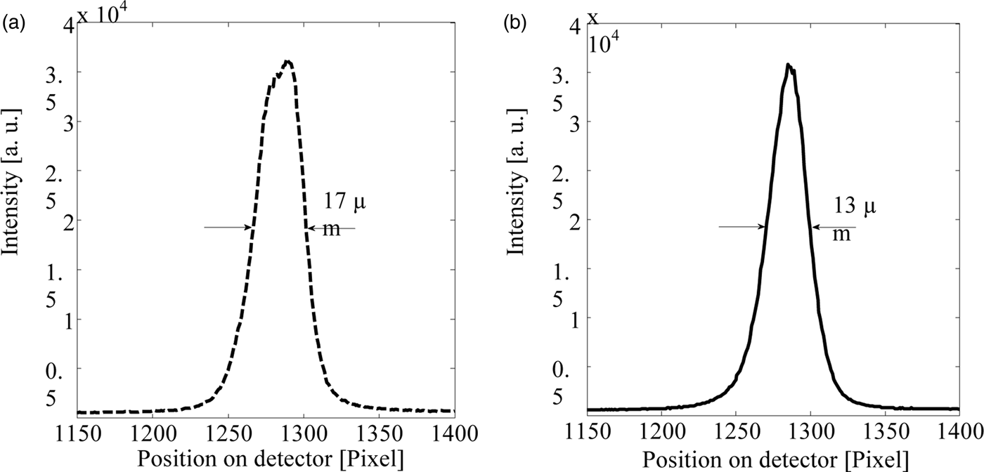
The RXPLs were tested at synchrotron sources as well as in a tube diffractometry experiment. Figure 9 shows, for example, the results obtained at ANKA, TopoTomo beamline in Karlsruhe, Germany. The lens was designed for a working distance of 150 mm for 9.9 keV. The graph in Figure 9(a) is the maximum intensity on a CCD detector (PCO 4000) for different distances between CCD and lens exit aperture. Figure 9(b) shows a cut perpendicular to the optical axis through the focus at the maximum intensity distance.
Figure 9. Results obtained at 9.9 keV at a synchrotron source: maximum intensity on detector versus distance between lens exit aperture and detector plane (a) and cut perpendicular to the optical axis through the focal spot at the position of maximum intensity along the optical axis (b).
The working distance of 145 mm is below the desired one of 150 mm. The full-width at half-maximum (FWHM) of the peak perpendicular to the optical axis in the vertical direction lies close to the theoretical minimum of about 10 µm because of the height of the prisms. The FWHM in the horizontal direction is larger (17 µm) due to the shape of the source. The overall spectral intensity enhancement, which is the ratio between the mean intensity on the detector within the FWHM area of the peak, with the lens and the mean intensity within the FWHM area without the lens, was about 60.
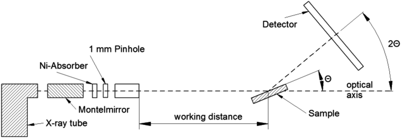
The diffractometry experiment was performed in an existing Bruker D8 Discover setup. Figure 10 shows the principal setup.
Figure 10. Principal setup of diffractometry experiment.
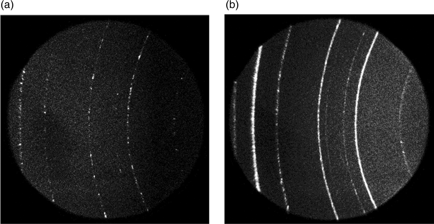
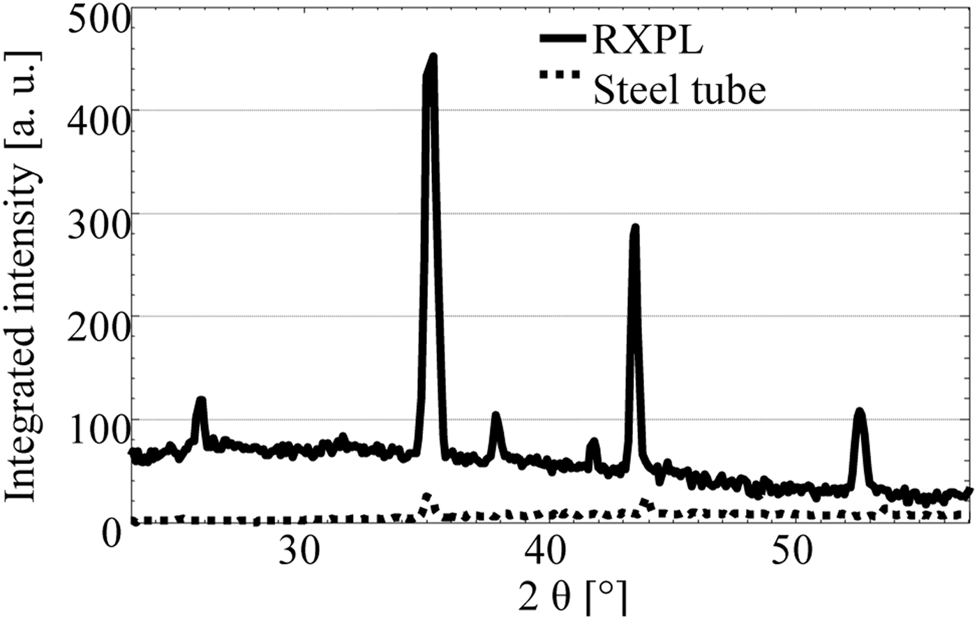
To create a semi-monochromatic source and to reduce divergence of the X-rays emitted by the X-ray tube (Siemens KFL-CU-2 K) a Montel mirror (Incoatec Montel-p) and a Ni-absorber foil was placed between the tube and the RXPL. The lens itself was then mounted on a tilt-stage to be adjusted to the beam. The sample used was a NIST-1976a corundum powder sample and the diffractogram was acquired using a VANTEC 500 detector. To acquire a figure of merit the diffractogram was once acquired using an RXPL and once using a steel-tube collimator, which yielded the same focal spot diameter on the sample. Figure 11 shows the resulting diffractograms: the diffractogram acquired using the steel-tube collimator (a) and the diffractogram acquired using the RXPL lens (b). The difference is apparent: the diffractogram with the RXPL shows additional lines as well as fewer speckles, but also more background toward the beam axis. In Figure 12, the integrated intensity values are plotted: the diffractogram with the lens shows up to an 18-fold enhanced intensity compared to that with the steel-tube collimator for the same acquisition times. The peak-shift between the integrated intensities results from a shifting of the position of the focal spot on the sample.
Figure 11. Two-dimensional diffractogram patterns acquired using a steel-tube collimator (a) and an RXPL (b).
Figure 12. Integrated intensities derived from diffraction patterns in Figure 11.
IV. CONCLUSION
RXPLs offer an alternative to existing illumination optics at synchrotron sources as well as for tube sources. The fabrication process does not require any time-consuming and cost-intensive high aspect ratio processes. Therefore RXPLs can be a low-cost alternative to other X-ray illumination optics. Experiments at synchrotron sources show the potential of the lenses (spectral intensity enhancement of 60) and diffractometry experiments show promising results (integrated intensity enhancement 18-fold compared to steel-tube collimator).
ACKNOWLEDGEMENTS
The authors acknowledge Synchrotron Light Source ANKA for provision of instruments at their beamlines and the authors thank Venera Altapova for assistance. The authors appreciate the support of the virtual institute “New X-ray analytic methods in material science” of the Helmholtz association.