


Introduction
Echinococcosis, caused by larval stage of the genus Echinococcus, is one of the most important zoonotic diseases worldwide (Alvarez Rojas et al., Reference Alvarez Rojas, Romig and Lightowlers2014). Echinococcus granulosus sensu lato (s.l.) and Echinococcus multilocularis are the most prevalent species and causal agents of human cystic echinococcosis (CE) and alveolar echinococcosis (AE), respectively (Eckert et al., Reference Eckert, Gemmell, Meslin and Pawlowski2001).
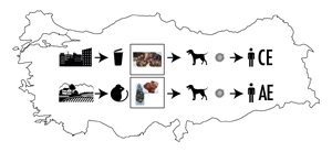
Echinococcus granulosus s.l. life cycle involves a carnivorous definitive host, the domestic dog in general and livestock as the intermediate host (Thompson, Reference Thompson2017). CE is a zoonotic disease of global importance and has socio-economic significance in communities where livestock farming is the main source of living. Echinococcus multilocularis has a predominantly sylvatic life cycle, with carnivorous species, such as foxes, wolves and coyotes, and to a lesser extent dogs, as definitive hosts and small rodent species serving as intermediate hosts (Vuitton et al., Reference Vuitton, Zhou, Bresson–Hadni, Wang, Piarroux, Raoul and Giraudoux2003). AE is a severe zoonotic disease that can lead to the death of the patient if left untreated or inadequately treated. Humans can be accidental dead-end intermediate hosts for both species. Human infection occurs through the ingestion of Echinococcus eggs with contaminated water or food or after direct contact with definitive hosts (Moro and Schantz, Reference Moro and Schantz2009).
E. granulosus s.l. is known to be endemic in all continents. However, E. multilocularis is mainly found in the northern hemisphere (Eckert et al., Reference Eckert, Gemmell, Meslin and Pawlowski2001). Both AE and CE are considered neglected zoonotic diseases. Whereas CE is globally distributed and highly prevalent, AE is more pathogenic, often resulting in mortality (Deplazes et al., Reference Deplazes, Rinaldi, Rojas, Torgerson, Harandi, Romig, Antolova, Schurer, Lahmar, Cringoli, Magambo, Thompson and Jenkins2017).
E. granulosus was previously considered a single species, but it is now recognized as an assemblage of cryptic species that have differences in morphology, development and host specificity, including infectivity and pathogenicity for humans (Romig et al., Reference Romig, Deplazes, Jenkins, Giraudoux, Massolo, Craig, Wassermann, Takahashi and de la Rue2017). E. granulosus s. l. currently includes five species, namely E. granulosus sensu stricto (s.s.) (G1/G3), E. equinus (G4), E. ortleppi (G5), E. canadensis (G6/7, G8 and G10) and E. felidis (Romig et al., Reference Romig, Deplazes, Jenkins, Giraudoux, Massolo, Craig, Wassermann, Takahashi and de la Rue2017; Vuitton et al., Reference Vuitton, McManus, Rogan, Romig, Gottstein, Naidich, Tuxun, Wen and Menezes da Silva2020). Phylogenetic studies based on sequencing of mitochondrial genes and microsatellite analysis in recent years have been successfully used to investigate the polymorphism within the E. multilocularis, which was previously considered to have relatively low genetic diversity (Knapp et al., Reference Knapp, Bart, Giraudoux, Glowatzki, Breyer, Raoul, Deplazes, Duscher, Martinek, Dubinsky, Guislain, Cliquet, Romig, Malczewski, Gottstein and Piarroux2009; Vuitton et al., Reference Vuitton, McManus, Rogan, Romig, Gottstein, Naidich, Tuxun, Wen and Menezes da Silva2020).
Diagnosis of Echinococcus infection in dogs is challenging in that the tapeworm eggs are shed irregularly and are indistinguishable from the eggs of other taeniids. The diagnosis of Echinococcus species in definitive hosts relies on techniques such as necropsy, arecoline purgation, copro-antigen ELISA and copro-PCR with faecal matter or isolated eggs (Craig et al., Reference Craig, Mastin, van Kesteren and Boufana2015).
To date, molecular studies on E. granulosus carried out in Turkey have reported several genotypes (G1−G3, G4, G6 and G7) in domestic livestock (Bowles et al., Reference Bowles, Blair and McManus1992; Vural et al., Reference Vural, Baca, Gauci, Bagci, Gicik and Lightowlers2008; Snabel et al., Reference Snabel, Altintas, D'Amelio, Nakao, Romig, Yolasigmaz, Gunes, Turk, Busi, Hüttner, Sevcová, Ito, Altintas and Dubinský2009; Simsek et al., Reference Simsek, Balkaya and Koroglu2010, Reference Simsek, Roinioti and Eroksuz2015; Simsek and Cevik, Reference Simsek and Cevik2014; Erdogan et al., Reference Erdogan, Ozkan, Mutlu, Karaca and Sahin2017) as well as in humans (G1−G3, G6 and G7) (Snabel et al., Reference Snabel, Altintas, D'Amelio, Nakao, Romig, Yolasigmaz, Gunes, Turk, Busi, Hüttner, Sevcová, Ito, Altintas and Dubinský2009; Eryildiz and Sakru, Reference Eryildiz and Sakru2012) from different endemic foci of Turkey. Only one genotype (G1) has been reported in dogs from different parts of Turkey (Utuk et al., Reference Utuk, Simsek, Koroglu and McManus2008; Kuru et al., Reference Kuru, Aypak and Aysul2013; Oge et al., Reference Oge, Oge, Gonenc, Sarimehmetoglu and Ozbakis2017; Oguz et al., Reference Oguz, Ozdal, Kilinc and Deger2018).
In Turkey, a highly endemic region for AE and CE (Deplazes et al., Reference Deplazes, Rinaldi, Rojas, Torgerson, Harandi, Romig, Antolova, Schurer, Lahmar, Cringoli, Magambo, Thompson and Jenkins2017), echinococcosis is a major public health problem, especially in the rural areas of eastern regions (Altintas, Reference Altintas2008). Erzurum province in the northeastern part of Turkey is a hyperendemic area for both human AE and CE, and the largest number of AE and CE patients nationwide was reported in the region (Altintas, Reference Altintas2008). Similarly, high prevalence rates of CE in cattle (Simsek et al., Reference Simsek, Balkaya and Koroglu2010) and sheep (Arslan and Umur, Reference Arslan and Umur1997) in Erzurum province and have been reported. Thus far, metacestodes of E. multilocularis in humans (Kurt et al., Reference Kurt, Avcioglu, Guven, Balkaya, Oral, Kirman, Bia and Akyuz2020) and rodents (Avcioglu et al., Reference Avcioglu, Guven, Balkaya, Kirman, Bia, Gulbeyen, Kurt, Yaya and Demirtas2017a), and E. multilocularis adults in red foxes (Avcioglu et al., Reference Avcioglu, Guven, Balkaya, Kirman, Bia and Gulbeyen2016, Reference Avcioglu, Guven, Balkaya, Kirman, Akyuz, Bia, Gulbeyen and Yaya2021) and lynx (Avcioglu et al., Reference Avcioglu, Guven, Balkaya and Kirman2018) have been reported in this region.
Data on the prevalence of echinococcosis in intermediate hosts (for CE and AE) and definitive hosts (for AE) is available for this region, which has provided valuable information on the geographical distribution of the parasites and the role of different animal species in parasite transmission. However, there is no available information on the presence and prevalence of Echinococcus species in dogs in Erzurum. The infection in dogs is important to estimate the relative infection pressure on intermediate hosts and humans, and to determine their roles in environmental contamination. Therefore, the purpose of the study was to determine the presence and prevalence of Echinococcus species in stray dogs in Erzurum province.
Materials and methods
Study area
The study was conducted from October 2015 to February 2016 in Erzurum (39°54′31″N, 41°16′37″E) province. The province is located in the eastern part of Turkey and has the fourth largest surface area (25 066 km2) in the country, 20 counties and a total population of 762 000 inhabitants. The province has an elevation of 1853 m above sea level. It receives an annual rainfall of 453 mm. The temperature range is −35 to 35°C. Agriculture and livestock raising constitute the principal economic activities of the province.
Sample collection
The animal shelter under the division of Erzurum Metropolitan Municipality regularly collected stray dogs and cats from all counties. Rehabilitation and medical services including sterilization, vaccination against rabies and praziquantel application for tapeworms were provided by the shelter. After the applications, the animals were ear tagged and may then be either set free or adopted. The stray dogs were housed in pens with concrete floor kennels individually during the application of praziquantel and collection of the faecal samples 24 h after the drug application. All investigated dogs were older than one year. None of the dogs were microchipped or wearing an identity tag. Therefore, we could not determine their collection area or medical history.
A total of 446 faecal samples were regularly collected from individual dogs after the praziquantel application. Faecal samples were placed in labelled Ziploc bags, stored at −86°C for at least seven days (Deplazes and Eckert, Reference Deplazes and Eckert1996) to reduce the risk of laboratory infection by inactivating any Echinococcus oncospheres, and stored at −20°C until further examination.
Isolation of taeniid eggs and DNA extraction
The sequential sieving and flotation method (SSFM) described by Mathis et al. (Reference Mathis, Deplazes and Eckert1996) was used for the concentration of taeniid eggs in the faecal samples. Briefly, flotation with zinc chloride (density 1.45 g mL−1) and sequential sieving through sieves of 40 and 21 μm mesh sizes were performed. The sediment accumulated in the 21 μm sieve was deposited in a flat-sided tube and examined under an inverted microscope to determine the presence of taeniid eggs. Positive samples were centrifuged at 1000 g for 10 min, and the pellet with taeniid eggs was stored in 2 mL microcentrifuge tubes.
Taeniid eggs were subjected to DNA extraction using a Qiamp DNA Mini Kit (DNeasy Tissue kit; Qiagen, Hilden, Germany) according to the manufacturer's instructions. The concentration of extracted DNA was measured with a spectrophotometer (NanoDrop One; Thermo Fisher Scientific, WI) and stored at −20°C until further step.
Molecular analyses and sequencing
Three sets of primers were used to amplify partial sequences of two mitochondrial genes, 12S rRNA and cytochrome c oxidase subunit 1 (COI), to detect E. multilocularis, E. granulosus s.s. and E. granulosus s.l. with conventional PCRs. Two PCR protocols with specific primers Egss1F/1R (254 bp, Dinkel et al., Reference Dinkel, Njoroge, Zimmermann, Walz, Zeyhle, Elmahdi, Mackenstedt and Romig2004) and Emnestfor/rev (204 bp, Dyachenko et al., Reference Dyachenko, Pantchev, Gawlowska, Vrhovec and Bauer2008), amplifying partial sequences of the 12S rRNA gene of E. granulosus s.s. and E. multilocularis, respectively, were performed. Only second step of the nested PCR protocol (Dyachenko et al., Reference Dyachenko, Pantchev, Gawlowska, Vrhovec and Bauer2008) was performed in E. multilocularis PCR. For all the samples, JB3/4.5 primers (446 bp), which amplified a part of the COI gene, were also used to amplify E. granulosus s. l. (Bowles et al., Reference Bowles, Blair and McManus1992). DNA of E. granulosus s.s. and E. multilocularis, which were previously confirmed by molecular analysis as positive control and distilled water as negative control, were included in each PCR run. The PCR products were analysed using 1.5% agarose gel electrophoresis, stained with SYBR Safe (Invitrogen, USA) and visualized using UV transillumination (Vilbert Lourmat, Quantum ST4, 1100/20M, France).
Bidirectional sequencing of all amplicons obtained in three PCRs was performed commercially with an ABI PRISM 310 genetic analyser (Applied Biosystems, Foster City, CA). Sequences were checked by eye (Finch TV), aligned using BioEdit 7.0 (http://www.mbio.ncsu.edu/BioEdit/bioedit.html) and compared with those on the GenBank database through the use of BLAST algorithms (http://www.ncbi.nlm.nih.gov/BLAST/) to determine the species. Pairwise calculations were obtained using BioEdit software.
Ethical approval
Ethical approval was obtained from the Atatürk University Animal Research Local Ethics Committee (Approval no: 2015/27).
Results
Among the 446 faecal samples, 119 (26.68%, Table 1) were positive for taeniid eggs by microscopy. The positive samples were subjected to molecular analysis, and Echinococcus spp. were detected in 63/446 (14.13%) of the faecal samples. E. granulosus s.s. was obtained in 41 (9.19%) of the samples, whereas E. multilocularis was found in 16 (3.58%) samples by 12S rRNA-PCRs.
Table 1. Echinococcus species in stray dogs in Erzurum.

E.g.s.s.: E. granulosus s.s.
E.e.: E. equinus
E.o.: E. ortleppi
E.c.: E. canadensis
E.m.: E. multilocularis
All the taeniid egg positive samples subjected to COI PCR were also positive. Sequence analysis of the COI PCR amplicons confirmed 28 of the 41 E. granulosus s.s. PCR positive samples and 10 of the 16 E. multilocularis PCR positive samples to be E. granulosus s.s. and E. multilocularis, respectively. To obtain longer sequences for E. multilocularis, we repeated the egg isolation and DNA extraction section for 6 of the 16 E. multilocularis PCR positive samples and performed COI PCR and sequencing until we succeeded.
Thereafter, BLAST analysis of the sequences revealed that the stray dogs harboured five different Echinococcus spp., namely E. granulosus s.s. (G1/G3) (n = 41), E. equinus (G4) (n = 3), E. ortleppi (G5) (n = 1), E. canadensis (G6/G7) (n = 3) and E. multilocularis (n = 16) (Table 1). E. granulosus s.s. was the most abundant species in stray dogs. Taenia spp. identified in 64 samples (details not reported here). DNA sequencing was not achieved in 12/119 of the samples due to the low yield of PCR products.
Multiple infections were recorded in some faecal samples: E. canadensis (G6/G7) and E. multilocularis in one sample, E. granulosus s.s. and Taenia spp. in 13 samples and E. multilocularis and Taenia spp. in five samples.
Isolated partial sequences were then deposited into GenBank with the following accession numbers: E. granulosus s.s. (G1/G3): MN732801-MN732821, E. equinus (G4): MN737094-MN737096, E. ortleppi (G5): MN737097, E. canadensis (G6/G7): MN737098-MN737100, E. multilocularis: MN732822-MN732837.
The COI nucleotide sequences of Erzurum isolates were compared with those of the references. Erzurum E. granulosus s.s. isolates showed 99.7–100% identity with each other and 100% identity with those reported from Turkey (MN990735, HM598451, KM100574, KX874711, EU178104), Brazil (KT382540, HF947571), Iran (MW350099, KJ162568, MT786855) and India (JX854029). Erzurum E. equinus isolates showed 100% identity with each other and were identical with those reported from Turkey (MK616473, KC953029, KM525658), Namibia (KP161210) and Uzbekistan (MK975893). Erzurum E. ortleppi isolates had 100% identity with the isolates reported from Japan (AB235846), Namibia (KU743926), Brazil (KT382535), Bosnia and Herzegovina (MG976769), France (KC430087) and Egypt (MK492625). Erzurum E. canadensis isolates were 100% identical with each other, and also had 100% identity with isolates from Iran (KU359038, KU220241), Sudan (MH300947), Nigeria (MN025264, KY996491) and France (MH823709). Erzurum E. multilocularis showed 99.5–100% identity with each other, also 100% identical with the isolates from Switzerland (MT461411), Canada (MK843308, MT461409, KC550004), USA (LC380931), China (MN251849, MH259774), South Korea (AB780998), Poland (KY205679, MW255909), Slovakia (DQ979365), Kyrgyzstan (MN829539), Russia (AB777915, AB688134) and Japan (AB385610).
Discussion
Echinococcus granulosus s.l. and E. multilocularis are two of the most widespread zoonoses as they cause disease in both humans and animals which are responsible for serious health and economic problems. In Turkey, a highly endemic region for AE and CE (Deplazes et al., Reference Deplazes, Rinaldi, Rojas, Torgerson, Harandi, Romig, Antolova, Schurer, Lahmar, Cringoli, Magambo, Thompson and Jenkins2017), echinococcosis is a major public health problem, especially in the rural areas of eastern regions (Altintas, Reference Altintas2008). Erzurum province in the northeastern part of Turkey is a hyperendemic area for both human AE and CE. However, to date, there have been no data available on the presence and prevalence of Echinococcus spp. in the dogs of this province. This study reports the prevalence of Echinococcus spp. (14.1%) in dogs based on faecal samples in Erzurum province. The prevalence of E. granulosus s.l. and E. multilocularis was 10.8 and 3.6%, respectively. The presence of E. granulosus s.s. (G1/G3), E. equinus (G4), E. ortleppi (G5) and E. canadensis (G6/G7) was reported. Notably, E. multilocularis from dogs and E. ortleppi (G5) in Turkey were identified for the first time.
Most studies in Turkey have been performed on CE in humans and livestock animals but limited in dogs (Altintas, Reference Altintas2008; Simsek et al., Reference Simsek, Balkaya and Koroglu2010; Deplazes et al., Reference Deplazes, Rinaldi, Rojas, Torgerson, Harandi, Romig, Antolova, Schurer, Lahmar, Cringoli, Magambo, Thompson and Jenkins2017; Avcioglu et al., Reference Avcioglu, Guven, Balkaya, Bia, Kirman, Gulbeyen, Yaya and Akyuz2017b; Kurt et al., Reference Kurt, Avcioglu, Guven, Balkaya, Oral, Kirman, Bia and Akyuz2020). This is thought to be due to the difficulties in fieldwork of definitive hosts, such as obtaining dogs and wild canids, and contamination risk with zoonotic infections such as rabies and echinococcosis.
In Turkey, several studies on E. granulosus infection in dogs indicated endemicity across the country, ranging from 0.8 to 40.5%, and varying according to the geographical location and diagnostic methods (Umur and Arslan, Reference Umur and Arslan1998; Oter et al., Reference Oter, Bilgin, Tınar and Tuzer2011; Kuru et al., Reference Kuru, Aypak and Aysul2013; Oge et al., Reference Oge, Oge, Gonenc, Sarimehmetoglu and Ozbakis2017). Determination of the prevalence of Echinoccoccus spp. in definitive hosts in an endemic area is essential to understand the transmission dynamics of the parasite and to design effective control programmes. Although there is a lack of data on the presence and prevalence of E. granulosus in dogs, Erzurum is known as an endemic region for CE based on human cases and the high prevalence in livestock animals. The study reports the overall prevalence of E. granulosus s.l. as 10.8% in dogs based on faecal samples in Erzurum province. The results of the study determined the presence and prevalence of E. granulosus s.l. in the stray dogs in the province for the first time. This information will serve as a source for control strategies in the region.
The prevalence in stray dogs reported here was lower than some studies and higher than others from different parts of the country. The lower prevalence in this study can be explained by the lower sensitivity of our detection method (eggs in faeces) compared to the previous studies that used copro-antigen, arecoline purgation and necropsy. Prevalence rates between these studies are therefore not comparable. The low prevalence could also be explained by the prepatent infections and periodic shedding of Echinococcus eggs during patent infections (Trachsel et al., Reference Trachsel, Deplazes and Mathis2007; Huttner et al., Reference Huttner, Siefert, Mackenstedt and Romig2009). However, for about two decades, antiparasitic applications on stray dogs periodically collected by the local government in Erzurum are thought to reduce the prevalence of the parasite. In connection with the results of this practice, three studies conducted approximately 10 years apart on the prevalence of CE in cattle in Erzurum province reported 46.4% (Arslan and Umur, Reference Arslan and Umur1997), 34.3% (Simsek et al., Reference Simsek, Balkaya and Koroglu2010) and 24% (Avcioglu et al., Reference Avcioglu, Guven, Balkaya, Bia, Kirman, Gulbeyen, Yaya and Akyuz2017b) prevalence rates. It has been observed that the prevalence decreases by about 10% every 10 years in cattle. The fact that the prevalence of CE in intermediate hosts has decreased in recent years compared to previous years undoubtedly has a negative effect on the prevalence in final host dogs.
Determination of the species and genotypes of Echinoccoccus in definitive and intermediate hosts in an endemic area is essential to understanding the transmission dynamics of the parasite and designing effective control programmes (Alvarez Rojas et al., Reference Alvarez Rojas, Romig and Lightowlers2014; Romig et al., Reference Romig, Ebi and Wassermann2015). To date, molecular studies on E. granulosus carried out in Turkey have reported several genotypes (G1/G3, G4, G6 and G7) in livestock (Bowles et al., Reference Bowles, Blair and McManus1992; Vural et al., Reference Vural, Baca, Gauci, Bagci, Gicik and Lightowlers2008; Snabel et al., Reference Snabel, Altintas, D'Amelio, Nakao, Romig, Yolasigmaz, Gunes, Turk, Busi, Hüttner, Sevcová, Ito, Altintas and Dubinský2009; Simsek et al., Reference Simsek, Balkaya and Koroglu2010, Reference Simsek, Roinioti and Eroksuz2015; Simsek and Cevik, Reference Simsek and Cevik2014; Erdogan et al., Reference Erdogan, Ozkan, Mutlu, Karaca and Sahin2017) and humans (G1/G3, G6 and G7) (Snabel et al., Reference Snabel, Altintas, D'Amelio, Nakao, Romig, Yolasigmaz, Gunes, Turk, Busi, Hüttner, Sevcová, Ito, Altintas and Dubinský2009; Eryildiz and Sakru, Reference Eryildiz and Sakru2012; Kurt et al., Reference Kurt, Avcioglu, Guven, Balkaya, Oral, Kirman, Bia and Akyuz2020) from different endemic foci. Among these, G1 has been considered the most prevalent in animals and human CE cases in Turkey. The G1 genotype was first reported in a dog by Utuk et al. (Reference Utuk, Simsek, Koroglu and McManus2008) and later among 100 dogs by three other studies (Kuru et al., Reference Kuru, Aypak and Aysul2013; Oge et al., Reference Oge, Oge, Gonenc, Sarimehmetoglu and Ozbakis2017; Oguz et al., Reference Oguz, Ozdal, Kilinc and Deger2018) from different parts of Turkey using limited molecular studies.
Based on COI sequence analysis, the majority of Erzurum samples (n = 41) were identified as G1 genotype (E. granulosus s.s.); three were G4 genotype (E. equinus), one was G5 (E. ortleppi) and three were G6/G7 genotypes (E. canadensis). It was found that E. granulosus s.s. was the most abundant species in the study, confirming the results of the studies conducted in humans and livestock in Turkey, as indicated earlier. In Erzurum, traditional animal husbandry practices are typically used. Poor abattoir conditions and home slaughtering (especially sheep) are common in rural regions of the city, and there is a high population of stray dogs with insufficient public health education. The metacestodes of E. granulosus s.s. are able to reach fertility in sheep, which increases the infection risk of dogs. The combination of home slaughter, lack of thorough meat inspection, poor abattoir conditions, high cyst fertility and the high numbers of roaming dogs may explain the abundance of E. granulosus s.s. identified in the present study.
Apart from E. granulosus s.s., E. canadensis (G6/G7) has been estimated to be responsible for 12.2 and 9.6% of global human CE infections, respectively (Cucher et al., Reference Cucher, Macchiaroli, Baldi, Camicia, Prada, Maldonado, Avila, Fox, Gutiérrez, Negro, López, Jensen, Rosenzvit and Kamenetzky2016). The presence of G6 and G7 genotypes in livestock and humans was reported in Turkey (Snabel et al., Reference Snabel, Altintas, D'Amelio, Nakao, Romig, Yolasigmaz, Gunes, Turk, Busi, Hüttner, Sevcová, Ito, Altintas and Dubinský2009; Simsek et al., Reference Simsek, Kaplan and Ozercan2011; Eryildiz and Sakru, Reference Eryildiz and Sakru2012). The G6/G7 genotypic cluster was first reported in cyst sample of a sheep in Elazig province of Turkey by Mehmood et al. (Reference Mehmood, Simsek, Celik, Kesik, Kilinc and Ahmed2020). The camel population is fairly lower in Turkey than in southeast neighbour countries, which have a large camel population. In Erzurum, camel breeding is not carried out or even available. Pig production is very limited due to religious reasons but wild boar population is very common in the country. In rural areas of the Erzurum, unofficial wild boar hunting is practiced because of their harmful effects on farming activities. Illegal dog transport from the border or access of stray dogs to dead wild boars can explain the existence of G6 and G7 genotypes. Reported G6/G7 genotype both from sheep in Elazig and from stray dogs in Erzurum province indicate that this genotype may have a wider distribution than previously thought in Turkey.
E. equinus (G4) is known to be a specific parasite of equids and non-pathogenic for humans (Romig et al., Reference Romig, Dinkel and Mackenstedt2006). However, in a study conducted on the molecular characterization of E. granulosus s.l in humans, E. equinus was reported for the first time (Romig et al., Reference Romig, Deplazes, Jenkins, Giraudoux, Massolo, Craig, Wassermann, Takahashi and de la Rue2017). Further, there have been many reports indicating the presence of E. equinus in different intermediate hosts worldwide (Thompson and McManus, Reference Thompson and McManus2002; Boufana et al., Reference Boufana, Stidworthy, Bell, Chantrey, Masters, Unwin, Wood, Lawrence, Potter, McGarry, Redrobe, Killick, Foster, Mitchell, Greenwood, Sako, Nakao, Ito, Wyatt, Lord and Craig2012). E. equinus has been detected in humans, mules and donkeys in Turkey (Simsek and Cevik, Reference Simsek and Cevik2014; Kesik et al., Reference Kesik, Kilinc, Simsek and Gul2019; Macin et al., Reference Macin, Orsten, Samadzade, Colak, Cebeci and Fındık2021). This study is the first report of E. equinus from stray dogs in Turkey.
Echinococcus ortleppi (G5) is particularly well adapted to cattle as intermediate hosts. The morphology and developmental features of E. ortleppi show substantial differences compared with those of E. granulosus s.s. and other taxa (Romig et al., Reference Romig, Ebi and Wassermann2015). Although it was formerly known as almost exclusively found in cattle as intermediate hosts, in recent years, many countries have detected infections in sheep (Mbaya et al., Reference Mbaya, Magambo, Njenga, Zeyhle, Mbae, Mulinge, Wassermann, Kern and Romig2014; Addy et al., Reference Addy, Wassermann, Banda, Mbaya, Aschenborn, Aschenborn, Koskei, Umhang, De La Rue, Elmahdi, Mackenstedt, Kern and Romig2017), pigs (Dinkel et al., Reference Dinkel, Njoroge, Zimmermann, Walz, Zeyhle, Elmahdi, Mackenstedt and Romig2004; Pednekar et al., Reference Pednekar, Gatne, Thompson and Traub2009; Tigre et al., Reference Tigre, Deresa, Haile, Gabriel, Victor, Pelt, Devleesschauwer, Vercruysse and Dorny2016; Addy et al., Reference Addy, Wassermann, Banda, Mbaya, Aschenborn, Aschenborn, Koskei, Umhang, De La Rue, Elmahdi, Mackenstedt, Kern and Romig2017), goats (Mbaya et al., Reference Mbaya, Magambo, Njenga, Zeyhle, Mbae, Mulinge, Wassermann, Kern and Romig2014; Addy et al., Reference Addy, Wassermann, Banda, Mbaya, Aschenborn, Aschenborn, Koskei, Umhang, De La Rue, Elmahdi, Mackenstedt, Kern and Romig2017), camels (Ahmed et al., Reference Ahmed, Eltom, Musa, Ali, Elamin, Grobusch and Aradaib2013; Amer et al., Reference Amer, Helal, Kamau, Feng and Xiao2015; Addy et al., Reference Addy, Wassermann, Banda, Mbaya, Aschenborn, Aschenborn, Koskei, Umhang, De La Rue, Elmahdi, Mackenstedt, Kern and Romig2017; Ebrahimipour et al., Reference Ebrahimipour, Sadjjadi, Darani and Najjari2017), monkeys (Pednekar et al., Reference Pednekar, Gatne, Thompson and Traub2009), oryx (Addy et al., Reference Addy, Wassermann, Banda, Mbaya, Aschenborn, Aschenborn, Koskei, Umhang, De La Rue, Elmahdi, Mackenstedt, Kern and Romig2017) and spotted deer (Boufana et al., Reference Boufana, Stidworthy, Bell, Chantrey, Masters, Unwin, Wood, Lawrence, Potter, McGarry, Redrobe, Killick, Foster, Mitchell, Greenwood, Sako, Nakao, Ito, Wyatt, Lord and Craig2012). Human infections with E. ortleppi occur less frequently than with other species, such as E. granulosus s.s. (Alvarez Rojas et al., Reference Alvarez Rojas, Romig and Lightowlers2014), with only 12 human cases reported in various parts of the world (Alvarez Rojas et al., Reference Alvarez Rojas, Romig and Lightowlers2014; Shi et al., Reference Shi, Wan, Wang, Li, Jiang and Yang2019). To our knowledge, only four reports of the presence of E. ortleppi in dogs are available, including those from Argentina (Kamenetzky et al., Reference Kamenetzky, Gutierrez, Canova, Haag, Guarnera, Parra, García and Rosenzvit2002; Soriano et al., Reference Soriano, Pierangeli, Pianciola, Mazzeo, Lazzarini, Saiz, Kossman, Bergagna, Chartier and Basualdo2010), Brazil (de la Rue et al., Reference de la Rue, Takano, Brochado, Costa, Soares, Yamano, Yagi, Katoh and Takahashi2011) and Kenya (Mulinge et al., Reference Mulinge, Magambo, Odongo, Njenga, Zeyhle, Mbae, Kagendo, Addy, Ebi, Wassermann, Kern and Romig2018). E. ortleppi was detected in one dog faecal sample and was reported for the first time in Turkey by this study. Cattle are most frequently infected with E. granulosus (G1/G3), but the majority of the cysts are infertile (Latif et al., Reference Latif, Tanveer, Maqbool, Siddiqi, Kyaw-Tanner and Traub2010), which may explain the rare presence of E. ortleppi in the study (Avcioglu et al., Reference Avcioglu, Guven, Balkaya, Bia, Kirman, Gulbeyen, Yaya and Akyuz2017b). Home slaughtering is uncommon in cattle, unlike sheep, which restricts the access of the local dog populations to cattle offal.
AE has been recognized as an emerging zoonosis in Turkey, with an annual incidence of 100 cases (Torgerson et al., Reference Torgerson, Keller, Magnotta and Ragland2010). Most human AE cases have occurred in Eastern Anatolia, particularly in Erzurum (Gurler et al., Reference Gurler, Bolukbas, Acıcı and Umur2019). The occurrence of E. multilocularis in definitive hosts is used to describe its endemicity in areas of Europe and North America. However, human cases were considered the most reliable source of data concerning AE in Turkey (Deplazes et al., Reference Deplazes, Rinaldi, Rojas, Torgerson, Harandi, Romig, Antolova, Schurer, Lahmar, Cringoli, Magambo, Thompson and Jenkins2017). Until the last few years, the occurrence or prevalence of E. multilocularis has not been studied in detail in wild or domestic canids. Recently, E. multilocularis was confirmed morphologically and molecularly in red foxes (Avcioglu et al., Reference Avcioglu, Guven, Balkaya, Kirman, Bia and Gulbeyen2016, Reference Avcioglu, Guven, Balkaya, Kirman, Akyuz, Bia, Gulbeyen and Yaya2021) and lynx (Avcioglu et al., Reference Avcioglu, Guven, Balkaya and Kirman2018) in Erzurum. E. multilocularis in fox faecal samples from Central Anatolia and the European part of Turkey was reported by Gurler et al. (Reference Gurler, Gori, Bolukbas, Umur, Açıcı and Deplazes2018). Avcioglu et al. (Reference Avcioglu, Guven, Balkaya, Kirman, Akyuz, Bia, Gulbeyen and Yaya2021) reported that the prevalence of adult E. multilocularis in the fox intestines and environmental faecal contamination with E. multilocularis eggs were 42% (21/50) and 10.5% (63/600), respectively. Additionally, the prevalence of the infection was found to be higher in the urban (32.1%) than the rural (5.5%) in that study.
The dog is a suitable host for the development of adult E. multilocularis (Kapel et al., Reference Kapel, Torgerson, Thompson and Deplazes2006). In endemic areas, roaming dogs that have access to infected rodents are considered to have a high risk of intestinal E. multilocularis infection. They are also a potential source of infection for humans (Gottstein et al., Reference Gottstein, Saucy, Deplazes, Reichen, Demierre, Busato, Zuercher and Pugin2001). Dogs are also a source of concern for non-endemic areas, as infected companion animals may carry the parasite across country borders (Hojgård et al., Reference Hojgård, Sundstrom, Christensson, Hallgren, Hjertqvist, Wallensten, Vagsholm and Wahlstrom2012). These indicate that dogs cannot be ignored as potential sources of AE for humans. Sixteen (3.6%) E. multilocularis-positive stray dogs were found in the study, resulting in the report of E. multilocularis infection in dogs for the first time in Turkey. There have been several reports of E. multilocularis infection in dogs from highly endemic regions in some countries. Many studies in China have reported prevalence rates in the range of 3–36% (Zhang et al., Reference Zhang, Bart, Giraudoux, Craig, Vuitton and Wen2006; Yang et al., Reference Yang, McManus, Huang and Heath2009). Epidemiological data showed 5% in Kazakhstan (Torgerson et al., Reference Torgerson, Rosenheim, Tanner, Ziadinov, Grimm, Brunner, Shaiken, Shaikenov, Rysmukhambetova and Deplazes2009), 18% in Kyrgyzstan (Ziadinov et al., Reference Ziadinov, Mathis, Trachsel, Rysmukhambetova, Abdyjaparov, Kuttubaev, Deplazes and Torgerson2008), 0.2–1.1% in Japan (Morishima et al., Reference Morishima, Sugiyama, Arakawa and Kawanaka2006; Yamamoto et al., Reference Yamamoto, Morishima, Kon, Yamaguchi, Tanno, Koyama, Maeno, Azuma, Mizusawa, Kimura, Sugiyama, Arakawa and Kawanaka2006) and 7% in Iran (Beiromvand et al., Reference Beiromvand, Akhlaghi, Fattahi Massom, Mobedi, Meamar, Oormazdi, Motevalian and Razmjou2011). Despite the high prevalence of E. multilocularis in foxes in European countries, a relatively low prevalence of E. multilocularis in dogs was determined (Oksanen et al., Reference Oksanen, Siles-Lucas, Karamon, Possenti, Conraths, Romig, Wysocki, Mannocci, Mipatrini, La Torre, Boufana and Casulli2016), for example, 1.5% in Poland (Karamon et al., Reference Karamon, Sroka, Dąbrowska, Bilska-Zając, Zdybel, Kochanowski, Różycki and Cencek2019), 2.8% in Slovakia (Antolová et al., Reference Antolová, Reiterová, Miterpaková, Dinkel and Dubinský2009), Lithuania 0.8% (Bruzinskaite et al., Reference Bruzinskaite, Sarkunas, Torgerson, Mathis and Deplazes2009), 0.5% in eastern France (Umhang et al., Reference Umhang, Comte, Raton, Hormaz, Boucher, Favier, Combes and Boué2014) and 0.24% in Germany (Dyachenko et al., Reference Dyachenko, Pantchev, Gawlowska, Vrhovec and Bauer2008). The prevalence of E. multilocularis (3.6%) in stray dogs reported in Turkey was lower than reports from Asian countries but higher than European countries.
E. multilocularis was once considered a problem unique to rural areas due to the habitat requirements of its hosts. However, Deplazes et al. (Reference Deplazes, Hegglin, Gloor and Romig2004) documented the presence of E. multilocularis near urban areas. This situation could be related to the anthropogenic food resources for foxes as they adapt to synanthropic life (Deplazes et al., Reference Deplazes, Hegglin, Gloor and Romig2004). Regarding dog infections, the E. multilocularis peridomestic cycle is also known to exist through dogs preying on infected rodents in close proximity to human settlements in endemic areas (Kamiya et al., Reference Kamiya, Lagapa, Nonaka, Ganzorig, Oku and Kamiya2006; Vaniscotte et al., Reference Vaniscotte, Raoul, Poulle, Romig, Dinkel, Takahashi, Guislain, Moss, Tiaoying, Wang, Qiu, Craig and Giraudoux2011). Avcioglu et al. (Reference Avcioglu, Guven, Balkaya, Kirman, Bia, Gulbeyen, Kurt, Yaya and Demirtas2017a) previously reported that rodents captured from Erzurum's urban areas also exhibited E. multilocularis positivity. Recently, Avcioglu et al. (Reference Avcioglu, Guven, Balkaya, Kirman, Akyuz, Bia, Gulbeyen and Yaya2021) reported a relatively higher prevalence of E. multilocularis in fox carcasses and faecal samples from Erzurum's central district counties, suggesting that the foxes in these counties have adapted to the human environment. The higher prevalence of E. multilocularis in intermediated host (rodent) and definitive host (fox) is higher in urban areas close to the settlements and will inevitably be a source of infection for stray dogs in the city. Despite the low prevalence of E. multilocularis in stray dogs, they may be an important source of infection for humans due to their close contact. The risk of transmission from infected dogs to humans by shedding parasitic eggs remains a significant concern. In endemic areas, data of the infection prevalence among dogs is essential for understanding the risk for human AE and guiding recommendations for the prevention of infections in dogs.
Conclusion
The study has clarified Echinococcus spp. infection in stray dogs, verifying the extensive knowledge of the definitive host in a region endemic for AE and CE. Our study confirmed that both E. granulosus s.l. and E. multilocularis were present in stray dogs in Erzurum province. The occurrence of E. multilocularis in dogs was revealed for the first time in Turkey. Notably, the presence of E. ortleppi was reported for the first time in Turkey. Dogs may be regarded as epidemiologically important components of the E. multilocularis life cycle because of their closer relationship with humans than sylvatic final hosts. The results of the study indicate a significant public health risk for human AE and CE and provide important baseline data on Echinococcus spp. infection in dogs for the design of control strategies.
Author contribution
Conceptualization and methodology: HA, EG; Investigation: HA, EG, IB, RD, MA, MMB, HG, SY; Writing – Original draft: HA, EG; Writing – Review & editing: HA, EG. All authors approved the final version of the manuscript before submission.
Financial support
This work was supported financially by the Scientific and Technical Research Council of Turkey (TUBITAK) (grant number: 115S420).
Conflicts of interest
We declare no competing interests.
Ethical standards
Ethical approval was obtained from the Atatürk University Animal Research Local Ethics Committee (Approval no: 2015/27).

