


Introduction
Blood parasites and rickettsia infections are important tick-borne pathogens (TBPs) that affect canine and may cause death. Hepatozoon canis, Anaplasma platys and Ehrlichia canis are common TBPs detected in dogs in many countries around the globe including Thailand (Solano-Gallego et al., Reference Solano-Gallego, Trotta, Carli, Carcy, Caldin and Furlanello2008; Baneth, Reference Baneth2011; Santamaria et al., Reference Santamaria, Calzada, Saldaña, Yabsley and Gottdenker2014). Hepatozoonosis is a disease caused by H. canis which are Apicomplexa protozoans transmitted by ingestion of the Rhipicephalus sanguineus (brown dog ticks) containing mature oocyst. This parasite invades leucocytes causing fever, weakness, anaemia, emaciation, myositis and death (Baneth and Weigler, Reference Baneth and Weigler1997; Valenciano, Reference Valenciano2014). Canine thrombocytic anaplasmosis is an infectious disease caused by the rickettsial Gram-negative pathogens, A. platys and transmitted also by R. sanguineus ticks. This pathogen affects dog's platelet and the infection causes fever, weight loss, cyclic thrombocytopenia, epistaxis, haemorrhage and possibly death (Abarca et al., Reference Abarca, López, Perret, Guerrero, Godoy, Veloz, Valiente-Echeverría, León, Gutjahr and Azócar2007). Canine monocytic ehrlichiosis (CME) is caused by E. canis which is a common pathogenic rickettsia of domestic dogs worldwide, transmitted by R. sanguineus ticks. Ehrlichia canis can affect dog's monocytes and macrophages (Sainz et al., Reference Sainz, Roura, Miró, Estrada-Peña, Kohn, Harrus and Solano-Gallego2015). CME is characterized by clinical signs which are similar and usually more severe than those of A. platys infection (Abarca et al., Reference Abarca, López, Perret, Guerrero, Godoy, Veloz, Valiente-Echeverría, León, Gutjahr and Azócar2007).
The diagnostic method for canine TBP infections through direct microscopic examination of the pathogens in Giemsa-stained blood manifests low sensitivity in case of low parasitaemia (Aktas et al., Reference Aktas, Özübek, Altay, Ipek, Balkaya, Utuk, Kırbas, Şimsek and Dumanlı2015). Serological tests are an alternative approach for detection, commonly used by vets with the rapid tests commercially available. But antibodies take a few weeks to appear. The molecular method by polymerase chain reaction (PCR) is reliable and widely used in diagnosing the infections especially in laboratory providing high sensitivity and specificity in case of low parasitaemia or early stage of infection in domestic animals (Harrus and Waner, Reference Harrus and Waner2011; Aktas et al., Reference Aktas, Özübek and Ipek2013). In Thailand, little is known about the prevalence of TBPs (Piratae et al., Reference Piratae, Pimpjong, Vaisusuk and Chatan2015, Reference Piratae, Senawong, Chalermchat, Harnarsa and Sae-Chue2019). Hence, this work aimed to investigate the occurrence and genetic diversity of three pathogens in shelter dogs from the north and central regions of Thailand. As well, haplotype diversity and entropy analysis among the isolated sequences identified in this study and those from different countries are presented. The results are expected to provide further information on the genetic structure of these pathogen populations.
Materials and methods
Study areas and sample sizes
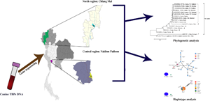
This study was conducted during October 2019 to September 2020 in the north and central regions of Thailand (Fig. 1). A total of 275 blood samples were collected from 200 dogs from Worldwide Veterinary Services (WVS) in Hang Dong district in Chiang Mai province and 75 dogs from dog Island Shelter in Phutthamonthon district in Nakhon Pathom province. The sample sizes were calculated to determine the appropriate number of samples from infinite population using the recipe based on the equation, n = t 2 × p(1 − p)/m 2, inserting the following values: number of animals to be sampled (n), the prevalence (p) of the canine tick-borne diseases among shelter dogs in Thailand, a 95% of confidence level (t) and 5% of margin of error (m). The prevalence of the canine tick-borne diseases in shelter dogs was calculated by the number of positive samples divided by the number of total samples multiplied by 100.

Fig. 1. Geographical location of Chiang Mai and Nakhon Pathom provinces where dog blood samples were obtained. Legends indicate the distribution of tick-borne pathogens (Hepatozoon canis, Anaplasma platys and Ehrlichia canis) Thailand strains identified in dogs from Hang Dong (HD) distinct in Chiang Mai province and Phutthamonthon (PM) district in Nakhon Pathom province.
Sample collection
Three mL of blood samples were collected directly from the cephalic or lateral saphenous veins of animals. After collection, half of the blood volume was transferred to sterile tubes with Ethylene Diamine Tetraacetic Acid (EDTA) (BD Vacutainer®, USA) as an anticoagulant and citrate salt to preserve the blood for PCR experiment. The remaining blood was transferred to clotted blood sterile tubes (BD Vacutainer®, USA) and then centrifuged at 5000 g at room temperature for 10 min and the serum samples were collected. All blood and serum samples were stored at −20°C for long-term preservation until further experiments. In addition, all steps of animal restraints and blood sample collection were proceeded by expert veterinarians.
DNA extraction
Genomic DNA of canine TBPs (H. canis, A. platys and E. canis) was extracted from 250 μL of dogs’ blood samples using a Tissue DNA Extraction Kit (OMEGA, bio-tex, USA) following the method of Watthanadirek et al. (Reference Watthanadirek, Chawengkirttikul, Poolsawat, Junsiri, Boonmekam, Reamtong and Anuracpreeda2019) and Junsiri et al. (Reference Junsiri, Watthanadirek, Poolsawat, Kaewmongkol, Jittapalapong, Chawengkirttikul and Anuracpreeda2020) with some modifications. Briefly, 250 μL of each blood sample was transferred to sterile microcentrifuge tubes. Twenty-five mL of OB Protease buffer and 250 μL of BL buffer were added. Then, samples were incubated at 70°C for 10 min, and 250 μL of absolute ethanol was added. Samples were transferred to HiBind® DNA Mini Column (Omega Bio-tek, USA) and centrifuged at maximum speed for 1 min. After removing the flow-through, 500 μL of HBC buffer was added and centrifuged at maximum speed at room temperature for 30 s. After discarding the flow-through, 700 μL of DNA washing buffer was added and centrifuged at maximum speed at room temperature for 30 s. Subsequently, the DNA samples were eluted in 50 μL MiliQ water and kept at −20°C until further use. Finally, the concentration and purity of DNA were defined with NanoDrop™ 2000 Spectrophotometers (Thermo Scientific™, USA) at the 260/280 and 260/230 ratios.
Molecular amplification and detection of canine tick-borne pathogens
All of the specific primer pairs designed from the H. canis 18S rRNA gene, A. platys 16S rRNA gene and E. canis 16S rRNA gene submitted in Genbank database under accession numbers MT107096.1, KY010669.1 and MT896774.1, respectively, were used to amplify DNA fragments of the rRNA genes. Hepatozoon canis 18S rRNA gene was amplified by single PCR using specific primer (HCF 5′ ATACATGAGCAAAATCTCAAC 3′ and HCR 5′ CTTATTATTCCATGCTGCAG 3′), while A. platys and E. canis 16S rRNA genes were amplified by nested PCR using 2 pairs of specific primers. In the first step of amplification, universal primers for rickettsia (F 5′ AGAACGAACGCTGGCGGCAAGCC 3′ and R 5′ CGTATTACCGCGGCTGCTGGCA 3′) were used. In the second step, the specific primers: PLATYS F 5′ TTTGTCGTAGCTTGCTATG 3′ and GA1UR 5′ GAGTTTGCCGGGACTTCTTCT 3′ were used for A. platys, and CANIS F 5′ CAATTATTTATAGCCTCTGGCTATAGG A 3′ and RHE3 5′ TATAGGTACCGTCATTATCTTCCCTAT 3′ were used for E. canis.
PCR reaction mixtures containing approximately 50 ng of DNA template, 0.2 μ m each of the primers, 200 μ m of each deoxynucleoside triphosphate (dNTPs), 1 × standard Taq reaction buffer, nuclease-free water and 1.25 U Taq DNA polymerase (BioLabs®, USA) were performed in a thermal cycler (Bio-Rad, USA) with the following conditions: 35 cycles of denaturation at 95°C for 45 s, annealing at 60°C and 63°C for the 1st and 2nd steps of A. platys, annealing at 60°C and 53°C for the 1st and 2nd steps of E. canis, annealing at 43°C of H. canis for 45 s, extension at 72°C for 90 s and a final extension at 72°C for 5 min. PCR products were analysed by 1.2% agarose gels stained with FluoroStain™ DNA Fluorescent Staining Dye (SMOBIO, Taiwan) and visualized under ultraviolet (UV) transilluminator.
Cloning of the rRNA genes from canine tick-borne pathogens DNA
Hepatozoon canis 18S rRNA gene, A. platys and E. canis 16S rRNA genes were cloned into vector with following specific primers: HCF 5′ CACCATACATGAGCAAAATCTCAAC 3′ and HCR 5′ CTTATTATTCCATGCTGCAG 3′ for H. canis, APF 5′CACCTTTGTCGTAGCTTGCTATG 3′ and APR 5′ GAGTTTGCCGGGACTTCTTCT 3′ for A. platys as well as ECF 5′ CACCCAATTATTTATAGCCTCTGGCTA 3′ and ECR 5′ TATAGGTACCGTCATTATCTTCCCTAT 3′ for E. canis. The 4 nucleotides (CACC) were added at 5′ end of forward primer with the overhang sequence (GTGG) in pET100/D-TOPO® vector (Invitrogen, USA) to empower directional cloning. The PCR reaction was performed with the following conditions as described previously in section ‘Molecular amplification and detection of canine tick-borne pathogens’. A 100 bp DNA Ladder M (MolBio™ HIMEDIA®, India) was used as a standard for defining the molecular mass of PCR products. Positive PCR products were purified using GenepHlow™ Gel/PCR Kit (Geneaid, Taiwan) following the manufacturer's instructions for cloning. The 20 ng of the blunt-end PCR product was used as an insert in the pET100/D-TOPO® vector (Invitrogen Life Technologies, USA). Chemically competent Escherichia coli host strain TOP10 cells (Invitrogen, USA) was then transformed with the ligation product. Subsequently, 200 μL of transformed bacterial culture was spread on the agar plates containing 100 μg of ampicillin and incubated at 37°C for overnight. Then, the positive clones were selected and grown in Luria Bertani (LB) medium containing ampicillin overnight. Finally, the plasmid extraction was performed by Presto™ Mini Plasmid Kit (Geneaid, Taiwan) following the manufacturer's instructions and analysed for precisely sized inserts by agarose gel electrophoresis.
Sequence analysis
Purified PCR products were approved by Sanger sequencing. All sequences were analysed by BLAST (The National Center for Biotechnology Information, NCBI, http://www.ncbi.nlm.nih.gov/ BLAST). All DNA sequences were submitted and deposited in the GenBank database (Table 1).
Table 1. The tick-born pathogens rRNA nucleotide sequences amplified in Thailand strain were deposited in the GenBank database

Phylogenetic analysis
The sequences of H. canis 18S rRNA gene, A. platys and E. canis 16S rRNA genes were aligned with Clustal W algorithm and genetic inference was analysed by neighbour-joining (NJ) phylogenetic tree with a small number of gap-free positions using MEGA software version 7.0.26 (Saitou and Nei, Reference Saitou and Nei1987; Kumar et al., Reference Kumar, Stecher and Tamura2016). Bootstrap analysis with 1000 repetitions was used to assess the confidence of the branching pattern of the trees (Felsenstein, Reference Felsenstein1985). The evolutionary distances were computed using the Kimura 2-parameter method (Kimura, Reference Kimura1980). The similarity was defined with the pairwise-distance method (Nei and Kumar, Reference Nei and Kumar2000).
Haplotype diversity
The obtained alignment of the E. canis and A. platys 16S rRNA gene sequences was used to evaluate the nucleotide diversity (π), diversity of haplotypes (Dh), number of haplotypes and the average number of nucleotide differences (K), using the DnaSP version 6.0 software (Librado and Rozas, Reference Librado and Rozas2009). In addition, all nucleotide sequences were submitted to the Population Analysis with the Reticulate Trees (popART) program (Leigh and Bryant, Reference Leigh and Bryant2015) for analysis of the TCS Network construction.
Results
Occurrence of tick-borne pathogen infections in canine blood samples
The TBPs detected by PCR were shown in Table 2. The sizes of PCR products of H. canis, A. platys and E. canis were 633, 373 and 356 bp, respectively. The representative positive PCR products for each of the pathogens examined were shown in Fig. 2. The PCR results exhibited that 71/275 (25.82%) of shelter dog blood samples were infected with TBPs. Of the 200 blood samples from Chiang Mai province, A. platys (17.0%) was the most prevalent single infection, followed by E. canis (5.0%) and H. canis (2.0%). In addition, 75 blood samples from Nakhon Pathom province, A. platys (14.67%) and E. canis (14.67%) were the most prevalent single infection, followed by H. canis (1.33%).

Fig. 2. PCR assay showing H. canis-infected sample (lane 1), H. canis-uninfected sample (lane 2), A. platys-infected sample (lane 3), A. platys-uninfected sample (lane 4), E. canis-infected sample (lane 5) and E. canis-uninfected sample (lane 6).
Table 2. Summary of tick-borne pathogen infections in dogs from the North and Central regions as analysed by PCR using the specific primers for rRNA genes

Phylogenetic and similarity analysis of tick-borne pathogen rRNA gene sequences
The phylogenetic tree based on the alignment of the 2 sequences of H. canis 18s rRNA gene obtained in this work with 9 other sequences obtained from the GenBank was classified as 8 clades. The sequences detected in this study were positioned in 1st clade and showed phylogenetic proximity. Clades 2–4, 6 and 7 were composed of the canine sequences from Malta, Nigeria, Malaysia, China and Iraq, while clades 5 and 6 comprised the sequences of feline and Pampas fox hosts from Brazil, respectively. The last clade was found to be a sequence observed in coyote host from the USA (Fig. 3). The similarity of 1st clade was 99.9–100% among the Thailand H. canis sequences, while the similarity of the sequences within other clades was 99.8–100% (2nd clade), 97.6–100% (3rd clade), 97.5–100% (4th clade), 98.2–100% (5th clade), 95.2–100% (6th clade), 94.0–100% (7th clade) and 95.1–100% (8th clade) (Table 3). The nucleic acid substitution rate in 18S rRNA sequences among H. canis was evaluated under the Tamura and Nei (Reference Tamura and Nei1993) mode as demonstrated in Table 4.

Fig. 3. Phylogenetic analysis of Hepatozoon canis 18S rRNA gene sequences using the neighbour-joining method. The numbers on each node correspond to the bootstrap analysis of 1000 replicates. The sequences amplified in this study are highlighted in boldface type letters. The GenBank accession numbers of the sequences used in the phylogenetic analysis are also shown. One gene sequence of the other strain was used as an outer group.
Table 3. Similarity of the H. canis 18S rRNA gene sequences as examined in canine samples in Thailand and other countries

Table 4. The nucleic acid substitution rate in H. canis 18S rRNA, A. platys and E. canis 16S rRNA sequences

Each entry is the probability of substitution (r) from one base (row) to another base (column). Rates of different transitional substitutions are shown in bold and those of transversional substitutions are shown in italics. The maximum Log likelihood for this computation was −3176.748, −2912.336 and −2614.490, respectively.
The A. platys 16S rRNA gene sequences were divided into 9 clades in the phylogram. The sequences assigned to 1st to 6th clade exhibited phylogenetic proximity representing the genetic variability of A. platys 16S rRNA sequence from Chiang Mai and Nakhon Pathom provinces of Thailand, while some sequence of cattle host from the USA was classified to clade 5. A sequence from the USA detected in white tail deer was positioned in clade 7, while two canine sequences from India and China were found in 8th and 9th clade, respectively (Fig. 4). The similarity of the Thailand A. platys sequences from 1st to 6th clades were 96.3–100%. The similarity of the sequences within other clades was 96.6–100% (7th clade), 96.2–100% (8th clade) and 96.4–100% (9th clade) as shown in Table 5. The nucleic acid substitution rate in 16S rRNA sequences among A. platys was estimated under the Tamura and Nei (Reference Tamura and Nei1993) mode (Table 4).

Fig. 4. Phylogenetic analysis of Anaplasma platys 16S rRNA gene sequences using the neighbour-joining method. The numbers on each node correspond to the bootstrap analysis of 1000 replicates. The sequences amplified in this study are highlighted in boldface type letters. The GenBank accession numbers of the sequences used in the phylogenetic analysis are also shown. Two gene sequences of the other strains were used as outer groups.
Table 5. Similarity of the A. platys 16S rRNA gene sequences as examined in canine sampled in Thailand and other countries.

Ehrlichia canis 16S rRNA gene sequences obtained in this study were divided into 6 clusters. Seven Thailand 16S rRNA sequences were detected in 1st clade together with sequences from Nigeria, Brazil and India. Four sequences of the canine host from China, Malaysia, Greece and Taiwan were found in clades 2, 4, 5 and 6, respectively, while 2nd and 7th clade were found to be the sequences detected in deer and feline hosts, respectively (Fig. 5). The total similarity among Thailand E. canis sequences was 98.3–100% (1st clade). The per cent similarity of the sequences within each clade was 94.0–100% (2nd clade), 95.3–100% (3rd clade), 95.3–100% (4th clade), 95.3–100% (5th clade), 95.3–100% (6th clade) and 95.3–100% (7th clade) (Table 6). The nucleic acid substitution rate in 16S rRNA sequences among E. canis was detected under Tamura and Nei (Reference Tamura and Nei1993) mode as shown in Table 4.

Fig. 5. Phylogenetic analysis of Ehrlichia canis 16S rRNA gene sequences using the neighbour-joining method. The numbers on each node correspond to the bootstrap analysis of 1000 replicates. The sequences amplified in this study are highlighted in boldface type letters. The GenBank accession numbers of the sequences used in the phylogenetic analysis are also shown. One gene sequence of the other strain was used as an outer group.
Table 6. Similarity of the E. canis 16S rRNA gene sequences as examined in canine sampled in Thailand and other countries

Haplotype diversity
The analysis of haplotype diversity based on 16S rRNA gene sequences of A. platys and E.canis found in Chiang Mai and Nakhon Pathom provinces of Thailand was diverse when compared to worldwide sequences. The haplotype networks of this gene were obtained from the TCS Network tool (Fig. 6). For haplotype analysis of A. platys 16S rRNA gene, 12 haplotypes shown in TCS network exhibited that haplotypes #1 to #10 were detected in canine in Chiang Mai and Nakhon Pathom provinces. The rest of the haplotypes was found in canine, cattle and deer hosts from other countries worldwide as shown in Table 7 and Fig. 6A. The haplotype network of E. canis 16S rRNA gene showed that all of the sequences of canine hosts from Thailand and other countries were detected in haplotype #1, while one sequence of feline host from Portugal was classified as haplotype #2 (Table 7 and Fig. 6B).

Fig. 6. TCS network of haplotypes based on the 16SsRNA gene sequences of A. platys (A) and E. canis (B) examined in Thailand and worldwide (small traits between a haplotype and another stand for mutational occurrence).
Table 7. Polymorphism and genetic diversity of A. platys and E. canis 16S rRNA gene sequences in canine samples in Thailand and other countries

N, number of analysed sequence; VS, number of variable sites; GC, G × C content; h, number of haplotypes; Dh, diversity of haplotypes; s.d., standard deviation; π, nucleotide diversity (per site); K, average number of nucleotide differences.
Discussion
Canine tick-borne diseases including hepatozoonosis, anaplasmosis and ehrlichiosis are important diseases that cause clinical infections in dogs in many areas of the globe, depending on the widespread distribution of the vector ticks (R. sanguineus). There are several conventional methods for the detection of TBPs in dogs, such as not pathognomonic, haematological findings, blood smear examination, serological test and molecular PCR tests. Molecular assay, i.e. PCR, is a sensitive diagnostic tool and required for the detection and characterization of TBPs.
In the present study, A. platys (16.36%) was the most common canine tick-borne pathogen. The positive animals in this study were similar to the 11% (17/157) reported in dogs in the USA (Kelly et al., Reference Kelly, Xu, Lucas, Loftis, Abete, Zeoli, Stevens, Jaegersen, Ackerson, Gessner, Kaltenboeck and Wang2013), and were lower than those investigated by previous works; for example, 28% (42/150) in Pakistan (Malik et al., Reference Malik, Qamar, Ain, Hussain, Dahmani, Ayaz, Mahmood, Davoust, Shaikh and Iqbal2018) and 12.12% (36/297) in China (Mengfan et al., Reference Mengfan, Lixia, Ying, Yan, Kuojun, Jinsheng, Zaichao, Weiwei, Yelong, Xuepeng, Chongyang, Jun and Qingling2020). In Thailand, our results have exhibited that the occurrences of canine TBP infections of dogs were similar to those reported by previous studies. For instance, Rucksaken et al. (Reference Rucksaken, Maneeruttanarungroj, Maswanna, Sussadee and Kanbutra2019) showed that H. canis, A. platys and E. canis infections in dogs from Buriram province were 4.08% (2/49), 30.6% (15/49) and 36.73% (18/49), respectively. In addition, the occurrences of A. platys and E. canis infections in dog from Kalasin province were reported to be 29.4% (20/68) and 25% (17/68), respectively (Piratae et al., Reference Piratae, Senawong, Chalermchat, Harnarsa and Sae-Chue2019). By contrast, in Songkha province, H. canis, A. platys and E. canis were found in 18.8% (34/181), 4.4% (80/181) and 3.9% (7/181) of sampled dogs, respectively (Liu et al., Reference Liu, Ruttayaporn, Saechan, Jirapattharasate, Moumouni, Cao, Inpankaew, Ybañez, Suzuki and Xuan2016). Our study is the first report that showed a molecular occurrence of canine tick-borne infections in shelter dogs in Chiang Mai and Nakhon Pathom provinces. Shelter dogs usually receive less, especially anti-tick treatments medical care, which is a concern because they are vector-borne pathogens. In addition, the season and the level of tick infestation may impact the rate of infection by TBPs.
The genetic diversity of H. canis, A. platys and E. canis strains based on the sequences of 18S and 16S rRNA genes has been investigated in many countries (Hsieh et al., Reference Hsieh, Lee, Tsang and Chung2010; Aktas et al., Reference Aktas, Özübek, Altay, Ipek, Balkaya, Utuk, Kırbas, Şimsek and Dumanlı2015; Zhang et al., Reference Zhang, Kelly, Guo, Xu, Wei, Jongejan, Loftis and Wang2015). Even though several genes have been used to detect H. canis, A. platys and E. canis in Thailand, i.e. 18S rRNA, 16S rRNA and gltA (Suksawat et al., Reference Suksawat, Xuejie, Hancock, Hegarty, Nilkumhang and Breitschwerdt2001; Piratae et al., Reference Piratae, Pimpjong, Vaisusuk and Chatan2015; Liu et al., Reference Liu, Ruttayaporn, Saechan, Jirapattharasate, Moumouni, Cao, Inpankaew, Ybañez, Suzuki and Xuan2016), little is known concerning the diversity of H. canis, A. platys and E. canis Thailand strains. In this study, the 18S and 16S rRNA genes in dog population sampled in the north and central areas of Thailand were employed to discriminate the genetic diversity of H. canis, A. platys and E. canis in these regions. The phylogenetic analysis of canine H. canis 18S rRNA gene and E. canis 16S rRNA gene Thailand isolates revealed only 1 clade, while canine A. platys 16S rRNA gene Thailand strain revealed 6 clades. Our findings exhibited that the genetic diversity observed in the phylogram was confirmed by the high similarity value for H. canis 18S rRNA gene (99.9–100%), A. platys 16S rRNA gene (96.3–100%) and E. canis 16S rRNA gene (98.3–100%).
In the present work, the 16S rRNA gene sequences in blood samples of dogs in the north and central areas of Thailand were investigated. The results exhibited that the population of E. canis was highly conserved in Thailand, while A. platys was more diverse, with the presence of probably more than one obvious haplotype. The genotypes of this gene were identified in the haplotype networks. They were carried out with the sequences observed in this study together with other host sequences from GenBank database that was found in various regions of the globe. The result indicated that there was some genetic diversity in 16S rRNA genes observed in the different haplotype networks in Thailand and worldwide when compared to other sequences. Furthermore, these sequences shared genetic traits with all sequences detected previously. On the contrary, E. canis 16S rRNA gene sequence was allocated in one haplotype.
Conclusions
This work is the first report indicating a molecular occurrence of TBP infections (H. canis, A. platys and E. canis) in canine blood samples in both north and central regions of Thailand. Our findings showed that H. canis 18S rRNA and E. canis 16S rRNA genes are conserved, while A. platys 16S rRNA gene is genetically diverse. These results could be used to ameliorate the insight of phylogeny and genetic diversity among 18S rRNA and 16S rRNA genes of TBP Thailand strain. Hence, the periodical appraisal of the occurrence of the canine tick-borne diseases is a necessitate to monitor the affectivity of the effective prevention and control strategies throughout the country to mitigate the infections of canine vector-borne pathogens.
Acknowledgements
We are grateful to Sutthida Minsakorn, Parasitology Research Laboratory (PRL), Institute of Molecular Biosciences, Mahidol University, for providing some equipment.
Author contributions
Napassorn Poolsawat: conceptualization, methodology, validation, investigation, writing original draft, visualization, project administration. Keiichiro Tazawa, Witchuta Junsiri, Amaya Watthanadirek, Nitipon Srionrod: Resources, Runglawan Chawengkirttikul: resources. Panat Anuracpreeda: conceptualization, methodology, validation, investigation, data curation, writing – review and editing, visualization, supervision, project administration, funding acquisition.
Financial support
This work was financially supported by Research Grants from National Research Council of Thailand (NRCT) to Panat Anuracpreeda, and Royal Golden Jubilee Ph.D. (RGJ-PHD) Scholarship [grant number PHD/0055/2561] to Napassorn Poolsawat.
Conflict of interest
None.
Ethical standards
All experimental procedures involving animals were approved by the Animal Care and Use Committee (IMBMU-ACUC), Institute of Molecular Biosciences, Mahidol University, Thailand.


