INTRODUCTION
Viscoelastic gels are a common form of strong, temporary adhesive used by invertebrates (Smith, 2002), including limpets (Smith, Quick & Peter, 1999), periwinkles (Smith & Morin, 2002), echinoderms (Flammang, 1996) and a variety of worms (Hermans, 1983; Whittington & Cribb, 2001). Knowledge of glues in parasitic flatworms is limited by the small quantities of adhesive produced. A considerable body of data is available on the morphology of the secretions involved (Whittington & Cribb, 2001) and preliminary chemical characterization has been achieved for a variety of monopisthocotylean monogenean flatworms (Hamwood et al. 2002), showing that the adhesive has some similar characteristics to other invertebrate groups. Little is known, however, about the actual mechanism of adhesion and detachment in Monogenea.
Monopisthocotylean monogeneans are small ectoparasites principally of teleosts and elasmobranchs. Temporary adhesion in this group is referred to as tissue adhesion because it involves attachment to the epithelium of a living host, but attachment may also be induced on artificial surfaces such as glass (Whittington & Cribb, 2001). Although considerable differences in the morphology of the anterior adhesive region and secretions have been found across monopisthocotylean Monogenea (see Whittington & Cribb, 2001), the mechanism of attachment has only been addressed in detail for one species, the capsalid Entobdella soleae (see Kearn & Evans-Gowing, 1998). Morphology of the secretions in this species differs from the arrangement in other monogeneans since there is no electron-dense spheroidal secretion paired with rod-shaped bodies (see Whittington & Cribb, 2001) and, therefore, it is unwise to extrapolate this arrangement to other species. Furthermore, a number of questions remain unanswered after the study on E. soleae. Detachment in E. soleae is still not fully understood (Kearn & Evans-Gowing, 1998). Also, the mechanism within the gel-like adhesive that achieves adhesion is not yet appreciated. Gels are composed of a dilute polymer network and can be held together either through entanglement of molecules (as in the giant molecules of mammalian mucus) or by cross-linking of molecules that are usually shorter (such as in agar and gelatin) (Smith, 2002). Although some description of monogenean adhesives has been provided (Kearn & Evans-Gowing, 1998; Hamwood et al. 2002), details of the type of bonding are still unavailable. Here, we investigate adhesion and detachment in Merizocotyle icopae (Monocotylidae), a species that differs markedly from E. soleae in terms of the morphology of the anterior adhesive regions, secretion morphology and host species.
MATERIALS AND METHODS
Shovelnose rays, Rhinobatos typus (Rhinobatidae), were obtained from Heron Island (23°27′S, 151°55′E) and Moreton Bay adjacent to Dunwich (27°30′S, 153°25′E), Queensland, Australia. The nasal tissue was excised and live adult Merizocotyle icopae were removed for observation, manipulation and fixation. Oncomiracidia were obtained from eggs laid by adults left in Petri dishes containing filtered seawater. Eggs were incubated at 25 °C in a LD 12[ratio ]12 illumination regime until hatching occurred using methods outlined by Chisholm & Whittington (2000). Excised epidermis from the nasal fossae of 2 R. typus and 6 oncomiracidia were preserved for electron microscopy. Samples for transmission electron microscopy (TEM) were fixed using Protocol 1, sectioned, stained and viewed following Cribb, Armstrong & Whittington (2004). Thirty adult M. icopae were preserved at different stages of adhesion and detachment by timing application of fixative to coincide with the required attachment behaviour when under observation with a stereo-dissecting microscope in a fume hood. A glass substrate replaced host tissue as the site for attachment since specimens attached readily to glass and its use aided the manipulation of specimens. Although adult M. icopae temporarily attach by the anterior end in normal movement across a substrate, anterior attachment is usually too brief to allow application of fixative before detachment has been initiated. However, by lifting the posterior attachment organ (haptor) with a fine needle, adults could be induced to attach by the anterior end for longer. Specimens were preserved when in the following phases: attached by the haptor with the body unextended; attached by the haptor with the body extended, searching the substrate with the anterior end (a prelude to anterior attachment); haptor unattached and anterior end attached to the glass substrate; and newly detached at the anterior end but reattached by the haptor. After fixation, attached specimens were peeled away carefully from the substrate. Fixation, sample processing and observation procedures for adult M. icopae follow Protocols 1, 2 and 3 as described by Cribb et al. (2004). Briefly, fixation protocols used glutaraldehyde in cacodylate buffer followed by osmium fixation (Protocol 1) or osmium tetroxide and glutaraldehyde applied simultaneously (Protocols 2 and 3), after which samples were prepared for scanning (SEM) and TEM. Data are presented as the mean±standard error of the mean with the number of samples in parentheses, unless otherwise stated.
Since a separate study (Whittington et al. 2004) describes, compares and statistically analyses anterior secretions in larval and adult M. icopae, these details are not repeated here, but are referred to where necessary in summary form (see Table 1).
Table 1. Summary of data on types of anterior adhesive region and secretions from Merizocotyle icopae and Entobdella soleae
(ED, electron dense; EL, electron lucent.)
RESULTS
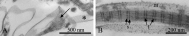
The nasal tissue of R. typus is the natural surface to which M. icopae attach. While some ciliated structures are encountered, it generally conforms to a uniform structure of small protrusions (Fig. 1).
Fig. 1. Transmission electron micrograph of the epidermis of a nasal fossa of the Shovelnose ray Rhinobatos typus. Arrows indicate the surface to which the monogenean, Merizocotyle icopae, attaches.
In adult M. icopae, the process of anterior attachment involved changes to the 3 pairs of ventrolateral apertures. The sequence involved opening and closing of the apertures as well as retraction and eversion of the duct endings that terminate within these apertures. These morphological changes related to the behavioural phase under study (Fig. 2A–H; Table 2). Changes were also seen in the morphology of the anterior ‘horns’, on which were found numerous putatively sensory, ciliated sensilla (Table 2). In live specimens observed using a stereomicroscope, these horns were observed to contact the substrate before anterior attachment occurred.
Fig. 2. (A–H) Scanning electron micrographs of the anteroventral region of adult Merizocotyle icopae. White arrows indicate putative sensory ‘horns’; black arrowheads point to 3 of the 6 adhesive apertures; d, duct endings; a, adhesive; r, aperture rim. (A) Specimen unattached at anterior end. (B) Higher magnification of unattached aperture with rim closed. (C) Specimen with anterior end elongated in searching mode. Note the extended sensory ‘horns’. (D) Higher magnification of aperture from searching specimen showing partially open aperture rim and duct endings free of adhesive. (E) Specimen attached by 4 of the 6 anterior apertures: two apertures are folded over (f) with adhesive exuded at the edges and 2 apertures have recently begun detachment (asterisks). (F) Higher magnification of adhering aperture showing the rim at the same level as the everted duct endings and adhesive mat obscuring many duct endings: some duct endings lie horizontally. (G) Specimen shortly after anterior detachment. (H) Higher magnification of an anterior aperture that has just detached, showing strands of adhesive over vertical duct endings which now extend beyond the aperture rim.
Separate ducts within the anterior apertures carry 3 different secretion types (Whittington et al. 2004: Table 1). In detached and searching worms, all secretions had an electron-dense appearance (Fig. 3A,B), but in attached worms the large spheroidal S3 secretion was often swollen and had lost electron-density in the duct endings (Fig. 3C). The rod-shaped secretion was more abundant than either of the 2 spheroidal secretion types, but ducts carrying the smaller spheroidal secretion outnumbered those carrying the larger secretions in most samples across all behavioural phases. During the processes of searching, attachment and detachment among specimens, the number of full ducts varied, indicating a sequence of filling and emptying (Table 3; Fig. 3A,C). In calculating percentages of filled ducts, data for the spheroidal secretions (S2 and S3) were pooled because variability was high and abundance of type did not correlate with behavioural phase.
Fig. 3. (A–H) Electron micrographs of anterior adhesive apertures and secreted adhesive from adult Merizocotyle icopae. (A) Duct endings (d) in a searching individual, showing that all ducts of each of 3 types of secretory bodies (S1, S2 and S3) are filled. (B) Duct endings in a searching individual, shown at higher magnification, detailing small spheroidal secretory bodies (S2) and rod-shaped bodies (S1). (C) Duct endings in an individual attached by anterior adhesive showing swelling of large spheroidal secretory bodies (S3) and empty ducts (asterisks) as well as ducts containing S1 and S2 secretions. (D) Adhesive matrix (asterisk) at duct endings in an attached individual. (E) Adhesive matrix showing few banded fibres (b) and multiple, narrower fibres (arrows) as well as electron-dense clusters (c) and vesicles (v). (F) Adhesive matrix showing multiple banded fibres (b) and fewer narrow fibres (arrows) as well as electron-dense clusters (c) and S1 membranes (arrowheads). (G) Scanning electron micrograph (SEM) of adhesive matrix covering duct endings and showing patches of homogeneous material (arrows) and microvilli (m). (H) SEM of adhesive matrix over duct endings covered with microvilli (m), showing swollen S1 bodies and S1 membranes (asterisks) and banded fibres (b) with spheroidal clusters (c) attached.
A dense matrix overlayed the duct endings of adult M. icopae in the attached phase. This is the anterior adhesive (Fig. 3D). Within it, a number of structural components could be seen: S1 body membranes, clusters of banded electron-dense fibres, multi-directional narrower fibres without banding, electron-dense clusters and vesicles. The quantities and distribution of these components differed between regions (Fig. 3E,F). The S1 membranes, fibres and electron-dense clusters could also be seen using SEM (Fig. 3G,H). Banded fibres appeared to be coalesced bundles that aligned in such a way as to produce distinct patterning of 2 types (Fig. 4A). There was a large (84±1 nm (45)) and a small (21±0·3 nm (46)) banding periodicity (Fig. 4B). Measurements of the periodicity varied to a greater extent between different bundles of fibres than within fibres. For 10 bundles of fibres, the large banding varied from 77±1 nm (5) to 92±1 nm (10), but the small banding varied less with mean values per fibre from 20±0·4 nm (10) to 24±1 nm (5). The length of the fibre bundles was variable and measurement was constrained by the angle at which they were sectioned but attained a maximum of 1·2 μm (664±78 nm (15) (range 0·2–1·2 μm)). Narrow fibres showed no banding and a shorter maximum length of 335 nm (190±14 nm (15) (range 118–335 nm)). The electron-dense clusters (Fig. 3F) had a diameter of 62±2 nm (15) (range 52–68 nm).
Fig. 4. (A,B) Transmission electron micrographs of banded fibres. (A) Banded fibre showing separate strands splayed at one end (arrow): note adjacent S1 membrane with nano-banding (asterisk). (B) Banded fibre in S1 membrane (m) showing large (l) and small (s) periodicity.
Fibres and electron-dense clusters originate from the rod-shaped S1 bodies. There was a progression that could be followed morphologically. Within the duct, the S1 bodies were electron-dense and showed a nano-banding (Fig. 5A). Upon secretion, the S1 bodies swelled, lost their nano-banding and their inner contents became less electron dense (Fig. 5B) and fibrous (Fig. 5C). The fibrous contents left the S1 body, presumably by traversing the bounding membrane as well as through any apical or basal opening (Fig. 5C). Some fibres remained within the S1 body membrane and aligned to form banded structures whereas others formed banded fibres outside the membranes (Fig. 5D). Alternatively, depleted material within the S1 membrane appeared as small electron-dense clusters with the same periodicity as the large banding (Fig. 5D). These electron-dense clusters also occurred outside the S1 membranes, either as single bodies or as chains (Fig. 5D), both associated with S1 membranes and fibres (Fig. 3E,F). The clusters were noticeable on the surface of the banded fibres when observed with SEM (Fig. 3H).
Fig. 5. (A–D) Transmission electron micrographs of progression of S1 secretory bodies and banded fibres during adhesion in adult Merizocotyle icopae. (A) S1 body in a duct ending showing nano-banding (arrows). (B) Secreted S1 body showing swelling and internal loss of density. (C) Fibres from within the S1 body shown dispersing (arrow). (D) Banded fibres (b) and chains of electron-dense clusters (c) both within and outside S1 membranes.
When the worms detached themselves from the glass substrate, the majority of the adhesive matrix remained on the substrate and duct endings were covered by a thin layer of newly secreted adhesive material (Fig. 2H). This adhesive was rapidly lost (Fig. 2G), so that apertures prior to attachment showed negligible or no adhesive (Fig. 2D). Sections obtained from newly detached worms showed ribbons or clumps of adhesive matrix. This was diffuse and often contained vesicles and cytoplasmic material from the S1 ducts and, occasionally, recognizable S3 secretion (Fig. 6A). Ribbons extending from duct endings showed a progressive increase in density further from the body with fewer banded fibres close to the duct openings (Fig. 6B).
Fig. 6. (A,B) Transmission electron micrographs of anterior adhesive region during detachment of adult Merizocotyle icopae: d, duct endings; m, microvilli; t, tegument surrounding duct endings. (A) Secretion of S1 and S3 bodies as the ducts are pulled away from the overlying adhesive matrix. (B) Strand of adhesive matrix flowing away from a duct ending (d) showing a change in structure along the material.
Oncomiracidia (the free-swimming larval stage that must initially attach to the host) also possessed anterior secretory bodies in discrete ducts (Fig. 7A) which opened through a single pair of anterior apertures (Whittington et al. 2004: Table 1). While 3 secretory types were present, there was some difference in morphology from the adult: the large spheroidal type (S4) was larger than the adult S3 type, more elongate and less electron-dense; the small spheroidal type (S2) was also less electron dense than the adult S2 type (Whittington et al. 2004). However, the S1 bodies appeared similar. A matrix appeared to be formed from the extrusion of these larval secretions. Some specimens that were preserved showed a small amount of adhesive across the duct endings despite being fixed unattached to a substrate. Short individual fibres (82±2·7 nm (21)) as well as S1 membranes could be seen within this matrix, but no banded fibres were visible (Fig. 7B).
Fig. 7. (A,B) Transmission electron micrographs of anterior adhesive aperture and adhesive in the oncomiracidium of Merizocotyle icopae. (A) Anterior aperture (arrow) and 3 types of secretory bodies (S1, S2, S4) contained in ducts which exit into the aperture. (B) Anterior adhesive showing S1 membranes (m), short fibres without banding (arrows) and transverse sections of locomotory cilia (asterisks).
DISCUSSION
This study is the first to compare extruded anterior adhesive from the larva and from the adult of a monogenean parasite. It is the oncomiracidium that must locate and then attach to the specific host fish whereas the adult remains with the host for the duration of its life-span (Whittington, Chisholm & Rohde, 2000). Monogeneans are usually strictly host specific and initial and then continuing attachment may provoke an immunological response of some sort by the host (Buchmann, 1999). Although host specificity is not fully explained, an association between host mucus and the anterior adhesives of monogeneans may play a role (Whittington et al. 2000). It is interesting, therefore, that the oncomiracidial and adult anterior adhesive secretory types (Whittington et al. 2004) and the secreted anterior adhesive matrix (present study) of M. icopae differ in morphology. These differences may reflect a change in chemistry that perhaps enables the parasite to be tolerated by the host, and this deserves further investigation.
The mechanism of adhesion was studied in detail for adults of M. icopae. The adult adhesive is synthesized from the 3 secretory types extruded into the anterior apertures. All secretory types are present before attachment occurs. Since no mechanism was identified that would allow individual ducts to control the flow of secretion, it appears that all secretory types exit together and mix in the ratio present to produce the final adhesive matrix. This process depletes duct contents, with the larger S1 ducts releasing more contents than ducts containing spheroidal secretions. Detachment involves elongation of the duct endings and secretion of additional secretory bodies as the worm pulls away from the substrate. Due to the uneven depletion of duct contents, there is a shift in the composition of secretion upon detachment where a proportionately greater concentration of spheroidal secretion occurs. Also, some S3 secretion is seen to swell before release. This change in ratio may modify the properties (physical and/or chemical) of the secreted matrix, resulting in a material that is easier for the worm to detach from than that which forms the initial matrix. After detachment from a substrate, body movements are likely to contribute to the mechanism for refilling duct contents, which are seen to be refilled rapidly before the next act of attachment. This is the first study to present evidence that eversion and retraction of duct endings within the anterior adhesive apertures is part of the mechanism of anterior attachment and detachment in monocotylid monogeneans.
The adhesive mechanism of only one other monogenean species has been investigated in detail: Kearn & Evans-Gowing (1998) studied the capsalid monogenean, Entobdella soleae (see Table 1 for summary of secretory bodies). Its anterior attachment mechanism differs from that reported here for M. icopae in a number of respects. In E. soleae there is evidence that the electron-lucent spheroidal secretion (absent in M. icopae) spreads over the flat, pad-like anteroventral adhesive regions much as a fluid would, before the rod-shaped secretion intermingles with it to form the adhesive. However, the bulk of the adhesive appears to be composed of rod-shaped secretion and its membranes. Since the spheroidal secretion is depleted after attachment, this is unlikely to play a role in detachment of E. soleae. Instead, the authors suggested that detachment may involve secretion from the tegument. In contrast, detachment of M. icopae appears to rely on mechanical action coupled with possible changes in physical and/or chemical properties resulting from a shift in the ratio of secretions present. No tegumental secretions appear to be involved. Such variation between capsalids and monocotylids indicates that the adhesive mechanism may be taxon specific, at least at the level of family, within the Monogenea. Differences are already known in the general morphology of the anterior adhesive areas as well as in the anterior secretions associated with the adhesive areas (Whittington & Cribb, 2001).
The ultrastructural appearance of the final adhesive matrix or gel also differs between M. icopae and E. soleae. No banded fibres were observed in secreted adhesive of E. soleae (see Kearn & Evans-Gowing, 1998). However, in other respects, the appearances were similar, with numerous membranes from the S1 bodies present as well as fibres and an electron-dense finely grained matrix. Although fibrous components are common to such gels, banding of fibres within the adhesive has not been noted for other invertebrate systems (Flammang, 1996; Rieger et al. 1991; Whittington & Cribb, 2001). However, the banding reported here is reminiscent of one protein that is well known for its polymer networks and tensile strength. Collagen monomers form native-type fibrils with a banding periodicity of about 67 nm (slightly shorter than the 82 nm periodicity seen in M. icopae) that are visible using TEM; the dark bands apparently represent periodic protrusions or thickenings of the fibrils as determined using atomic force microscopy (Lin & Goh, 2002). Like collagen, the banded fibres reported in the adhesive matrix of adult M. icopae are collections of narrower fibres. It is likely that the banding represents a similar alignment of component parts. Also, single fibres (maximum length 335 nm) from the anterior adhesive of adult M. icopae appear to encompass the size of collagen fibres in their monomeric form (300 nm). From the images we present, it is not possible to discern individual molecules, but this might be achieved with atomic force microscopy since it is possible to study biological molecules in buffer solutions (Radmacher et al. 1992).
The adhesive matrix found in oncomiracidia of M. icopae does not show banding. Instead, short fibres are present, having the same length as the large banding periodicity seen in adult samples. Since S1 secretory bodies are morphologically the same in the oncomiracidium but adult and spheroidal secretions differ (Whittington et al. 2004), it can be concluded that contents of the adult spheroidal secretions may control the process involved in producing banded fibres and may even control cross-linking within the matrix. Determination of their composition would provide a greater understanding of the process. Although adhesive composition has only been studied for E. soleae adults, it is noteworthy that the oncomiracidia of this species also show a difference in secretory morphology from that seen in the adult system. Molecular length and branching of molecules can be important in determining the characteristics of an adhesive gel (Smith, 2002). Length and branching determine the degree of entanglement of proteins and modifications in the concentration of components, ionic environments, pH, and possibly even the types of proteins present can alter the nature of the adhesive (Smith, 2002). Smith et al. (1999) have shown that the gliding mucus and attachment mucus of a species of limpet differ in protein composition and Smith & Morin (2002) found similar differences in the marsh periwinkle indicating that the mixing of different secretions may lead to changes in the adhesive properties of the same basic gel. It appears that the components in the adhesive of M. icopae may function in a similar way. However, it is worth noting that while the viscoelastic gels reported in other invertebrate groups are generally composed of proteins and carbohydrates (Smith, 2002), there is no evidence yet for carbohydrates in the adhesive secretions of M. icopae and other Monogenea (see Hamwood et al. 2002).
The present study investigated ultrastructure of adhesion to glass surfaces rather than host tissue, but it would also be valuable to preserve specimens attached to host tissue to ensure conservation of detail in worms attached to their natural substrate. Observations on live worms (unpublished) did not suggest a difference in behaviour during adhesion and detachment from that seen on glass but, without careful handling, the parasite becomes detached from the host tissue after fixation. Application of fixative to most flatworms usually causes an immediate and profound contractile response. However, we avoided this response in M. icopae selected for ultrastructural study by specific manipulation and fixation regimes and were able to obtain individuals that remained attached to glass surfaces during and after fixation (see Cribb et al. 2004). It is possible that some fixation regimes result in a degradation of the bond between the host tissue and adhesive matrix. This needs further study.
We thank the Directors of the Centre for Microscopy and Microanalysis, Heron Island Research Station and Moreton Bay Research Station, The University of Queensland, for access to facilities. Financial support has been provided by Australian Research Council (ARC) large grant A00104635 for 2001–2003.