INTRODUCTION
Trypanosoma cruzi, a flagellate protozoan parasite, is able to infect a wide range of cell types. Parasite persistence in the nerve, cardiac and skeletal muscle tissues is characteristic of Chagas’ disease. In most of the cases infection is not lethal in immunocompetent hosts, being asymptomatic throughout life, but 30–40% of the individuals will develop severe chronic symptoms. Chagas’ disease remains a health problem, considering that 8–10 million people are infected. Chemotherapy is still problematic and no effective vaccine is available. Parasite adhesion to the host cell surface is one of the necessary steps leading to internalization of T. cruzi, with the parasite exploiting a series of its own molecules as the Tc-85 family members (Giordano et al.Reference Giordano, Fouts, Tewari, Colli, Manning and Alves1999; Marroquin-Quelopana et al. Reference Marroquin-Quelopana, Oyama, Pertinhez, Spisni, Juliano, Juliano, Colli and Alves2004), gp82 (Neira et al. Reference Neira, Silva, Cortez and Yoshida2003), and the active and inactive trans-sialidase (TS) (Previato et al. Reference Previato, Andrade, Pessolani and Mendonca-Previato1985; Schenkman et al. Reference Schenkman, Jiang, Hart and Nussenzweig1991; Dias et al. Reference Dias, Fajardo, Graca-Souza, Freire-de-Lima, Vieira, Girard, Bouteille, Previato, Mendonca-Previato and Todeschini2008) that belong to a large superfamily of surface glycosylphosphatidylinositol (GPI)-anchored glycoproteins, the gp85/TS, encoded by more than 700 genes (El-Sayed et al. Reference El-Sayed, Myler, Bartholomeu, Nilsson, Aggarwal, Tran, Ghedin, Worthey, Delcher, Blandin, Westenberger, Caler, Cerqueira, Branche, Haas, Anupama, Arner, Aslund, Attipoe, Bontempi, Bringaud, Burton, Cadag, Campbell, Carrington, Crabtree, Darban, da Silveira, de Jong, Edwards, Englund, Fazelina, Feldblyum, Ferella, Frasch, Gull, Horn, Hou, Huang, Kindlund, Klingbeil, Kluge, Koo, Lacerda, Levin, Lorenzi, Louie, Machado, McCulloch, McKenna, Mizuno, Mottram, Nelson, Ochaya, Osoegawa, Pai, Parsons, Pentony, Pettersson, Pop, Ramirez, Rinta, Robertson, Salzberg, Sanchez, Seyler, Sharma, Shetty, Simpson, Sisk, Tammi, Tarleton, Teixeira, Van Aken, Vogt, Ward, Wickstead, Wortman, White, Fraser, Stuart and Andersson2005).
Data in the literature show that members of gp85/TS superfamily are targets of the antibody response (Burns et al. Reference Burns, Shreffler, Rosman, Sleath, March and Reed1992; Ribeirão et al. Reference Ribeirão, Pereira-Chioccola, Renia, Augusto Fragata, Schenkman and Rodrigues2000; Pitcovsky et al. Reference Pitcovsky, Mucci, Alvarez, Susana Leguizamón, Burrone, Alzari and Campetella2001), CD4+ and CD8+ T cells (Martin et al. Reference Martin, Weatherly, Laucella, Cabinian, Crim, Sullivan, Heiges, Craven, Rosenberg, Collins, Sette, Postan and Tarleton2006; Singh et al. Reference Singh, Buscaglia, Wang, Levay, Nussenzweig, Walker, Winzeler, Fujii, Fontoura and Nussenzweig2007; Alvarez et al. Reference Alvarez, Postan, Weatherly, Albareda, Sidney, Sette, Olivera, Armenti, Tarleton and Laucella2008; Dias et al. Reference Dias, Fajardo, Graca-Souza, Freire-de-Lima, Vieira, Girard, Bouteille, Previato, Mendonca-Previato and Todeschini2008), being particularly prominent targets of T. cruzi-specific CD8+T cells (Martin et al. Reference Martin, Weatherly, Laucella, Cabinian, Crim, Sullivan, Heiges, Craven, Rosenberg, Collins, Sette, Postan and Tarleton2006; Tarleton, Reference Tarleton2007). In addition, TS is known to (1) increase macrophage invasion by T. cruzi through mitogen-activated protein (MAP) kinase signalling (Chuenkova and Pereira, Reference Chuenkova and Pereira2001), (2) induce lymphocyte apoptosis (Leguizamon et al. Reference Leguizamon, Mocetti, Garcia Rivello, Argibay and Campetella1999), (3) increase cell responses in vitro through activation of accessory cell functions and, finally, (4) bind to and co-stimulate T cells through interaction with CD43 in vivo (Todeschini et al. Reference Todeschini, Nunes, Pires, Lopes, Previato, Mendonca-Previato and DosReis2002). Moreover, while surface TS is involved in the escape of the parasite from the parasitophorous vacuole (Lopez et al. Reference Lopez, Huynh, Andrade, Pypaert and Andrews2002; Rubin-de-Celis et al. Reference Rubin-de-Celis, Uemura, Yoshida and Schenkman2006), but not in the invasion step (Yoshida et al. Reference Yoshida, Dorta, Ferreira, Oshiro, Mortara, Acosta-Serrano and Favoreto1997), soluble TS acts as a virulence factor increasing parasitaemia and mortality in T. cruzi-infected mice (Chuenkova and Pereira, Reference Chuenkova and Pereira1995). TS and Tc-85 proteins are also released by the infective trypomastigotes as membrane vesicles. An increase in heart parasitism with an intense inflammatory response was observed in vesicle-primed mice before infection with T. cruzi (Torrecilhas et al. Reference Torrecilhas, Tonelli, Pavanelli, Da Silva, Schumacher, de Souza, Cunha-e-Silva, Abrahamsohn, Colli and Alves2009).
Although the virulence-enhancing effect described for TS was associated to the sialic acid-binding site of the enzyme, the observed activity may also occur through a site distinct from the catalytic region. The conserved sequence (VTV×NV×LYNR, herein called FLY) localized upstream from the carboxyl terminus in all members of the gp85/trans-sialidase family is a potential good candidate for a virulence factor in vivo, since FLY- synthetic peptide activates ERK 1/2 signalling pathways in vitro leading to cytokeratin 18 dephosphorylation and potentiation of T. cruzi infection (Magdesian et al. Reference Magdesian, Giordano, Ulrich, Juliano, Juliano, Schumacher, Colli and Alves2001, Reference Magdesian, Tonelli, Fessel, Silveira, Schumacher, Linden, Colli and Alves2007).
In order to clarify whether FLY has any role in vivo a well-studied mouse model was employed. Here it is shown that FLY increases parasitaemia and tissue parasitism, as well as the mortality rate of experimental animals (BALB/c). More intense cardiac inflammation, with the prevalence of CD4+ T cells and an increase of CD4+ T cells expressing the transcription factor FoxP3 were detected in FLY- primed animals. Depletion of CD4+CD25+ T cells with anti-CD25 antibodies resulted in a significant decrease of parasitaemia, suggesting the relevance of these cells to the establishment of T. cruzi infection in FLY-primed animals.
MATERIALS AND METHODS
Materials
Hoechst 33342 was purchased from Molecular Probes (Oregon, USA), Tissue-Tek® OCT compound from Sakura Finetek, and Superfrost Plus Micro Slide for frozen tissue sections were obtained from VWR (West Chester, PA, USA), and 3,3′-diaminobenzidine (D4418) and paraformaldehyde (P6148) from Sigma (St Louis, MO, USA). Normal goat serum (S-1000), biotinylated anti-mouse IgG (BA-1300), anti-goat IgG (BA-5000) or anti-rabbit IgG (BA-1000) and avidin (A-2000) were purchased from Vector Laboratories Inc., (Burlingame, CA, USA). Goat anti-mouse CD4 polyclonal antibody (RM4-5 sc-19643); goat anti-FoxP3 polyclonal antibody (N-12, sc 21072); rabbit anti-mouse CD8 monoclonal antibody (H-160, sc 7188); mouse mAb anti-CD14 (UCH-M1, sc-1182) and anti-iNOS polyclonal antibody (C-11 sc-7271) were obtained from Santa Cruz Biotechnology, (Santa Cruz, CA, USA). RPMI medium was purchased from Life Technologies (Grand Island, NY, USA) and Opteia kit for Elisa assays from B&D (Franklin Lakes, NJ, USA).
Experimental animals and parasites
Eight-week-old wild-type BALB/c female mice were housed under barrier conditions at the animal care facility at the Instituto de Química, Universidade de São Paulo (IQUSP, Brazil). All procedures were in accordance with international guidelines for the use of animals and received prior approval from the animal ethics committee of IQ-USP. Mice were routinely anesthetized by intraperitoneal (i.p.) injection of ketamine hydrochloride (100 mg/kg) plus xylazine hydrochloride (10 mg/kg). Trypanosoma cruzi parasites (Y strain) were routinely maintained by weekly i.p. infection of BALB/c mice with 500 parasites. Blood trypomastigotes were obtained from infected blood by cardiac puncture of anesthetized mice, as described (Torrecilhas et al. Reference Torrecilhas, Tonelli, Pavanelli, Da Silva, Schumacher, de Souza, Cunha-e-Silva, Abrahamsohn, Colli and Alves2009).
Parasite inoculation and parasitaemia and mortality determination
BALB/c mice were intraperitoneally (i.p) inoculated with 1 μg/animal of the FLY peptide (GKKSVTVTNVFLYNRPLN), PBS or the control peptide FAY (SVTVTNVFAYNRPLN), 7 days before infection (i.p.) with 500 blood trypomastigotes (5 mice per group). Parasitaemia was evaluated under a phase-contrast microscope (40× objective) by counting motile parasites in 10 μl blood samples drawn from lateral tail veins from infected mice (Brener, Reference Brener1962). For the determination of parasitaemia in CD25-depleted animals, BALB/c mice were injected i.p. with 0·4 mg of a purified anti-CD25 (mAb) or GL113 (rat IgG1 anti-Escherichia coli ˜βgalactosidase) 1 day before peptide injection, followed by parasite infection 7 days later. The anti-CD25 antibody (rat mAb, clone PC61, IgG1 isotype), purified by 45% ammonium sulfate precipitation from ascites liquid followed by protein G affinity chromatography, was kindly provided by Dr José Maria Alvarez Mosig (Universidade de São Paulo). Parasitaemia data were compared by ANOVA test and the Tukey's multiple comparison test. Mortality was monitored daily and was followed for up to 30 days after infection. Three independent experiments were performed, with 5 mice per experimental group. Statistical analysis of mortality was made using the Mantel-Cox test.
Histopathological analysis
Tissue specimens from anesthetized and infected mice previously inoculated with FLY or FAY peptides or PBS were collected on Day 15 after infection, fixed in neutral buffered formalin and paraffin-embedded for further processing. Three non-consecutive slides from tissue sections of each mouse (5 μm thick) were stained with haematoxylin-eosin (H&E) and analysed, in a blind manner, by optical microscopy for parasite nest quantification. The degree of heart inflammation (atria and ventricles) was evaluated at the same experimental points where the tissue parasitism was quantified. Considering the presence of inflammatory infiltrates, inflammation was graded as 0 (1 inflammatory focus or absent); 1 (1–2 inflammatory foci) and 2 (more than 3 inflammatory foci).
Immunohistochemical staining
Half of the hearts from experimental and control mice (Day 15 after infection) were covered with Tissue-Tek® OCT compound, snap-frozen in liquid nitrogen, and stored at −80°C until analysis. Cryostat sections (5 μm) were dried on Superfrost Plus Micro Slide glass slides and fixed in cold acetone. Subsequently, the sections were incubated in 3% normal goat serum, followed by one of the following antibodies: goat anti-mouse CD4 polyclonal antibody, goat anti-FoxP3 polyclonal antibody, rabbit anti-mouse CD8 mAb antibody, mouse mAb anti-CD14 or anti-iNOS diluted in phosphate-buffered saline (PBS). The sections were then incubated with biotinylated anti-mouse IgG, anti-rabbit IgG or anti-goat IgG, followed by incubation with avidin and then in a solution of 3,3′-diaminobenzidine (0·7 mg/ml). After washing, the slides were counter-stained with Mayer's haematoxylin. Negative controls were obtained by the omission of primary antibodies. The number of positively stained cells for each antibody was counted per 5 consecutive microscopic high-power fields (20×).
Assessment of cytokine and nitrite production by resident peritoneal cells
Peritoneal-derived cells were harvested from normal BALB/c mice by washing the peritoneal cavity with cold FCS-free RPMI medium. The suspension was centrifuged, re-suspended in RPMI-10% FCS and cultured in 24-well flat-bottomed plates at 5×10⁶cells/ml. Thereafter, cultured cells were incubated in the presence or absence of the FLY peptide (1, 10 and 100 μg/ml) for 24 h. IL10 was quantified by chemiluminescent enzyme-linked immune-assay (ELISA) using the Opteia kit. Nitrite quantities were measured indirectly by the Griess reaction, as described (Chuenkova and Pereira, Reference Chuenkova and Pereira2001).
Analysis of peritoneal cell infection with Trypanosoma cruzi
Peritoneal-derived cells plated onto cover slips were pre-incubated with 1, 10 or 100 μg from the FLY or FAY synthetic peptides for 30 min and then infected for 2 h at 37°C with 10 trypomastigotes/cell. Cover slips were washed 10 times with PBS, the cells were fixed in 4% paraformaldehyde and the nuclei stained with Hoechst dye. For each experimental condition 20 photos of each cover slip were made enabling the counting of approximately 500 cells in each sample. The data were compared for statistical significance using unpaired Student´s t-test.
Statistics
Statistical analysis and data representation were performed with GraphPad Prism 5.01 software (GraphPad Software, La Jolla, CA, USA) or Origin 8.0 software (OriginLab, Northampton, MA, USA). One-way ANOVA using Tukey's multiple comparison test was used for comparison of groups. Statistical analysis of mortality was made using the Mantel-Cox test. In all cases P values <0·05 were considered statistically significant.
RESULTS
FLY increases parasitaemia and mortality of Trypanosoma cruzi-infected mice
The effect of the peptide FLY on in vivo infection with T. cruzi was tested by monitoring the parasitaemia and the mortality of BALB/c mice pre-inoculated with 1 μg/animal of FLY (GKKSVTVTNVFLYNRPLN), FAY (SVTVTNVFAYNRPLN) or PBS, and challenged, 7 days later, with trypomastigote bloodstream forms. The FAY peptide was used as a control since it does not bind to the LLC-MK2 epithelial cell line nor increases parasitaemia in vitro, in contrast to FLY. The fact that the FLY peptide employed in the experiment (GKK-FLY) is 3 amino acids longer than FAY is not relevant since the effect of FLY is independent of the presence of GKK, as previously described (Magdesian et al. Reference Magdesian, Giordano, Ulrich, Juliano, Juliano, Schumacher, Colli and Alves2001). The peak of parasitaemia was detected on Day 9 after infection, independently of pre-inoculation with FLY, FAY or PBS (Fig. 1A). Consistently, a significantly 2-fold increase in the number of circulating trypomastigotes was quantified in the peripheral blood of mice inoculated with FLY when compared to the controls (ANOVA test, P<0·003) (Fig. 1A). Similar results were obtained with a 10-fold higher dose (10 μg FLY/animal) or when 1 or 10 μg FLY/animal were inoculated 2 h before the challenge with T. cruzi (data not shown). Mortality also increased in mice inoculated with FLY (1 μg/animal) with animals dying within 20 days post-infection (Fig. 1B) (ANOVA test, P<0·03).

Fig. 1. Parasitaemia and mortality increases in FLY-primed mice infected with Trypanosoma cruzi. Parasitaemia and mortality assays were performed on BALB/c mice administered intraperitoneally (i.p) with PBS, FLY or FAY peptides (1 μg/animal) 7 days before challenge with 500 bloodstream trypomastigote forms of T. cruzi, Y strain. (A) Parasitaemia level is represented by arithmetic means±s.e.m. of individual values from 5 mice. Differences are statistically significant when analysed by one-way ANOVA and the Tukey´s Multiple Comparison test (P<0·003). (B) Survival curves from 15 mice per group (Mantel-Cox test, P<0·05). Data are representative of 3 independent experiments.
FLY increases Trypanosoma cruzi tissue parasitism in vivo
We next examined whether FLY can potentiate the in vivo dissemination of T. cruzi infection. For that purpose, the number of parasite nests was determined in tissue sections from animals inoculated with FLY, FAY or PBS (3 animals per group) and infected, 7 days later, with T. cruzi. On Day 15 after infection, in all experimental groups, mainly amastigotes were observed individually or in small nests in the bladder (epithelium, connective tissue and muscle cells), heart (atria and ventricles), small intestine (in the proximity of the Auerbach plexus) (Fig. 2) and in the red and white pulp of the spleen (not shown). Although FLY does not influence parasite dissemination, higher parasitism in FLY-treated animals was observed when compared to the controls, being approximately 7·6-fold higher in the bladder (Student's t-test, P< 0·005) (Fig. 2D), 3·6-fold higher in the small intestine (Student's t-test, P<0·003) (Fig. 2G) and 3-fold higher in the heart (Student's t-test, P<0·0001) (Fig. 2A). An approximately 10-fold higher number of amastigotes/nest was found in the heart when compared to the bladder and small intestine. Amastigote nests were consistently more abundant in the atria than in the ventricles in all experimental groups, although approximately 3-fold higher in FLY-primed mice, as pointed out previously (Fig. 2A–I).

Fig. 2. Number of parasite nests increases in infected mice pre-administered with FLY. Thin sections of haematoxylin-eosin stained tissues from mice administered with FLY, FAY or PBS and infected with 500 bloodstream trypomastigotes from the Y strain were analysed on Day 15 for the determination of the number of parasite nests in the hearts (A–C), bladder (D–F) and small intestine (G–I). Results are arithmetic means±standard deviation of 20 HPF (High Power Fields, objective 40×). Arrows indicate parasite nests in organ sections from mice previously treated with FLY (B,E,H) or PBS (C,F,I).
The magnitude of the inflammation process in the experimental groups was then evaluated. Inflammatory foci were detected only in infected animals and were more intense in the atria than in the ventricles in all groups (Fig. 3). In keeping with the previous observations, inflammation was higher in infected mice pre-treated with FLY (mean scores: atria=2·6, ventricles=1·7) than in those pre-inoculated with FAY or PBS (mean scores: atria=1·6, ventricles=1).

Fig. 3. Analysis of the inflammation in atria and ventricles. Haematoxylin-eosin sections from the atria (A,C,E) and ventricles (B, D, F) of hearts from BALB/c mice administered with PBS (A and B), FLY (C and D) and FAY (E and F) on Day 15 post-infection with 500 trypomastigotes from the Y strain. Scale bars=20 μm.
CD4+CD25+ T cells are involved in the potentiation of Trypanosoma cruzi infection of FLY-treated mice
Nitric oxide (NO) produced by macrophages and other cells is an important microbicide to many intracellular pathogens, including T. cruzi. To address the question of whether NO production could be impaired in cardiomyocytes from FLY-treated animals, hearts from mice inoculated or not with the synthetic peptides were harvested on Day 15 after infection and analysed by immunohistochemistry with an antibody against inducible nitric oxide synthase enzyme (iNOS). As shown in Fig. 4A-C animals infected with T. cruzi presented higher levels of iNOS expression when compared with non-infected control mice. Nevertheless, no quantitative differences regarding iNOS expression were observed among the experimental groups. These data suggest that mechanisms other than induction/repression of iNOS expression may be involved in the potentiation of infection and/or amastigote survival and replication in FLY-treated animals.

Fig. 4. Analysis of iNOS and CD4+FoxP3+ T cells in the hearts of FLY-primed and infected mice. Number of labelled fields for iNOS (A) CD4+ T (D, white bars), FoxP3 (D, grey bars) and positive cells in the hearts of mice administered with PBS, FLY or FAY, not infected or infected with Trypanosoma cruzi on Day 15 after infection. Examples of fresh-frozen heart sections from FLY (B, E) and PBS (C, F) labelled for iNOS (A, B) and FoxP3 (E, F). The sections were incubated with anti-iNOS mouse mAb or with polyclonal specific anti-CD4+ mouse IgG or anti-FoxP3 goat IgG and the reactivity was developed with biotinylated secondary antibody – avidin as described in the Materials and Methods section. Twenty fields were counted under the 40× objective in 2 independent experiments. Scale bars=50 μm.
Evaluation of the infiltrating cell profile (CD4+ T, CD8+ T and CD14+) in the hearts of infected mice was performed by immunohistochemistry. The same number of labelled fields for CD8+ T and CD14+ cells was detected in the cardiac tissue of infected mice in all experimental groups (data not shown). In contrast, CD4+ T cells were approximately 1·7-fold more abundant in FLY- than in FAY- or PBS-treated and infected mice (Student's t-test, P<0·002) (Fig. 4D).
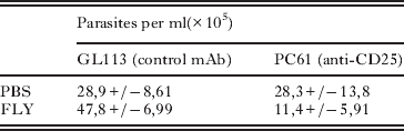
Although the role of CD4+ CD25+ T cells (Treg) in the persistence of T. cruzi infection is controversial in the literature (Kotner and Tarleton, Reference Kotner and Tarleton2007; Mariano et al. Reference Mariano, Gutierrez, Pavanelli, Milanezi, Cavassani, Moreira, Ferreira, Cunha, Cardoso and Silva2008; Sales et al. Reference Sales, Golgher, Oliveira, Vieira, Arantes, Lannes-Vieira and Gazzinelli2008), the expression of FoxP3 (a transcription factor specifically expressed by Tregs in mice) was assessed in heart sections by immunohistochemistry. Approximately 10% of the CD4+ T cell population in the cardiac tissue of the infected mice inoculated with FLY was positive for FoxP3 in comparison to 2% of positive cells in FAY or PBS pre-treated animals (Student's t-test, P<0·001) (Fig. 4D–F). CD25-inactivation by the administration of anti-CD25 mAb (PC61 clone) prior to FLY and T. cruzi inoculation reduces the number of circulating trypomatigotes to half of the control parasitaemia, injected with an unrelated antibody (GL113 mAb) (Table 1), pointing to a possible role of CD4+CD25+FoxP3+ T cells in the FLY-induced increment of T. cruzi infection.
Table 1. Anti-CD25 mAb, but not control IgG (GL113), abolishes the FLY-induced rise in parasitaemia in Trypanosoma cruzi-infected mice

FLY peptide increases infection and IL10 production by resident peritoneal cells
Previous studies suggest that the suppressor activity of Treg cells may also be induced by the presence of soluble factors such as TGFβ and IL10 (Murai et al. Reference Murai, Turovskaya, Kim, Madan, Karp, Cheroutre and Kronenberg2009; Scholzen et al. Reference Scholzen, Mittag, Rogerson, Cooke and Plebanski2009) both produced during infection by T. cruzi (Reed et al. Reference Reed, Brownell, Russo, Silva, Grabstein and Morrissey1994; Mariano et al. Reference Mariano, Gutierrez, Pavanelli, Milanezi, Cavassani, Moreira, Ferreira, Cunha, Cardoso and Silva2008).
The production of IL10 was then determined in the supernatant of resident-peritoneal cells (cells present in the locality of T. cruzi infection in our experimental model) incubated or not with FLY. As shown in Fig. 5A, the production of IL10 is dose dependent with levels of this cytokine being almost 1·8-fold higher in FLY-stimulated cells (at a concentration of 1 μg/well) than in FAY-stimulated cells. Interestingly, when the levels of NO were determined in the supernatants, no differences were observed amongst the experimental groups (Fig. 5B), showing that FLY does not affect NO production, in line with our results on the quantification of iNOS synthase in the heart (Fig. 4A–C). In addition, FLY may contribute to the increment of mouse parasitaemia by the enhancement of local infection of peritoneal cells. Although the clearance of FLY in the peritoneal cavity was not determined in our experimental conditions, it is worth noting that previous incubation of peritoneal cells with increasing concentrations of FLY in vitro resulted in a significant rise in the number of infected cells (Fig. 6), being 3·6 times higher at 10 μg of FLY (Student's t-test, P <0·002, P <0·0002) in relation to FAY or PBS, respectively.

Fig. 5. IL10 and NO production by peritoneal cells stimulated with different concentrations of FLY. Peritoneal cells were treated with the indicated amounts of FLY, FAY or culture medium (control) for 24 h. After incubation, the supernatants were removed and used to measure IL10 (A) and NO (B) by ELISA. Results represent mean±standard deviation (s.d.) from triplicate experiments.

Fig. 6. Resident peritoneal cells were incubated for 30 min with PBS, FLY or FAY peptides and then infected with 100 trypomastigotes/cell for 1 h. Non-associated trypomastigotes were removed, the cells were fixed with methanol and DNA stained. Fifty photos of each experiment were made, enabling the counting of parasites/100 cells. Each open square represents the number of parasites/100 cells present in each photo. Black lines represent the average number of parasites/100 cells. Significant differences were obtained between the infection in the presence of 1 μg of FLY or FAY (P<0·0002) or 10 μg of FLY and FAY (P<0·002). There were no significant differences between the controls (P>0·5).
DISCUSSION
It has previously been shown that the conserved VTVXNVFLYRP sequence (FLY), present on the gp85/TS glycoproteins contains a cell-binding motif and that substitution of the leucine residue for alanine (VTVXNVFAYRP, FAY sequence) abrogates its binding ability (Magdesian et al. Reference Magdesian, Giordano, Ulrich, Juliano, Juliano, Schumacher, Colli and Alves2001). Moreover, it was demonstrated that FLY, but not FAY, synthetic peptide phosphorylates ERK1/2, dephosphorylates cytokeratin 18 and increases infection of LLC-MK2 cells by T. cruzi (Magdesian et al. Reference Magdesian, Tonelli, Fessel, Silveira, Schumacher, Linden, Colli and Alves2007).
In the present study, the potentiating effect of the peptide FLY on the infection by T. cruzi previously described in vitro, is now confirmed in vivo. BALB/c mice administered intraperitoneally with a single dose of FLY have increased parasitaemia, higher mortality and an increment in tissue parasitism in the bladder (7·6-fold), small intestine (3·6-fold) and heart (3-fold), suggesting that FLY may be partially responsible for the virulence of trans-sialidase, previously described in the literature (Chuenkova and Pereira, Reference Chuenkova and Pereira1995), which is explained by the sialylation of CD8+ T cell surface, dampening the antigen-specific response and favouring the persistence of the parasite in the host (Freire-de-Lima et al. Reference Freire-de-Lima, Alisson-Silva, Carvalho, Takiya, Rodrigues, DosReis, Mendonca-Previato, Previato and Todeschini2010). However, the virulence of FLY has to occur by a different mechanism, since it lacks trans-sialidase activity. It is tempting to speculate that the host immune response to different segments of distinct members of the gp85/TS superfamily expressed by the parasite, may be relevant for the final outcome of the parasitaemia – inhibition or enhancement – during infection. In this context, it is important to mention that protection against T. cruzi infection was described for other members of the gp85/TS superfamily, such as the gp82 and gp90 proteins (Santori et al. Reference Santori, Paranhos-Bacalla, Franco DA Silveira, Yamauchi, Araya and Yoshida1996), devoid of trans-sialidase activity.
Since the heart is a main target organ in Chagas´ disease, this study was concentrated on that organ. In agreement with the increment of tissue parasitaemia, a more intense inflammatory response was detected in the heart of both FLY-primed and infected animals. The higher parasitaemia in FLY-treated animals, as compared to controls, could not be explained by differences in NO production, since similar amounts of labelled fields for NO synthase were detected among the groups. Interestingly, a 5-fold increment in the number of CD4+CD25+FoxP3+ T cells (T regulatory cells) was detected in the hearts of both FLY-primed and infected animals, which could be due to either local expansion of Tregs or their preferential homing at the inflammatory site. The latter possibility may be favoured by the more intense inflammation observed in FLY-treated mice and by the previous observation that the production of CCL4, a potent chemoattractant of CD4+CD25+ T in mice, overlaps with maximum tissue parasitism in T. cruzi (Sales et al. Reference Sales, Golgher, Oliveira, Vieira, Arantes, Lannes-Vieira and Gazzinelli2008). Additionally, FLY-induced IL10 production by resident peritoneal cells, in association with the inflammatory environment, may contribute to the increment of Tregs in heart tissues. It is now established that immune mediators (such as IL10 and TGFβ) drive the induction and expansion of Tregs, albeit with quite distinct phenotypes (Collison et al. Reference Collison, Pillai, Chaturvedi and Vignali2009). IL10 produced by cells other than T cells was required to maintain the expression of FoxP3 in Treg cells, despite their own ability to produce IL10, as shown recently in a murine model of colitis (Murai et al. Reference Murai, Turovskaya, Kim, Madan, Karp, Cheroutre and Kronenberg2009). Also, IL10, IL2 and TGFβ mediate Treg cell induction displaying high expression of FoxP3, independently of direct TCR stimulation, in infection by Plasmodium falciparum (Scholzen et al. Reference Scholzen, Mittag, Rogerson, Cooke and Plebanski2009). Whatever the mechanism might be, the increment of the Treg population may contribute to the local tolerance to infection by T. cruzi, as described also for Plasmodium yoelii (Hisaeda et al. Reference Hisaeda, Maekawa, Iwakawa, Okada, Himeno, Kishihara, Tsukumo and Yasutomo2004; Chen et al. Reference Chen, Liu, Wang, Wu, Feng, Zheng, Guo, Li, Wang and Cao2009) and Leishmania major (Belkaid et al. Reference Belkaid, Piccirillo, Mendez, Shevach and Sacks2002a,Reference Belkaid, Von Stebut, Mendez, Lira, Caler, Bertholet, Udey and Sacksb; Rouse and Suvas, Reference Rouse and Suvas2004; Belkaid and Rouse, Reference Belkaid and Rouse2005). Moreover, depletion of CD4+CD25+ T with anti-CD25 antibody abolished the FLY-induced rise in parasitaemia.
The role of Treg in the infection by T. cruzi is controversial, with data suggesting that depletion of Treg cells by anti-CD25 antibodies does not alter the outcome of the immune response (Kotner and Tarleton, Reference Kotner and Tarleton2007) or confer more resistance to infection in mice (Sales et al. Reference Sales, Golgher, Oliveira, Vieira, Arantes, Lannes-Vieira and Gazzinelli2008). In contrast, the involvement of Treg cells in the acute phase of T. cruzi infection has been described, with migration of Treg cells to the heart of infected mice and an increased mortality of anti-CD25-treated animals (Mariano et al. Reference Mariano, Gutierrez, Pavanelli, Milanezi, Cavassani, Moreira, Ferreira, Cunha, Cardoso and Silva2008). The conflicting results may be due to experimental protocols, distinct strains of mice and/or differences in the parasites employed, as pointed out (Mariano et al. Reference Mariano, Gutierrez, Pavanelli, Milanezi, Cavassani, Moreira, Ferreira, Cunha, Cardoso and Silva2008). In addition, Tregs were also implicated in the human immune response against T. cruzi. A higher frequency of circulating Tregs was found in patients with the indeterminate form in relation to the cardiac form of the disease, with the majority of CD4+CD25+ cells expressing FoxP3 (Araújo et al. Reference Araújo, Gomes, Rocha, Williams-Blangero, Pinheiro, Morato and Correa-Oliveira2007; Sathler-Avelar et al. Reference Sathler-Avelar, Vitelli-Avelar, Teixeira-Carvalho and Martins-Filho2009).
Taken together, our data suggest the importance of Treg cells in relation to the specific effect of FLY in the exacerbation of the infection by T. cruzi, although other activated CD25-expressing CD4+ T cells (Curotto de Lafaille and Lafaille, Reference Curotto de Lafaille and Lafaille2009) may concur to the same effect.
How FLY potentiates T. cruzi infection in vivo is not clear. FLY may act locally and/or systemically by independent, and not exclusive, mechanisms. A possible mechanism involves the dissemination of highly infected peritoneal cells to other organs/tissues, allowing the parasite to subvert the host immune system and to propagate. One example of this strategy was described for Toxoplasma gondii, where the adoptive transfer of Toxoplasma-infected dendritic cells in mice resulted in more rapid dissemination of the parasites to distant organs and exacerbation of infection when compared with intraperitoneal incubation with free parasites (Lambert et al. Reference Lambert, Hitziger, Dellacasa, Svensson and Barragan2006).
Phagocytic removal of apoptotic cells, which appear in the course of the immune response, promotes the secretion of cytokines that increase, in the particular case of T. cruzi, intracellular replication of the parasite and parasitaemia in infected mice (Freire-de-Lima et al. Reference Freire-de-Lima, Nascimento, Soares, Bozza, Castro-Faria-Neto, de Mello, DosReis and Lopes2000). In addition, the uptake of the parasites is enhanced in the presence of cytokines (Aliberti et al. Reference Aliberti, Machado, Souto, Campanelli, Teixeira, Gazzinelli and Silva1999) and the binding to T. cruzi of CCL2, one of the murine β-chemokines produced by macrophages and cardiomyocytes infected with the parasite, induces chemoattraction and morphogenesis of the infective form of T. cruzi. In this model, chemokines could attract and facilitate T. cruzi uptake, with implications for the migration of the parasite to different tissues (Yamauchi et al. Reference Yamauchi, Aliberti, Baruffi, Portela, Rossi, Gazzinelli, Mineo and Silva2007).
Incubation of resident peritoneal cells with FLY results in a higher number of cells infected by T. cruzi. This is a similar biological effect to that previously obtained employing an epithelial cell line, where an increased number of infected cells, as well as a higher number of parasites/cell, were observed, suggesting that FLY facilitates infection of the same cell, by a mechanism not fully understood (Magdesian et al. Reference Magdesian, Tonelli, Fessel, Silveira, Schumacher, Linden, Colli and Alves2007).
The FLY peptide can exert its function through a cell surface receptor as previously suggested (Magdesian et al. Reference Magdesian, Giordano, Ulrich, Juliano, Juliano, Schumacher, Colli and Alves2001) and/or inside the cells after internalization by the same mechanism described for the cell-penetrating peptides (CPPs) (Patel et al. Reference Patel, Zaro and Shen2007a,Reference Patel, Zaro and Shenb), since FLY contains a small cluster of 2 lysines and 1 arginine, essential residues for the internalization of CCPs.
One point to be addressed relates to how the FLY domain is exposed to the host in a natural infection. We hypothesized that membrane vesicles shed by Trypanosoma cruzi in vitro and in vivo (Gonçalves et al. Reference Gonçalves, Umezawa, Katzin, de Souza, Alves, Zingales and Colli1991) may act as cargo for the delivery of the FLY sequence. FLY is a conserved domain of the gp85/TS glycoproteins, abundant in membrane vesicles, which are internalized by cells, including macrophages, enhancing T. cruzi infection in vitro and heart parasitism in mice and IL10 production (Torrecilhas et al. Reference Torrecilhas, Tonelli, Pavanelli, Da Silva, Schumacher, de Souza, Cunha-e-Silva, Abrahamsohn, Colli and Alves2009). However, since FLY is not apparently exposed on the surface of these molecules, as judged by the structure of TS (Buschiazzo et al. Reference Buschiazzo, Amaya, Cremona, Frasch and Alzari2002) and Tc-85 (Marroquin-Quelopana et al. Reference Marroquin-Quelopana, Oyama, Pertinhez, Spisni, Juliano, Juliano, Colli and Alves2004) a molecular modification by proteolysis or conformational changes have to be hypothesized in order to envisage a role for FLY in vivo.
In summary, the results herein presented suggest that the T. cruzi-borne FLY domain, may act as a virulence factor in the animal model by altering the host cell-mediated immune response critical to combat the infection by the parasite.
ACKNOWLEDGEMENTS
The helpful critical comments on the manuscript by Drs José Maria Mosig and Marcello Barcinski are deeply acknowledged. This work was supported by Fundação de Amparo à Pesquisa do Estado de São Paulo (FAPESP grant #2004/03303-5) and CNPq. RRT and ACT were Post-doctoral Fellows supported by FAPESP.