


Introduction
The climate conditions and the territorial size are peculiarities that define an important characteristic of livestock in Brazil: a large part of the herd is raised on pastures, thus making possible one of the lowest cattle production costs in the world (EMBRAPA, 2019). In addition, Brazil offers a product with the potential to conquer more demanding markets, the so-called ‘green ox’ or ‘grass-fed beef’ (Dias-Filho, Reference Dias-Filho and Dias-Filho2016). However, pasture-reared ruminant populations are exposed to several gastrointestinal nematodes that are a cause of significant losses in animal production worldwide (Charlier et al., Reference Charlier, Rinaldi, Musella, Ploeger, Chartier, Vineer, Hinney, von Samson-Himmelstjerna, Băcescu, Mickiewicz, Mateus, Martinez-Valladares, Quealy, Azaizeh, Sekovska, Akkari, Petkevicius, Hektoen and Claerebout2020; Szwec et al., Reference Szwec, De Waal and Zintl2021). At the same time, difficulties encountered in the chemical control of nematodosis (Avramenko et al., Reference Avramenko, Redman, Windeyer and Gilleard2020) have encouraged researchers to develop new ecologically appropriate and sustainable technologies in pasture production systems (Velde et al., Reference Velde, Charlier and Claerebout2018; Szwec et al., Reference Szwec, De Waal and Zintl2021). Among them, the use of nematophagous fungi in the biological control of gastrointestinal nematodes in ruminants has been explored in several studies around the world, as it contributes directly to reduce the use of chemotherapy in cattle production systems (Braga and Araújo, Reference Braga and Araújo2014). In animal production, biotechnology can be used to increase food production, product quality and system sustainability (Carnevali et al., Reference Carnevali, Franchini, Otranto, Giangaspero, Di Bello, Ciccarelli, Szpila, Valastro and Van der Esch2019; Yuan et al., Reference Yuan, Zheng, Jiao, Cheng, Feng, Du and Liu2019; Baker et al., Reference Baker, Rice, Leemon, Godwin and James2020). The product Bioverm®, with a commercialization licence granted by the Ministério da Agricultura Pecuária e Abastecimento (MAPA, 2019), contains structures of the fungus Duddingtonia flagrans and enters the Brazilian market as a new tool for nematodosis control programmes and thus contributes to improving the efficiency of animal production in a way that is non-toxic to the environment.
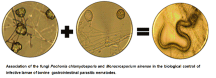
Nematophagous fungi are naturally present in the environment and comprise a variety of species with the ability to prey on, or render unviable, nematodes by enzyme production and then to use the nematodes as a source of nutrients (Van Ooij, Reference Van Ooij2011). The species Monacrosporium sinense predates nematode larvae through adhesive networks (Campos et al., Reference Campos, Araújo, Assis, Gandra and Guimarães2007). The species Pochonia chlamydosporia is an optional parasite of eggs and females of nematodes (Dallemole-Giaretta et al., Reference Dallemole-Giaretta, Freitas, Cavallin, Marmentini, Faria and Resende2013) and belongs to the group of nematophagous fungi called ‘ovicides’. The joint use of different isolates of nematophagous fungi may have a complementary and synergistic action in the biological control of helminths (Vieira et al., Reference Vieira, Oliveira, Campos and Araújo2019). For this, the introduction of a fungus with excellent interaction capacity and adaptation to the microenvironment is relevant to the success of a parasite control programme.
This research aimed to evaluate the compatibility of joint growth of the fungi M. sinense and P. chlamydosporia and the joint nematicidal activity of these fungal isolates on infective larvae of bovine nematodes.
Materials and methods
The fungal isolates M. sinense (SF53) and P. chlamydosporia (VC4) are part of the collection of the Parasitology Laboratory in the Veterinary Department of the Federal University of Viçosa, Brazil, where they are preserved on rice grains in silica gel tubes at 4°C in the dark. The rice grains containing structures of the fungus isolates were placed to grow separately in plates containing 2% potato dextrose agar medium (2% PDA).
Antagonism test in direct confrontation
Discs of potato dextrose agar medium of 5 mm diameter and containing P. chlamydosporia mycelia were placed at a distance of 1 cm from the edge of Petri dishes (9 cm in diameter) containing 2% potato dextrose agar medium (2% PDA). The plates were stored in the dark for 10 days at 26°C in an incubator chamber with biochemical oxygen demand (BOD). After this period, mycelium discs of M. sinense were placed on the agar opposite the colony of P. chlamydosporia. For the control group, colonies of the same fungus were compared. The confronted colonies were incubated in the dark for 8 days at 26°C in an incubator chamber (BOD). For the evaluations, the adapted scale of notes, proposed by Bell et al. (Reference Bell, Wells, Markhabell and Wells1982) was used: (1) complete colonization of the plaque by P. chlamydosporia; (2) colonization of 2/3 of the plaque by P. chlamydosporia; (3) colonization of 50% of the plaque per fungus; (4) colonization of 2/3 of the plaque by M. sinense; (5) complete colonization of the plaque by M. sinense. Ten repetitions were performed per treatment.
Antibiosis test
Dialysis membrane discs (SIGMA®) were placed on the surface of the 2% PDA culture medium in Petri dishes of 9 cm diameter (Fig. 1A). Subsequently, 5 mm diameter agar discs of the mycelia of P. chlamydosporia (VC4) and M. sinense (SF53) were placed separately in the centre of distinct plates (Fig. 1B). The colonies were incubated in the dark for 96 h at 26°C in an incubator chamber (BOD). The growth area of the colonies was demarcated externally at the bottom of the plates and the dialysis membrane together with the respective colony was removed (Fig. 1C). Then, the plates were inverted and 1 mL of chloroform was added to the bottom in order to eliminate possible structures of the fungus (Fig. 1D). After the product had evaporated, the plates were left for 30 min under direct irradiation of ultraviolet light in a laminar flow chamber (Fig. 1E). Then, an aqueous suspension containing mycelia of P. chlamydosporia was added to the surface of the culture medium in the plates where the fungus M. sinense grew and a suspension of M. sinense where P. chlamydosporia grew (Fig. 1F). These suspensions were obtained from colonies previously grown in 2% PDA culture medium for 10 days in the dark at 26°C in an incubator chamber (BOD).

Fig. 1. Antibiosis test represented by diagram.
The control group consisted of the cultivation of P. chlamydosporia and a subsequent suspension culture containing mycelia of P. chlamydosporia, the same procedure being carried out for the control group of M. sinense. The plates were kept at 26°C in an incubator chamber (BOD) for 10 days in the dark and after this period it was observed whether there was a growth inhibition halo of P. chlamydosporia formed by M. sinense, and vice versa. In order to ensure that the method generated reliable information about the samples, 10 repetitions were performed per treatment.
Effect of volatile compounds
Petri dish covers of 9 cm in diameter containing 2% PDA culture medium were positioned one above the other according to the technique described by Bharat et al. (Reference Bharat, Singh and Singh1980). A 5 mm diameter disc containing M. sinense mycelia was added to the lower plate and a 5 mm diameter disc containing P. chlamydosporia mycelia was added to the top plate. In another treatment, a 5 mm diameter disc containing mycelia of P. chlamydosporia was added to the lower plate and a 5 mm diameter disc containing M. sinense mycelia was added to the top plate. For the control group, P. chlamydosporia was cultivated in the lower and top plate; the same procedure was carried out for M. sinense (Fig. 2A). The plates were laterally sealed with a plastic membrane and kept at 26°C in an incubator chamber (BOD) for 15 days and in the dark (Fig. 2B). For the evaluations, the colony area was measured and compared with the control (Fig. 2C). Ten repetitions were performed per treatment and Student's t-test was used to compare the areas of the colonies at a significance level of 5%.

Fig. 2. Effect of volatile compounds test represented by diagram.
Production of pellets in sodium alginate containing mycelia of the fungi M. sinense and P. chlamydosporia
For the induction of fungal mycelium formation, agar fragments of approximately 5 mm in diameter containing mycelia and fungal spores of the isolates VC4 and SF53 were transferred separately to Erlenmeyer flasks containing 150 mL liquid medium of glucose, peptone and yeast extract (GPY). The fungi were grown at pH 6.5, 120 rpm agitation for 24 h in the dark at 26°C for 21 days. After this period, the mycelia were harvested with a platinum loop and weighed on an analytical scale for the manufacture of pellets. The pellets were produced in a matrix of sodium alginate, according to the technique described by Walker and Connick (Reference Walker and Connick1983) and modified by Lackey et al. (Reference Lackey, Muldoon and Jaffe1993).
In vitro evaluation of nematicidal activity of fungi towards infective larvae of gastrointestinal nematodes
Forty Petri dishes of 9 cm diameter containing 2% water–agar (2% WA) were used, divided into four groups. In the first group, 0.2 g of pellets containing P. chlamydosporia were added to the plates; in the second group, 0.2 g of pellets containing M. sinense (SF53) were added to the plates; in the third group, 0.2 g of pellets containing the association of M. sinense and P. chlamydosporia were added to the plates; and in the fourth group, 0.2 g of pellets without fungus (control) were added to Petri dishes containing 2% WA. Into each plate were poured 1000 infective larvae (L3) of bovine nematodes, corresponding to the genera Haemonchus sp., Cooperia sp. and Oesophagostomum sp. in the proportions (±s.d.) of 50% (±11), 44% (±5) and 6% (±5), respectively. The plates were kept in an incubator chamber at 26°C in the dark for a period of 7 days. On the 7th day, the L3 were recovered by emptying the plate contents into a Baermann funnel, with water at 42–45°C and waiting for 12 h before decantation. Recovered infectious larvae were counted and identified according to Keith's criteria (Reference Keith1953) under a light microscope (100× magnification), obtaining the average of the non-predated larvae by genus in the control group, and the average of non-predated larvae by genus in the treated group. The percentage reduction of larvae in the treated group in relation to the control was calculated according to the following formulae:
Reduction(%) = (mean of control larvae – mean of treatment larvae)/mean of control larvae × 100.
The recovered L3 averages, the L3 percentages of each genus and the reduction percentages were transformed into log (x + 1) and compared by Tukey's test at a significance level of 5% using IBM SPSS 2.0 software.
Results
The growth of M. sinense and P. chlamydosporia in association and separately is shown in Fig. 3. According to the Bell scale adapted for this study, the isolates confronted in the in vitro antagonism test were classified as grade ‘3’, in which the growth of each fungus occupied 50% of the plate.

Fig. 3. Direct confrontation test: (A) Pochonia chlamydosporia (1) and Pochonia chlamydosporia (1); (B) Monacrosporium sinense (2) and Monacrosporium sinense (2); (C) Pochonia chlamydosporia (1) and Monacrosporium sinense (2).
The non-formation of an inhibition halo between the fungi M. sinense and P. chlamydosporia observed in the antibiosis test demonstrated that the isolates did not produce substances that would interfere with mycelial growth.
Table 1 shows the average values of the mycelial growth area in the upper parts of the plates of the fungi M. sinense and P. chlamydosporia submitted to the volatile metabolites effect test, in which no differences were observed in the mycelial growth area of any of the isolates tested together (P ⩾ 0.05).
Table 1. The average values and standard deviation of the mycelial growth area, in the upper parts of the Petri dishes, 9 cm in diameter, containing 2% potato-dextrose-agar medium (2% PDA), of the fungi Pochonia chlamydosporia (VC4 isolate) and Monacrosporium sinense (SF53 isolate) submitted to the volatile metabolite effect test

Different letters in the same column indicate the difference between the data (P ⩽ 0.05). SF53: Monacrosporium sinense; VC4: Pochonia chlamydosporia.
The nematicidal activities of the fungi were demonstrated by means of infective larvae (L3) of parasitic bovine nematodes recovered after 7 days. The percentage L3 reduction in the group containing the association of M. sinense and P. chlamydosporia (98.90%) was higher than the percentage of the group containing M. sinense alone (82.20%), which was higher than the percentage of the group containing P. chlamydosporia alone (34.70%), as shown in Fig. 4.

Fig. 4. The percentage reduction of infectious larvae (L3) of gastrointestinal parasitic nematodes of cattle recovered from plates with 2% water-agar medium (2% WA), after 7 days of interaction, of the fungi Monacrosporium sinense (SF53 isolate), Pochonia chlamydosporia (VC4 isolate) and the association of the fungi Monacrosporium sinense and Pochonia chlamydosporia (SF53 + VC4). Different letters indicate the difference between the values (P ⩽ 0.05).
Table 2 shows the average values of infectious larvae (L3) of cattle recovered from the plates with 2% WA medium, containing the association of the fungi M. sinense and P. chlamydosporia (SF53 + VC4), P. chlamydosporia (VC4) alone and M. sinense (SF53) alone, as well as the percentage distribution of recovered L3 genera. Compared to the control group without fungus, the three groups that contained nematophagous fungi showed lower values of recovered L3 (P ⩽ 0.05). The group that contained the association M. sinense and P. chlamydosporia showed the lowest recovery of L3 of all three groups. The group that contained M. sinense alone showed lower recovery of L3 compared to the group that contained P. chlamydosporia alone (P ⩽ 0.05).
Table 2. The mean values and standard deviations of the number of infective larvae (L3) recovered from plates with 2% water-agar medium (2% WA), after 7 days of interaction, containing the association of nematophagous fungi Monacrosporium sinense and Pochonia chlamydosporia (SF53 + VC4), P. chlamydosporia (VC4) and M. sinense (SF53), as well as the percentage distribution of recovered L3 genera

Different letters in the same column indicate the difference between the data (P ⩽ 0,05). SF53: Monacrosporium sinense; VC4: Pochonia chlamydosporia.
There was no significant variation (P ⩾ 0.05) in the percentages of L3 of the Haemonchus genus between the four groups. The percentage of L3 of the genus Cooperia was higher in the group containing only M. sinense than in the control and VC4 groups. The percentage of L3 of the genus Oesophagostomum recovered from the group containing the fungal association was lower compared to the control group, while the recovery of Oesophagostomum in the P. chlamydosporia group was higher than in the group containing only M. sinense.
Discussion
To multiply, microorganisms depend on the availability of nutrients, space and oxygen. In addition, they need to adapt to the existing competition with any microbiota. Thus, interactions between fungi can be positive when synergistic multiplication occurs, or negative when inhibition of one of the agents occurs. Thus, in this work, the viability of the association between M. sinense and P. chlamydosporia was evaluated in order to use this combination of fungi in the biological control of gastrointestinal nematodes in animals.
The present study showed that M. sinense and P. chlamydosporia in direct confrontation showed homogeneous growth without overlapping colonies. This result suggests that these isolates can present very promising results in field tests. In addition, no significant volatile compound production was found by any of the fungi, since no degree of antagonism was seen in the in vitro test for volatile metabolites. There was no inhibition halo between the fungi M. sinense and P. chlamydosporia in the antibiosis test, demonstrating that the isolates did not produce substances that would interfere with each other's mycelial growth. The performance of compatibility tests is a way of understanding the capacity for interaction between fungi and reinforces the possibility of the joint use of these isolates in further studies. The fungi Arthrobotrys cladodes and P. chlamydosporia evaluated by Vieira et al. (Reference Vieira, Oliveira, Campos and Araújo2019) also presented themselves as compatible species for joint growth, since, in direct confrontation, each fungus colonized 50% of the plate, which revealed the possibility of other isolates with joint survivability. On the other hand, in contrast to our results and to those previously described, Ayupe et al. (Reference Ayupe, Monteiro, Braga, Soares, Mello, Araujo, Freitas and Araújo2016) reported that Arthrobotrys robusta (I31) competes with and antagonizes D. flagrans (AC001), which reinforces the need to perform compatibility tests for the successful use of fungal associations.
The species M. sinense and P. chlamydosporia are commonly found in the soil, and although these isolates have already been tested for their ability to be included in nematode biological control strategies (Campos et al., Reference Campos, Araújo, Assis, Gandra and Guimarães2007; Vieira et al., Reference Vieira, Oliveira, Campos and Araújo2020a, Reference Vieira, Oliveira, Freitas, Campos and Araújo2020b), the combined use of these fungi had not yet been studied. In addition, the success of their use in the biological control of parasites requires detailed knowledge of the agents and their interactions in the environment.
Regarding the suppression of the high-density nematode population, the association of M. sinense and P. chlamydosporia proved to be more efficient than the other treatments, reducing by 98.90% the number of infective larvae of bovine gastrointestinal parasites, while the reductions in groups containing M. sinense or P. chlamydosporia alone were 82.20 and 34.70%, respectively. The predator fungi M. sinense modified its hyphae in adhesive networks holding the nematode (Campos et al., Reference Campos, Araújo, Assis, Gandra and Guimarães2007). Diversely, P. chlamydosporia produced extracellular enzymes which were capable of causing cuticle hydrolysis and death of nematode larvae (Yang et al., Reference Yang, Liang, Li and Zhang2013) and it produced toxins with nematicidal activity (Mukhtar and Pervaz, Reference Mukhtar and Pervaz2003) which made it possible to achieve a satisfactory nematicidal activity index (Vieira et al., Reference Vieira, Oliveira, Campos and Araújo2020a). This reinforces the hypothesis that the association of fungi with different nematicidal activities has a greater chance of success in controlling parasites than the use of a single species. This was also reported in the compatibility between P. chlamydosporia and A. cladodes determined by Vieira et al. (Reference Vieira, Oliveira, Campos and Araújo2019), which resulted in a higher percentage reduction in the number of bovine infective larvae when compared their use in isolation. Efficiency is essential for the success of a biocontrol and its market acceptance.
When testing the effect of the fungal association on larvae of the genus Haemonchus, there was no significant difference between the four groups. The combination of fungi studied by Vieira et al. (Reference Vieira, Oliveira, Campos and Araújo2019) showed greater selectivity for the genus Haemonchus. The fact that there were variations in the percentage reduction of larvae of other genera can be justified by the predominance of the nematode genus, motility and cuticle composition (Mendoza-de-Gives et al., Reference Mendoza-de-Gives, Davies, Clark and Behnke1999; Oliveira et al., Reference Oliveira, Carvalho, Vieira, Campos, Freitas, Araujo, Braga and Araújo2018; Vieira et al., Reference Vieira, Oliveira, Campos and Araújo2019).
This research made it possible to confirm the compatibility between the isolates M. sinense and P. chlamydosporia, which demonstrated their best efficiency when used together to control bovine infective larvae under in vitro conditions.
Acknowledgements
The authors thank ‘Coordenação de Aperfeiçoamento de Pessoal de Nível Superior’ (CAPES), ‘Conselho Nacional de Desenvolvimento Científico e Tecnológico’ (CNPq) and ‘Fundação de Amparo à Pesquisa do Estado de Minas Gerais’ (FAPEMIG) for support in this study, in the form of a doctoral scholarship.
Author contribution
Isabela and Jackson Victor conceived and designed the study. Isabela conducted data gathering. Ítalo performed statistical analyses. Isabela, Ítalo, Artur and Jackson Victor wrote the article.
Financial support
This research received no specific grant from any funding agency, commercial or not-for-profit sectors.
Conflict of interest
None.
Ethical standards
Not applicable.


