


Introduction
Microsporidia are obligate intracellular eukaryotic parasites that infect most animal taxa (Stentiford et al., Reference Stentiford, Bateman, Feist, Stone and Dunn2013; Cali and Takvorian, Reference Cali, Takvorian, Weiss and Becnel2014). More than 1500 microsporidia belonging to about 220 genera have been reported worldwide, among which around half genera are known to infect aquatic organisms, including fish, aquatic arthropods and aquatic non-arthropod invertebrates, as well as protists (Stentiford et al., Reference Stentiford, Bateman, Feist, Stone and Dunn2013; Vávra et al., Reference Vávra, Hyliš, Fiala, Sacherová and Vossbrinck2017; Park and Poulin, Reference Park and Poulin2021). Fish are the common hosts of aquatic microsporidia and some fish-infecting microsporidia are known to cause disastrous diseases in aquaculture (Vávra and Lukeš, Reference Vávra and Lukeš2013; Kent et al., Reference Kent, Shaw, Sanders, Weiss and Becnel2014). More than 160 species belonging to about 22 genera are known to infect fishes (Liu et al., Reference Liu, Stentiford, Voronin, Sato, Li and Zhang2019). Although a few fish-infecting microsporidia have a broad host range, the majority of species show a comparative strict host specificity (Summerfelt and Goodwin, Reference Summerfelt and Goodwin2010; Kent et al., Reference Kent, Shaw, Sanders, Weiss and Becnel2014). Recent evidence showed that phylogenetic relationships between microsporidia were linked to their host and the infected tissues (Smith, Reference Smith2009; Stentiford et al., Reference Stentiford, Bateman, Feist, Stone and Dunn2013; Vávra and Lukeš, Reference Vávra and Lukeš2013). Therefore, host and tissue information are thought to be important for the classification of Microsporidia.
Based on the morphological and molecular difference of Pleistophora mirandellae and P. ovariae from the fish trunk muscle-infecting Pleistophora species, Pekkarinen et al. (Reference Pekkarinen, Lom and Nilsen2002) erected a genus Ovipleistophora to accommodate these two microsporidia and defined that the genus Ovipleistophora had a tissue tropism for ovary of fish. However, the recently reported two Ovipleistophora species did not follow the definition. Among them, O. diplostomuri infects the liver of the bluegill sunfish Lepomis macrochirus and the metacercarial cyst wall of the digenean parasite Posthodiplostomum minimum (Lovy and Friend, Reference Lovy and Friend2017). O. arlo infects the musculature of the common prawn Palaemon serratus (Stentiford et al., Reference Stentiford, Ross, Minardi, Feist, Bateman, Gainey and Bass2018). Moreover, Bojko et al. (Reference Bojko, Behringer, Moler and Reisinger2020) found that O. diplostomuri can also infect the musculature of the crayfish Procambarus bivittatus. Therefore, based on the above findings it was proposed that Ovipleistophora species have a broad host range or utilize crustaceans in a multi-host lifecycle. Further elucidating the tissue tropism, host specificity and geographical distribution of the genus Ovipleistophora will undoubtedly deepen insights into the classification and evolutionary biology of this group of microparasites. As a part of an ongoing project to investigate the diversity of aquatic microsporidia in the middle and lower reaches of the Yangtze River, two fish-infecting microsporidia from the ovary of three different fishes were for the first time reported. Morphological and molecular characterization indicated that these two species are affiliated to the genus Ovipleistophora and this finding extends their host and geographical range. Remarkable intraspecific genetic variation referred from ITS and LSU molecular marker were distinct for between-host Ovipleistophora diplostomuri isolates.
Materials and methods
Sample collections
Culter alburnus and Xenocypris argentea were captured by gill nets from the Danjiangkou Reservoir located between the northwest of Hubei province and the southwest of Henan province, China (32°55′ 38.46″N, 111° 34′ 27.44″E) in April 2019. Parabramis pekinensis was captured by gill nets from the Yangtze Estuary, China (31°34′ 44.17″N, 121°37′50.89″E) in August 2019. Specimens were held on ice and transported immediately to the local laboratory for the preliminary parasitological examination. Fish were anaesthetized by an overdose of MS 222 (Sigma, Germany) and then subjected for necropsy. Microsporidian infections were screened by the presence of visible whitish cysts embedded in ovaries. The cysts were ruptured to prepare wet mounts which were observed at 1000× with an oil immersion objective to determine the microsporidian infection. The microsporidian cysts and cysts-containing tissue samples were preserved in 95% ethanol for further molecular characterization and in 2.5% glutaraldehyde in 0.1 M sodium cacodylate buffer (PH 7.4) for electron microscopic observation, as well as in 10% neutral buffered formalin for morphological and histological analysis, respectively. Formalin-fixed spores were used to capture spore images using an Olympus BX 53 microscope equipped with an Olympus DP72 digital camera (Olympus, Japan).
Histological examination
Cyst-containing tissue samples from the ovaries of the infected fish fixed in 10% neutral-buffered formalin were dehydrated through a series of graded concentrations of ethanol and embedded in paraffin wax. Tissue sections, 3 μm in width, were stained with haematoxylin and eosin (H&E) and examined under a light microscope (Olympus, Japan) (Liu et al., Reference Liu, Xu, Luo, Zhao, Zhang, Liu and Zhang2018).
Transmission electron microscopy (TEM) observation
Glutaraldehyde-fixed cysts were washed three times for 10 min in sodium cacodylate buffer and then fixed with 1% osmium tetroxide (OsO4) in the same buffer for 1 h. After dehydration through a gradually ascending series of ethanol and propylene oxide, samples were embedded in Spur resin. Ultrathin sections (70–90 nm) were mounted on an uncoated copper grid and stained with uranyl acetate and lead citrate (Liu et al., Reference Liu, Stentiford, Voronin, Sato, Li and Zhang2019). Sections of two cysts were examined using a Hitachi HT-7700 transmission electron microscope (TEM).
DNA extraction, polymerase chain reaction (PCR), and sequencing
Ethanol-fixed cysts were washed with distilled water three times to remove ethanol remnants. The genomic DNA was extracted using the Qiagen DNeasy Blood & Tissue Kit (Qiagen, Germany) with the assistance of a Fast Prep cell disruptor 1 min at 6 m s−1 (MP Biomedicals, USA) following the manufacturer's instructions. The primer sets used for rRNA gene amplification are shown in Table 1. PCR was carried out in a 50 μL reaction system, containing PCR buffer, 200 mm dNTP, 2 mm MgCl2, 1.25 units Taq polymerase, 20 pmol each primer, and 2 μL DNA template. The partial SSU rDNA was amplified using the primer pair V1f/1492r and the PCR cycle consisted of an initial denaturation step at 95°C for 4 min, followed by 35 cycles at 95°C for 1 min, 50°C for 30 s, 72°C for 2 min, and a final extension at 72°C for 10 min. For some taxa, for which the primer pair V1f/1492r did not work the primer pair V1f/1047r was applied, with thermocycler parameters as follows: an initial denaturation step at 94°C for 4 min, followed by 35 cycles at 94°C for 30 s, 48°C for 30 s, 72°C for 1 min and a final extension at 72°C for 10 min. The 3′ terminal partial SSU rDNA, complete ITS and the partial LSU rDNA sequence was amplified using the primer pair HG4F/ILSUR and the PCR cycle consisted of an initial denaturation step at 95°C for 4 min, followed by 35 cycles at 95°C for 30 s, 53°C for 30 s, 72°C for 2 min, and a final extension at 72°C for 10 min. The PCR products were excised from an agarose gel and purified using a PCR purification kit (CWBiotech, Beijing, China) and cloned into the PMD-18 T vector system (Takara, Tokyo, Japan). Then positive clones were randomly selected for sequencing in both directions with the ABI BigDye Terminator v3.1 Cycle Sequencing Kit and an ABI 3100 Genetic Analyzer.
Table 1. The primers used for amplifying and sequencing microsporidia rDNA

Phylogenetic analysis
The obtained all sequences of fragments were assembled by BioEdit (Hall, Reference Hall1999) to produce consensus sequences which were applied to determine their matching species by a BLAST search. Sequences with high similarity and those of interest were retrieved from the GenBank database. A total of 23 sequences were aligned with Clustal X by the default setting (Thompson et al., Reference Thompson, Gibson, Plewniak, Jeanmougin and Higgins1997). This alignment was corrected manually using the alignment editor function within MEGA 6.0 (Tamura et al., Reference Tamura, Stecher, Peterson, Filipski and Kumar2013). Dictyocoela berillonum (KM657354) was used as an outgroup. Pairwise genetic distances/similarities were calculated using the Kimura-2 parameter model distance matrix for transitions and transversions. Phylogenetic analyses were conducted using the maximum likelihood (ML) method in PhyML 3.0 and Bayesian inference (BI) in MrBayes 3.2.4, respectively. The optimal evolutionary model was determined to be GTR + I + G by ModelTest 3.7 using the Akaike information criteria. Two independent runs were conducted with four chains for one million generations for BI. Phylogenetic trees were sampled every 100 generations. The first 25% of the samples were discarded from the cold chain (burninfrac = 0.25). Bootstrap confidence values were calculated with 100 repetitions for ML. The tree was initially examined in Figtree v1.4.4 (http://tree.bio.ed.ac.uk/software/figtree/), edited and annotated in Adobe Illustrator (Adobe System, San Jose, CA, USA).
Results
Macroscopical and light microscopical observations
All sampled fish did not appear any external disease symptoms. After necropsy, macroscopic whitish cysts embedded in the ovary of one out of 14 (prevalence: 7.1%) C. alburnus (Fig. 1a), 1 out of 6 (prevalence: 16.7%) X. argentea (Fig. 1b) sampled from the Danjiangkou Reservoir and 1 of 2 (prevalence: 50%) P. pekinensis (Fig. 1c) sampled from the Yangtze Estuary. The cysts from C. alburnus were oval and whitish-opaque in colouration, measuring 612.3 (498.1–791.8) μm long and 419.6 (318.7–567.7) μm wide (N = 15) (Fig. 1a). Monomorphic spores enclosed within the cysts were oval (Fig. 1d), measuring 4.03 ± 0.23 (3.74–4.54) μm long and 2.63 ± 0.17 (2.40–2.94) μm wide (N = 40). Cysts from X. argentea were slightly larger than those from C. alburnus, which were pyriform and whitish-opaque in colouration, measuring 761.2 (691.1–815.1) μm long and 434.5 (371.78–490.9) μm wide (N = 15) (Fig. 1b). Monomorphic spores enclosed within the cysts from X. argentea were also oval and measured 4.03 ± 0.3 (3.43–4.60) μm long and 1.94 ± 0.14 (1.60–2.20) μm wide (N = 40). Cysts from P. pekinensis were of irregular shape (Fig. 1c) where were filled with a mass of dimorphic spores, both of which were oval. Microspores measured 4.17 ± 0.29 (3.70–4.67) μm long and 2.72 ± 0.21 (2.30–3.20) μm wide (N = 40) and macrospores measured 9.26 ± 0.36 (8.61–10.13) μm long and 4.90 ± 0.15(4.59–5.19) μm wide (N = 40) (Fig. 1e).

Fig. 1. Macroscopical observation and light microscopy of Ovipleistophora species isolated from the ovary of different fishes. (a) Cyst (arrow) isolated from the ovary of Culter alburnus was macroscopically visible. Scale bar = 5 mm. (b) Cyst (arrow) isolated from the ovary of Xenocypris argentea was macroscopically visible. Scale bar = 5 mm. (c) Cyst (arrow) isolated from the ovary of Parabramis pekinensis was macroscopically visible. Scale bar = 5 mm. (d) Fresh spores of O. diplostomuri isolated from the Culter alburnus observed under light microscopy, the posterior vacuole (arrow) distinctly located at the posterior end of spores. Scale bar = 20 μm. (e) Macrospores (white arrow) and microspores (black arrow) of O. ovariae isolated from the P. pekinensis were observed under light microscopy. Scale bar = 20 μm.
Histological examination
Histopathological observations showed that the cysts from the three fish hosts developed the connective tissue of ovary parenchyma between oocytes (Fig. 2). The wall of cysts from C. alburnus and X. argentea were significantly thicker than that of cysts from P. perkinensis (Fig. 2b, d and f). The somatic cells in the ovary were distinctly attached the connective tissue layer of the cyst wall (Fig. 2b and f). No remarkable inflammatory responses were observed in the infected ovaries.

Fig. 2. Histopathology of the ovary of 3 fish species with infection of Ovipleistophora spp. (a) Large parasite-filled cysts isolated from the ovary of Culter alburnus were surrounded by connective tissue. Scale bar = 50 μm. (b) Higher magnification of the boxed area in Fig. 2a showing the thickened cyst wall (black arrow) surrounded with a layer of connective tissue (white arrow) where was attached with massive somatic cells (*) were visible. Scale bar = 20 μm. (c) Large parasite-filled cysts isolated from the ovary of Xenocypris argentea developed in the connective tissue between the oocytes. Scale bar = 200 μm. (d) Higher magnification of the boxed area in Fig. 2c shows large amounts of microsporidian spores (white arrow) within the cyst and the thickened cyst wall (black arrow). Scale bar = 20 μm. (e) Parasite-filled cyst isolated from the ovary of Parabramis pekinensis was surrounded by a layer of connective tissue. Scale bar = 50 μm. (f) Higher magnification of the boxed area in Fig. 2e shows the cyst wall (black arrow) and a thin surrounding connective tissue layer (white arrow) where were attached with massive somatic cells (*). Scale bar = 20 μm.
TEM
Sporoblasts and mature spores were observed for both microsporidian species from both C. alburnus and P. pekinensis and only mature spores could be found for species from X. argentea under electron microscopy. Sporoblasts isolated from the ovary of C. alburnus were of irregular shape and electron-dense which were surrounded by a thick electron-dense membrane (Fig. 3a). Only monomorphic mature spores were found which were uninucleate. Mature spores were in direct contact with the host cell cytoplasm (Fig. 3g). A large posterior vacuole occupying the half volume of spores was visible in the posterior end of spores (Fig. 3b). Isofilar polar filaments coiled 8–10 coils and arranged in one row (Fig. 3b). The polar filament measured 112–122 nm in diameter and could be stratified with six discontinuous density concentric circles which included from outside to inside a 3 nm thick unit membrane, a 4 nm thick moderately electron-lucent layer, a 2 nm thick electron-dense layer, a 12 nm thick moderately dense layer, a 10 nm thick moderately electron-lucent layer and a wide lucent centre (Fig. 3c). The spore wall was trilaminar, including a 43–46 nm thick electron-dense exospore, a 61–68 nm thick electron-lucent endospore and a 4–8 nm thick plasma membrane. The exospore consisted of two layers, including an electron-dense layer (layer 1) and a moderately electron-dense layer (layer 2) (Fig. 3c). The polaroplast was bipartite with a narrow anterior lamella and a wide posterior lamella (Fig. 3d). An umbrella-shaped anchoring disc located in the apex of spores was surrounded by the polaroplast (Fig. 3d). Mature spores isolated from X. argentea ultrastructurally resemble with those from C. alburnus. Monomorphic spores were uninucleate. Isofilar polar filaments coiled 8–10 coils and arranged in one row (Fig. 3e). The spore wall consisted of a 56–60 nm thick electron-dense exospore, a 39–44 nm thick electron-lucent endospore and a 3–5 nm thick plasma membrane. The exospore constituted of two layers, including an electron-dense layer (layer 1) and a moderately electron-dense layer (layer 2) (Fig. 3f). Sporoblasts isolated from the ovary of P. pekinensis were also of irregular shape and surrounded by a trilaminar electron-dense membrane (Fig. 4a). The transition of uninucleate sporoblasts to mature spores involved the differentiation of typical spore structures, including the anchoring disk, trilaminar spore wall, polar filaments and polaroplast. Mature spores were dimorphic. The structures of macrospores and microspores are similar, including a bipartite polaroplast with anterior lamellae and posterior tubules, a large posterior vacuole, an umbrella-shaped anchoring disc located at the apex of spore and three layers of spore wall (Figs 4b–d and 5a–d). Mature spores were in direct contact with the host cell cytoplasm (Fig. 4e). The exospore of microspores and macrospores consisted of two layers, including an electron-dense layer (layer 1) and a moderately electron-dense layer (layer 2) (Figs 4c and 5c). Isofilar polar filaments of microspores coiled 6–10 coils and arranged in one row (Fig. 4b and c). The spore wall of microspores consisted of a 53–58 nm thick electron-dense exospore, a 46–52 nm thick electron-lucent endospore and a 6–8 nm thick plasma membrane (Fig. 4c). Isofilar polar filaments of macrospores coiled 19–34 coils and arranged in one to three rows (Fig. 5a and b). The polar filament of macrospores measured 162–178 nm in diameter and exhibited seven discontinuous density concentric circles which included from outside to inside a 4 nm thick unit membrane, a 4 nm thick moderately electron-lucent layer, a 3 nm thick electron-dense layer, a 9 nm thick moderately electron-lucent layer, a 10 nm thick moderately electron-dense layer, a 10 nm thick moderately electron-lucent layer and a wide lucent centre (Fig. 5c). The spore wall of the macrospore constituted of a 23–51 nm thick electron-dense exospore, a 53–96 nm thick electron-lucent endospore and a 5–8 nm thick plasma membrane (Fig. 5c). Given the similar ultrastructural features and typical infection site (ovary), these 3 species herein can be assigned to the genus Ovipleistophora (Microsporidia: Pleistophoridae).

Fig. 3. Electron microscopy of Ovipleistophora diplostomuri. (a) Irregular sporoblasts of O. diplostomuri isolated from Culter alburnus surrounding by a thick electron-dense membrane (arrow). Magnification of the thick electron-dense membrane (inset in a). Scale bar = 500 nm. (b) A mature spore of O. diplostomuri isolated from Culter alburnus with typical microsporidian features of internal structure, including the isofilar polar filaments (pf), a bipartite polaroplast, a vacuole (V), a mushroom-shaped anchoring disc (AD) and a trilaminar spore wall consisting of an electron-dense exospore, an electron-translucent endospore and a plasma membrane. Scale bar = 500 nm. (c) Magnification of the exospores (Ex) showing two distinct layers (layer 1 and layer 2). Trilaminar spores wall consisting of exospore (Ex), endospore (En) and plasma membrane (short black arrow). Transverse section of polar filament coils exhibiting six discontinuous density concentric circles. Scale bar = 200 nm. (d) Bipartite polaroplast consisting narrow lamellae (Pp1) and wide lamellae (Pp2). Scale bar = 200 nm. (e) A mature spore of O. diplostomuri isolated from Xenocypris argentea with eight coils of polar filament (pf) and anchoring disc (AD). Scale bar = 500 nm. (f) Magnification of the exospores (Ex) showing two distinct layers (layer 1 and layer 2). Scale bar = 200 nm. (g) Mature spores occur in direct contact with the host cell cytoplasm. Scale bar = 5 μm.

Fig. 4. Electron microscopy of Ovipleistophora ovariae. (a) Irregular sporoblasts of O. ovariae isolated from Parabramis pekinensis surrounding by three-layered electron-dense membrane (arrow). Magnification of the three-layered electron-dense membrane (inset in a). Scale bar = 1 μm. (b) A mature microspore of O. ovariae with typical microsporidian features of internal structure, including isofilar polar filaments (pf), bipartite polaroplast (Pp1, Pp2), vacuole (V) and mushroom-shaped anchoring (AD). Scale bar = 1 μm. (c) Trilaminar spores wall including an electron-dense exospore, an electron-translucent endospore and a plasma membrane. Exospore showing two distinct layers (layer 1 and layer 2). Scale bar = 200 nm. (d) Bipartite polaroplast (Pp1, Pp2) and mushroom-shaped anchoring disc (AD). Scale bar = 500 nm. (e) Mature spores occur in direct contact with the host cell cytoplasm. Scale bar = 10 μm.

Fig. 5. Electron microscopy of Ovipleistophora ovariae. (a) A mature macrospores of O. ovariae shows bipartite polaroplast (Pp1, Pp2), a vacuole (V) and isofilar polar filaments. Scale bar = 1 μm. (b) A mature macrospore of O. ovariae contains a mushroom-shaped anchoring disc (AD), a large nucleus (N) and isofilar polar filament (pf). Scale bar = 2 μm. (c) Transverse section of polar filament coils exhibiting seven discontinuous density concentric circles. Trilaminar spores wall consisting of an electron-dense exospore, an electron-translucent endospore and a plasma membrane. Exospore showing an electron-dense layer (layer 1) and a moderately electron-dense layer (layer 2). Scale bar = 200 nm. (d) Bipartite polaroplast with anterior lamellae (Pp1) and posterior tubules (Pp2). Scale bar = 500 nm.
Molecular characterization
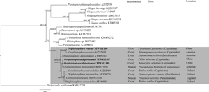
Three SSU sequences of the microsporidian species from C. alburnus, X. argentea and P. pekinensis were successfully amplified and the obtained consensus sequences were individually deposited in GenBank under accession numbers MW811110, MW811108 and MW811109, respectively. As such, their concatenated partial SSU, complete ITS and partial LSU was respectively assigned with accession numbers MW811107, MW811105 and MW811106. Blast searches and sequence comparison analysis showed the closest identity of these sequences with several Ovipleistophora species. SSU rDNA sequence obtained from C. alburnus and X. argentea is 99.68% identical, which is the highest identical to that of O. diplostomuri (MN515056 with 99.79% and 99.57% identity, respectively). Their next closest identities were to O. diplostomuri (KY809102 with 99.36% and 99.37% identity, respectively) and O. arlo (MH911630 with 98.93% and 99.04% identity, respectively). However, the SSU rDNA sequence obtained from P. pekinensis is most identical to O. ovariae (99.58% identity) and only 98.18% and 98.61% identical with that from C. alburnus and X. argentea, respectively. The pairwise distances/ similarities calculated by Kimura 2-parameter model between SSU rDNA sequences of Ovipleistophora species and 2 other microsporidian species of high sequence similarity ranged from 0.002/99.79% (between the species from C. alburnus MW811110 and O. diplostomuri MN515056) to 0.055/94.54% (between O. ovariae AJ252850 and Heterosporis anguillarum AF387331) (Table 2). Therefore, from the morphological and high SSU rDNA sequence identity, the species from C. alburnus and X. argentea can be determined to be conspecific to O. diplostomuri and species from P. pekinensis to be O. ovariae. To further explore the possible genetic variation among isolates of O. diplostomuri from different hosts, their full ITS and partial LSU rDNA sequences comparison analysis were performed. Results clearly indicated that the between-host genetic variation of the ITS sequences was distinct. ITS rDNA sequence of the isolates from C. alburnus and X. argentea was 97.9% similar, but only 85.7% and 87.8% similar to that of the isolate (KY809102) from the bluegill sunfish L. marcrochirus and its digenean parasite, P. minimum (Table 3). As such, between-host variations among the partial LSU rDNA sequences of three O. diplostomuri hosts were distinctly higher that those among their SSU rDNA sequences. LSU sequences of the isolates from C. alburnus and X. argentea were 99.0% identical, but only 96.2% identical with that from the typical host of O. diplostomuri (Table 3). Nineteen single polymorphism sites (SNPs) was found among the partial LSU rDNA gene sequences (about 440 bp) of three isolates of O. diplostomuri (KY809102) (Table 4). Bayesian and maximum likelihood analyses of the aligned SSU rDNA gene sequences of 22 microsporidian species generated highly similar topologies, although with different support values at some branch nodes. The results showed that the genus Ovipleistophora was monophyletic which grouped with Pleistophora and Heterosporis into an independent clade. The O. diplostomuri isolates infecting the ovary of C. alburnus and X. argentea clustered with the isolate infecting the musculature of P. bivittatus collected from the USA to form an independent branch, but excluding the L. marcrochirus isolate collected from the USA. The O. ovariae isolate from the ovary of P. pekinensis clustered with the isolate from the ovary of Notemigonus crysoleucas with a high support value (Fig. 6).

Fig. 6. The SSU rDNA-inferred phylogenetic relationships between Ovipleistophora species and the other aligned microsporidian species by Bayesian Inference (BI) method. The species names are followed by GenBank accession number. BI posterior probabilities were shown firstly, followed by ML support values on branch nodes. The present species was highlighted in bold. The infection site and host, as well as the geographical location of species belonging to Ovipleistophora spp. were presented.
Table 2. Pairwise nucleotide sequence identity (upper right) values and evolutionary distances (left bottom) among Ovipleistophora species/isolates and two other microsporidian species with high sequence similarity by Kimura-2 Parameter analysis based on SSU rDNA sequences

Table 3. Percentage of sequence similarity of the Ovipleistophora diplostomuri isolated from different hosts

Table 4. The partial LSU rDNA gene sequences (440 bp) of Ovipleistophora diplostomuri isolated from different hosts show the single nucleotide polymorphism sites (SNPs)

Discussion
To discriminate freshwater fish oocyte-infecting species originally assigned to the genus Pleistophora from other congeneric species, Pekkarinen et al. (Reference Pekkarinen, Lom and Nilsen2002) erected the genus Ovipleistophora. Four species, O. mirandellae, O. ovariae, O. diplostomuri and O. arlo have been so far formally described with morphological, ultrastructural and molecular features. Based on the definitive features of the genus Ovipleistophora and the high SSU rDNA sequence identity, the pathogen from C. alburnus and X. argentea can be identified to be O. diplostomuri and the parasite from P. pekinensis to be O. ovariae, although there are not all morphologically consistent with those of the previously reported isolates (Table 5). This is the first report of fish-infecting Ovipleistophora in Asia. In the present work, only monomorphic microspores of O. diplostomuri were found. However, dimorphic spores were firstly reported herein for O. ovariae. Dimorphic spores (macrospores and microspores) have been reported in several fish-infecting microsporidian genera, such as Pleistophora and Heterosporis (Al-Quraishy et al., Reference Al-Quraishy, Abdel-Baki, Al-Qahtani, Dkhil, Casal and Azevedo2012; Kent et al., Reference Kent, Shaw, Sanders, Weiss and Becnel2014; El-Garhy et al., Reference El-Garhy, Cali, Morsy, Bashtar and Al-Quraishy2017), which was also thought to be the definitive taxonomic feature of Ovipleistophora (Maurand et al., Reference Maurand, Loubes, Gasc, Pelletier and Barral1988; Pekkarinen et al., Reference Pekkarinen, Lom and Nilsen2002). At present, the mechanisms underlying the occurrence of two types of spores for some species and the respective functionality of microspores and macrospores remain unknown. Furthermore, the development of O. diplostomuri and O. ovariae herein were found to be in direct contact with the host cell cytoplasm, rather than within sporophorous vesicles which was one of the definitive taxonomic features of the genus Ovipleistophora and was present in the previously reported isolates of O. diplostomuri and O. ovariae (Kent et al., Reference Kent, Shaw, Sanders, Weiss and Becnel2014; Lovy and Friend, Reference Lovy and Friend2017). The absence of sporophorous vesicles was also previously reported for the development of O. ovariae infecting the fathead minnows, Pimephales promelas (Ruehl-Fehlert et al., Reference Ruehl-Fehlert, Bomke, Dorgerloh, Palazzi and Rosenbruch2005). So, the developmental pattern referred from the ultrastructural observations was suggested not to be the reliable taxonomic criterion of the genus Ovipleistophora for the plasticity derived from the habitat adaptation, including host, infection sites and host habitat environment.
Table 5. Morphological comparison of Ovipleistophora spp.

In terms of host, Ovipleistophora species were originally restricted to freshwater fish (Pekkarinen et al., Reference Pekkarinen, Lom and Nilsen2002). With the subsequent increasing diversity, the genus Ovipleistophora was supposed to have a broad host range or a complex lifecycle between fish and crustacean (Stentiford et al., Reference Stentiford, Ross, Minardi, Feist, Bateman, Gainey and Bass2018; Bojko et al., Reference Bojko, Behringer, Moler and Reisinger2020). Additionally, the cyst wall of the digenean parasite P. minimum of the bluegill sunfish was also found to be simultaneously infected by O. diplostomuri which further extended the host range of Ovipleistophora species. A wide host range has been previously reported for other microsporidian taxa including the genus Amblyospora (Andreadis et al., Reference Andreadis, Thomas and Shepard2018), Paranucleospora theridion (Nylund et al., Reference Nylund, Nylund, Watanabe, Arnesen and Karlsbakk2010), Hyalinocysta chapmani (Andreadis and Vossbrinck, Reference Andreadis and Vossbrinck2002) and Parathelohania anophelis (Avery and Undeen, Reference Avery and Undeen1990). Wide host range is one of the factors to derive the polymorphic development and the occurrence of between-host morphologically and functionally distinct spores for an individual microsporidian species. Interestingly, the developmental features of Ovipleistophora species were inconsistent with those of taxa with a wide host range which were generally characterized by the presence of diplokaryotic meronts and polysporoblastic sporogony and haploid meiospores. However, the presence of the isolated nuclei during the whole life cycle and a single sporulation sequence were the typical features of O. mirandellae, O. diplostomuri and O. ovariae. This can partially explain why Ovipleistophora possesses dimorphic spores with similar structures. Screening the possible crustacean host of O. diplostomuri and O. ovariae in the sympatric wetlands of this sampling will facilitate to extend their host range.
Originally, the genus Ovipleistophora was defined to be the obligate parasite of the fish ovary (Pekkarinen et al., Reference Pekkarinen, Lom and Nilsen2002). However, Lovy and Friend (Reference Lovy and Friend2017) found that O. diplostomuri infected both the liver of bluegill sunfish L. macrochirus and the cyst wall of the digenean parasite P. minimum. Furthermore, O. diplostomuri and O. arlo was subsequently recorded in muscle tissue of the freshwater crayfish and the common pawn, respectively (Stentiford et al., Reference Stentiford, Ross, Minardi, Feist, Bateman, Gainey and Bass2018; Bojko et al., Reference Bojko, Behringer, Moler and Reisinger2020). So, it can be supposed that the infection sites of Ovipleistophora species from vertebrate and invertebrate hosts are diverse with the high diversity being uncovered in future, although fish ovary is still the common infection site.
Ribosomal DNA genes have been widely applied into the species identification, classification and intraspecific discrimination of Microsporidia for it cover not only comparative loci but also highly variable loci (Haro et al., Reference Haro, del Águila, Fenoy and Henriques-Gil2003; Santín and Fayer, Reference Santín and Fayer2009; Krebes et al., Reference Krebes, Blank, Frankowski and Bastrop2010; Malcekova et al., Reference Malcekova, Valencakova, Luptakova, Molnar, Ravaszova and Novotny2011; Li et al., Reference Li, Chen, Wu, Peng, An, Schmid-Hempel and Schmid-Hempel2012a; Pombert et al., Reference Pombert, Xu, Smith, Heiman, Young, Cuomo, Weiss and Keeling2013; González-Tortuero et al., Reference González-Tortuero, Rusek, Maayan, Petrusek, Piálek, Laurent and Wolinska2016; Xu et al., Reference Xu, Wang, Jing, Cao, Zhang, Jiang, Yin, Cao and Shen2020). In the present study, we proved for the first time the between-host variation of ITS and LSU sequences of O. diplostomuri and the conservative SSU rDNA sequences of all three O. diplostomuri isolates. The intraspecific conservative SSU rDNA sequence of O. diplostomuri is similar with most of the fish-infecting microsporidian taxa, rather than insect-infecting microsporidia, such as Nosema ceranae (Sagastume et al., Reference Sagastume, del Aguila, Martín-Hernández, Higes and Henriques-Gil2011) and Dictyocoela spp. (Krebes et al., Reference Krebes, Blank, Frankowski and Bastrop2010; Wilkinson et al., Reference Wilkinson, Rock, Whiteley, Ovcharenko and Ironside2011). These results indicate that SSU rDNA sequence can be used as a suitable molecular marker for the identification of O. diplostomuri and ITS and LSU rDNA sequence are a good molecular marker to discriminate the intraspecific isolates of this pathogen.
Intraspecific genetic variations are common among Microsporidia, especially for insect-infecting species, such as Nosema ceranae (Sagastume et al., Reference Sagastume, del Aguila, Martín-Hernández, Higes and Henriques-Gil2011), N. bombi (Cordes et al., Reference Cordes, Huang, Strange, Cameron, Griswold, Lozier and Solter2012), N. apis (Maside et al., Reference Maside, Gómez-Moracho, Jara, Martín-Hernández, Rúa, Higes and Bartolomé2015), N. granulosis (Ironside, Reference Ironside2013) and N. bombycis (Ironside, Reference Ironside2013; Liu et al., Reference Liu, Pan, Luo, Li, Yang, Vossbrinck, Debrunner-Vossbrinck and Zhou2013). However, the mechanisms to cause the intra-isolate copy variation of SSU rDNA sequence and the inter-isolate variation of ITS or LUS rDNA sequence remain unclear. Host shift was thought to be one of the possible factors to derive species differentiation. Chaimanee et al. (Reference Chaimanee, Chen, Pettis, Cornman and Chantawannakul2011) found that N. ceranae isolated from different host species formed different branches. Maside et al. (Reference Maside, Gómez-Moracho, Jara, Martín-Hernández, Rúa, Higes and Bartolomé2015) found that the genetic variation of the N. apis occurred among isolates from the honey bee of different lineages. Few studies concern the possible phenotypic changes of isolates with genetic variation referred from the difference of ITS and LUS sequences. Between-host ultrastructural differences of Pleistophora hyphessobryconis were documented (Li et al., Reference Li, Chang, Wang, Liu, Liang and Wu2012b). Phylogenetic analysis indicated that O. diplostomuri isolated from the liver of bluegill sunfish L. macrochirus (Perciformes) and the digenean parasite P. minimum in North America was an independent branch, rather than clustering with O. diplostomuri isolated from C. alburnus (Cypriniformes) and X. argentea (Cypriniformes) in China, indicating that host affinity and geographical distribution provide the possible evolutionary signal for the species differentiation in some extents. As such, O. mirandellae isolated from the ovary of Gymnocephalus cernuus (Perciformes) clustered with O. arlo, rather than clustering with O. mirandellae isolated from Cypriniformes to form an independent branch. According to our speculation, the potential fish host of O. arlo possibly belongs to Cypriniformes.
Genetic variation of a parasite can cause different disease consequences (Branchiccela et al., Reference Branchiccela, Arredondo, Higes, Invernizzi, Martín-Hernández, Tomasco, Zunino and Antúnez2017; Shaw et al., Reference Shaw, Bilich, O'Brien, Cáceres, Hall, James and Duffy2021; Taggart-Murphy et al., Reference Taggart-Murphy, Alama-Bermejo, Dolan, Takizawa and Bartholomew2021). No inflammatory responses occurred in the ovary of C. alburnus, X. argentea and P. pekinensis for fish ovary is a general immune-privileged site. Given the possible more variable infection sites of O. diplostomuri and O. ovariae, ITS or LUS rDNA will be a feasible molecular marker for determining the virulent and avirulent isolates.
In summary, this present study reports for the first time two Ovipleistophora species infecting the ovary of three cyprinid fish in China which extends their host range and host geographical distribution. Distinct genetic variations were found in the between-host O. diplostomuri isolates.
Acknowledgements
We thank Yuan Xiao and Zhenfei Xing (Institute of Hydrobiology, Chinese Academy of Sciences) for their assistance with transmission electron microscopy analysis.
Author contributions
MQW collected fish samples, performed a parasitological examination and data analysis and helped write the manuscript; DRX, QQZ and AHL performed morphological comparisons and helped write the manuscript. JYZ designed this study and drafted the manuscript.
Financial support
This work was supported by grants from the Natural Sciences Foundation of China (31772411); National Key R&D Plan Blue Granary Science and Technology Innovation Project (2020YFD0900502); the ‘First ClassFishery Discipline’ programme in Shandong Province, China; the Talent plan ‘OneThing One Decision (Yishi Yiyi)’ in Shandong Province, China; Young experts of Taishan Scholars in Shandong Province and Initiative grant for high-level personnel recruitment in Qingdao Agricultural University awarded to JY Zhang.
Conflict of interest
The authors declare there are no conflicts of interest.
Ethical standards
Not applicable.


