


Introduction
Flukes (Platyhelminthes, Trematoda) are parasites that have a complex life cycle, involving several definitive and intermediate hosts. Traditionally, the identification of trematodes has been limited to the morphology of the adult in the definitive host (Jones et al., Reference Gibson, Jones and Bray2002). However, the continuous and fast growth of molecular databases offers the possibility of advance in the identification of parasite larvae found in different intermediate hosts, overcoming the taxonomic limitations of larval morphology studies or of assays that mimic the parasitic life cycle (Nolan and Cribb, Reference Nolan and Cribb2004; Blasco-Costa et al., Reference Blasco-Costa, Cutmore, Miller and Nolan2016). A molecular approach involving loci with different evolutionary rates (e.g. rRNA 28S gene or ribosomic Intergenic Spacer 1; Schulenburg et al., Reference Schulenburg, Englisch and Wägele1999; Tkach et al., Reference Tkach, Pawlowski and Mariaux2000; Olson et al., Reference Olson, Cribb, Tkach, Bray and Littlewood2003; Blasco-Costa et al., Reference Blasco-Costa, Cutmore, Miller and Nolan2016) has been helpful to identify trematode cryptic species (Miura et al., Reference Miura, Kuris, Torchin, Hechinger, Dunham and Chiba2005; Vilas et al., Reference Vilas, Criscione and Blouin2005; Razo-Mendivil et al., Reference Razo-Mendivil, Vázquez-Domínguez, Rosas-Valdez, de León and Nadler2010; Dellagnola et al., Reference Dellagnola, Montes, Martorelli and Vega2019a). Besides, integrated morphological and molecular approaches with ribosomal markers helped in the identification of fluke mixed larval infections (Dellagnola et al., Reference Dellagnola, Montes, Martorelli and Vega2019a).
Apple snails (Caenogastropoda, Ampullariidae) are distributed in tropical and subtropical wetlands worldwide (Berthold, Reference Berthold1991; Hayes et al., Reference Hayes, Cowie and Thiengo2009). The family Ampullariidae includes amphibious species with different degrees of air dependence (Seuffert and Martín, Reference Seuffert and Martín2009, Reference Seuffert and Martín2010; Hayes et al., Reference Hayes, Burks, Castro-Vazquez, Darby, Heras, Martin, Qiu, Thiengo, Vega, Wada, Yusa, Burela, Cardiemo, Cueto, Dellagnola, Dreon, Frassa, Giraud-Billoud, Godoy, Itualte, Koch, Matsukura, Pasquevich, Rodriguez, Seveanu, Seuffert, Strong, Sun, Tamburi, Tiecher, Turner, Valentine-Darby and Cowie2015; Rodriguez et al., Reference Rodriguez, Prieto, Vega and Castro-Vazquez2021), i.e. species with presumptively predominant gill respiration that lay gelatinous egg masses underwater and species that strictly depend on aerial lung respiration and lay calcareous eggs out of the water (Hayes et al., Reference Hayes, Cowie and Thiengo2009).
Pomacea is the most diverse genera of Ampullariidae (Hayes et al., Reference Hayes, Burks, Castro-Vazquez, Darby, Heras, Martin, Qiu, Thiengo, Vega, Wada, Yusa, Burela, Cardiemo, Cueto, Dellagnola, Dreon, Frassa, Giraud-Billoud, Godoy, Itualte, Koch, Matsukura, Pasquevich, Rodriguez, Seveanu, Seuffert, Strong, Sun, Tamburi, Tiecher, Turner, Valentine-Darby and Cowie2015). Pomacea canaliculata and Pomacea maculata are highly invasive species that affect different agricultural ecosystems of Southeast Asia, Pakistan, Hawaii, North America and Europe causing annual millionaires’ economic loss (Rawlings et al., Reference Rawlings, Hayes, Cowie and Collins2007; López-Soriano et al., Reference López-Soriano, Salgado and Tarruella2009; Oscoz et al., Reference Oscoz, Tomds and Duron2010; Yanygina et al., Reference Yanygina, Kirillov and Zarubina2010; Baloch et al., Reference Baloch, Memon, Burdi, Soomro, Tunio and Khatian2012; Hayes et al., Reference Hayes, Burks, Castro-Vazquez, Darby, Heras, Martin, Qiu, Thiengo, Vega, Wada, Yusa, Burela, Cardiemo, Cueto, Dellagnola, Dreon, Frassa, Giraud-Billoud, Godoy, Itualte, Koch, Matsukura, Pasquevich, Rodriguez, Seveanu, Seuffert, Strong, Sun, Tamburi, Tiecher, Turner, Valentine-Darby and Cowie2015; Joshi et al., Reference Joshi, Cowie and Sebastian2017). On the other hand, P. americanista (Ihering 1919) is an endemic snail to the Alto Paraná basin and Iguazú River in the rainforest of Argentina, Paraguay and Brazil, and is found on hard rocky substrates in swiftly flowing waters (Hylton Scott, Reference Hylton Scott1957). Unlike most ampullariids, P. americanista is a vulnerable species due to stringent habitat requirements and a narrow geographical range (Martín et al., Reference Martín, Burela and Gurovich2015; Gurovich et al., Reference Gurovich, Burela and Martín2017).
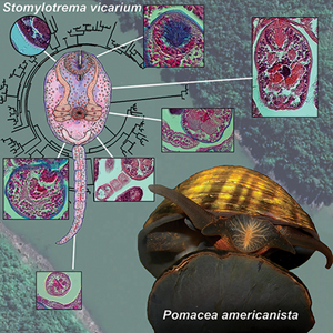
In the context of a broader program dealing with the symbiotic associations of apple snails, a stylet-bearing trematode was found hosted into the connective tissue and haemocoel spaces of the digestive gland in P. americanista. The aim of this study was to identify this parasite, plausibly from the suborder Xiphidiata (Plagiorchiida, Digenea), using a morphological and molecular approach. The trematode phylogeny inferred from the gene encoding the large subunit ribosomal RNA (28S rRNA) and the non-coding nuclear region of the ITS1 allowed identifying Stomylotrema vicarium Braun, Reference Braun1901 for the first time in an intermediate host; being the stomylotrematid flukes a small group of parasites from which there was a very scarce knowledge about their basic biology and early life cycle.
Material and methods
Collection site and host sampling
During a malacological survey carried out in the Bonito stream, Misiones rainforest, Argentina (27°26′39.60″S, 54°56′12.65″O; Supplementary Fig. 1A) in January 2014 southern summer, specimens similar to the ampullariid P. americanista (Ihering, 1919) were collected (Supplementary Fig. 1B and C). Only five individuals (range = 35.1–38.9 mm of shell length and 9.8–34.5 g) of this species were collected for different studies based on their scarcity, scant knowledge about their life history, and restricted geographic distribution (Martín et al., Reference Martín, Burela and Gurovich2015; Gurovich et al., Reference Gurovich, Burela and Martín2017).
Tissue sampling and histological procedures for light microscopy
Snails collected were submersed in an ice/water bath (~4°C for 15 min) for inducing relaxation and minimizing pain before careful shell cracking. Samples for light microscopy were obtained from sacrificed individuals by cutting 2–3 mm thick slices of the digestive gland with a razor blade from the gland surface, close to the kidney boundary. Tissues were then fixed in 4% paraformaldehyde in a buffered solution (43 mm NaCl, 1.8 mm KCl, 10.0 mm HEPES and 30 mm EDTA; pH 7.6; Cueto et al., Reference Cueto, Rodriguez, Vega and Castro-Vazquez2015) for 24 h at 20°C and kept in 70% ethanol. These were subsequently dehydrated in increasing concentrations of alcohol, embedded in a resin-paraffin mixture (Histoplast®, Argentina) and sectioned (5 μm). Sections were stained with haematoxylin-eosin and a trichrome stain (Alcian Blue, Nuclear Fast Red and eosin) as reported previously (Dellagnola et al., Reference Dellagnola, Vega and Castro-Vazquez2017). Digestive epithelial cells, intracellular symbiotic corpuscles and storage cells were morphologically identified as reported previously for Neotropical (Castro-Vazquez et al., Reference Castro-Vazquez, Albrecht, Vega, Koch and Gamarra-Luques2002; Vega et al., Reference Vega, Gamarra-Luques, Koch, Bussmann and Castro-Vazquez2005, Reference Vega, Giraud-Billoud, Koch, Gamarra-Luques and Castro-Vazquez2007; Giraud-Billoud et al., Reference Giraud-Billoud, Koch, Vega, Gamarra-Luques and Castro-Vazquez2008; Dellagnola et al., Reference Dellagnola, Rodriguez, Castro-Vazquez and Vega2019b) and Indomalayan apple snails (Meenakshi, Reference Meenakshi1955; Takebayashi, Reference Takebayashi2013).
Trematode larvae were also obtained from the digestive gland by the osmotic lysis of glandular cells, followed by sequential sedimentations (Dellagnola et al., Reference Dellagnola, Montes, Martorelli and Vega2019a). This fraction, enriched in trematode parasites, was used for morphometric and molecular (see next section) determinations. The micrographs were taken with a Nikon Eclipse 80i microscope, fitted with a Nikon DS-Fi1-U3 digital camera, using either Nomarski or bright field Differential Interference Contrast microscopy. The morphometry of the larval morphological features was made with ImageJ 1.51j8 (Rasband, Reference Rasband2012).
DNA extraction, PCR and sequencing
The isolated trematodes were washed and then centrifuged at 2300 g (5 min). The DNA was extracted using the CTAB method (Porebski et al., Reference Porebski, Bailey and Baum1997). Total DNA was suspended in sterile ultrapure water (Biodynamics) overnight at 4°C and then incubated for 20 min at 65°C for removing DNase activity. The DNA was quantified using a 260/280 absorbance ratio. The whole region of the large ribosomal RNA subunit (28S rRNA gene) and the complete ITS1 non-coding nuclear region (placed between the end of the 18S rRNA gene and the beginning of the 5.8S rRNA gene) were amplified by PCR and then sequenced for phylogenetic studies (Schulenburg et al., Reference Schulenburg, Englisch and Wägele1999; Van Steenkiste et al., Reference Van Steenkiste, Locke, Castelin, Marcogliese and Abbott2015). The primers and the reaction conditions (cycles of denaturation, annealing and extension) used for each PCR are shown in Tables 1 and 2, respectively. All PCRs were performed in a total volume of 20 μL, containing 10 ng of total DNA, 0.2 μ m primers, 0.8 mm nucleotides mix and 1 U of recombinant Taq polymerase (Invitrogen). The reactions were performed in a Mastercycler Gradient thermal cycler (Eppendorf). The size of both PCR amplicons was confirmed by 0.9% low-melting-point agarose gel electrophoresis. Amplicons were then purified according to the manufacturer's protocol of the PuriPrep-GP Kit (Inbio Highway®, Argentina) and sequenced by the dideoxynucleotide method at the Instituto de Biotecnología (INTA-Castelar, Argentina) and Macrogen Inc. (Korea). The electropherograms were analysed, assembled and corrected using Chromas 2.6.2 (Technelysium Pty Ltd., Tewantin Qld, Australia). Both sequences were deposited in GenBank® (accession numbers MW480895 for 28S rRNA and MW481318 for ITS1).
Table 1. Oligonucleotide primers

Table 2. PCR conditions for each genic region amplified

Trematoda phylogenetic relationships
A preliminary similarity analysis among the trematodes sequences obtained and those deposited in the GenBank® database was done using the BLASTn search (Zhang et al., Reference Zhang, Schwartz, Wagner and Miller2000). Two rRNA 28S gene sequences (KY982863 and MF155659) were found in the BLASTn analysis with an identity up to 95%. These sequences showed different coverage (KY982863 = 95%; MF155659 = 86%); thus, two independent rRNA 28S phylogenetic trees were run with the rest of the rRNA 28S sequences. Besides, sequences of the ITS1 region showed a percentage of coverage lesser than 95% and an identity lesser than 95% in the BLASTn analysis; thus, a unique tree was run.
Trematoda homologous sequences of both loci (rRNA 28S and ITS1) were downloaded, aligned (Clustal Omega; Sievers et al., Reference Sievers, Wilm, Dineen, Gibson, Karplus, Li, Lopez, McWilliam, Remmert and Söding2011) and ordered in an iterative process. The alignments were edited and trimmed to the shortest sequence length using MegaX (Kumar et al., Reference Kumar, Stecher, Li, Knyaz and Tamura2018). The final alignments were made with MAFFT version 7 using the iterative refinement method G-INS-I (Katoh et al., Reference Katoh, Rozewicki and Yamada2017). Parameters and nucleotide substitution models were calculated with Smart Model Selection (Lefort et al., Reference Lefort, Longueville and Gascuel2017) using AIC likelihood-based statistical criteria. Final Maximum Likelihood (ML) trees were inferred using PhyML 3.0 (Guindon et al., Reference Guindon, Dufayard, Lefort, Anisimova, Hordijk and Gascuel2010). For both rRNA 28S and ITS1 trees, the General Time Reversible model (γ distributed with invariant sites) was predicted as the best estimator by the Akaike information criterion.
For the large 28S ML tree, the estimated proportion of invariable sites was 0.339; the number of discrete γ categories was 4, and the γ shape parameter was 0.843. This tree was constructed using 103 taxa with 1221 positions in the last alignment dataset. For the short 28S ML tree, the estimated proportion of invariable sites was 0.332; the number of discrete γ categories was 4, and the γ shape parameter was 0.844. This tree was constructed using 104 taxa with 1068 positions in the last alignment dataset. For the ITS1 tree, the estimated proportion of invariable sites was 0.130; the number of discrete γ categories was 4, and the γ shape parameter was 1.147. This tree was constructed using 53 taxa with 1822 positions in the last alignment dataset.
A bootstrap analysis (100 replicates) was performed by each phylogenetic hypothesis. Sequences of the Schistosomatidae (Diplostomida) were used as the outgroup in all trees.
Results
The digestive gland of P. americanista
Figure 1 shows a non-parasitized digestive gland of P. americanista. This organ was mainly composed of elongated, irregular alveoli. The interstice is made up of narrow haemocoel spaces and vessels surrounded by storage tissue (Fig. 1A). Two digestive epithelial cells (pyramidal and columnar) lined an alveolar lumen of irregular width (Fig. 1B). Numerous symbiotic corpuscles ‘C’ and ‘K’ were found in the basal third of the epithelial cells. The symbiotic K corpuscles, black/brownish in colour and with an oval shape, were located in the acidophilic cytosol of pyramidal cells. Symbiotic C corpuscles were housed in vesicles of columnar cells and they reacted positively to Alcian Blue. Furthermore, cercariae-carrying sporocysts occupied huge interstitial spaces in the infected specimen of P. americanista (Fig. 1C). Secretions and the external epithelium of these larvae showed alcianophily. Both the location and size of the trematode appear to compress the tubuloacini and reduce their acinar lumen.

Fig. 1. Histology of the digestive gland of P. americanista. (A) Trichrome stain section of the digestive gland from a non-parasitized host, showing a typical tubuloacinar structure of the digestive gland of the ampullariid snails. (B) DIC micrograph of a trichrome stain section showing the haemocoel spaces, the columnar and pyramidal cells, and the symbiotic C and K corpuscles that form the digestive epithelium. (C) Trichrome stain section of the digestive gland from a parasitized host, showing a large number of sporocyst carrying cercariae in different stages of development; which occupied the haemocoel spaces and connective tissue between tubuloacini. cc, columnar cells; C cps, C corpuscles; hae, haemocytes; hsp, haemocoel space; K cps, K corpuscles; l, (tubuloacinar) lumen; lv, (trematode) larvae; pc, pyramidal cells; ta, tubuloacinus; st, storage tissue.
Morphology and morphometry of the trematode larvae
Stained sections of the digestive gland and isolated parasites were used for morphological and morphometric analysis, respectively. The xiphidiocercaria showed a conspicuous, sclerotized stylet in the oral sucker (Fig. 2A–C) which has a thickened base pointed anterior end that can exceed the anterior body margin, 36 μm (range = 25–43, n = 6) long and 5 μm (range = 3–7, n = 5) wide at the base (Fig. 2C). The body was ovoid, 213 μm long (range = 182–241, n = 17) and 147 μm wide (range = 132–164, n = 17) with a mean long/wide relationship of 1.5 (1.3–1.6, n = 16). The body had abundant cystogenic glands. The tail was 245 μm long (range = 195–300, n = 9) and 26 μm wide (range = 24–29, n = 9); fin-folds were not observed. The tail was larger than the body (T/B), 1.19 (range = 1.0–1.3, n = 6). Oral sucker was rounded, sub-terminal, 56 μm (range = 46–66, n = 19) long and 59 μm (range = 44–69, n = 19) wide. The digestive system was not observed possibly due to the presence of numerous cystogenic cells.

Fig. 2. Cercaria isolated from the digestive gland of P. americanista. (A) Scale drawing of a representative cercaria with their anatomical references. (B) Unstained xiphidiocercaria. (C) Detail of the oral sucker showing a well-developed, sclerotized stylet with a thickened base and pointed anterior end that exceeds the anterior body margin.
Abundant, Nuclear Fast Red-stained, cystogenic unicellular glands were found along the parasite's body (Fig. 3A–C). The parasite showed three pairs of large, eosinophilic penetration glands (Fig. 3A–D) at the mid-body, arranged laterally to the acetabulum. The first and second pair of penetration glands showed cells with finely granular cytoplasmic content (Fig. 3A, C and D); the third pair showed hyalinized contents (Fig. 3D). Ducts of the penetration glands opened on both sides of the stylet (Fig. 3A and B). No virgula organ and genital primordium were observed. Cells of the acetabulum appear basophilic in haematoxylin-eosin preparations (Fig. 3A) with red nuclei in trichrome preparations (Fig. 4C and D). The formula of the excretory system could not be recognized. The V-shaped excretory bladder was associated with the central duct of the tail (Fig. 3D–F). Tiny spines covered the body and tail surfaces. These spines were parallel to the tail's main axis (Fig. 3E and G).

Fig. 3. Anatomy of the cercaria from P. americanista. (A and E) Haematoxylin-eosin stained intramolluscan cercariae. (B–D and F) Trichrome stained intramolluscan cercariae. (G) Spines around the parasite's body and tail. act, acetabulum; dco, duct opening; exb, excretory bladder; pgd, penetration gland ducts; pgl, penetration glands; sty, stylet; tcd, tail's central duct; tor, tail ornamentation.

Fig. 4. Phylogeny of the trematode larval stages of P. americanista. (A) Large rRNA 28S ML tree based on 103 trematode taxa and 1221 homologous positions. The families of trematodes are compressed, except for those sequences of the superfamily Microphalloidea. (B) Short rRNA 28S ML tree based on 104 trematode taxa with 1068 homologous positions. The families of trematodes are compressed, being the exception of those sequences of Stomylotrematidae. For simplicity, only the Microphalloidea subtree is shown. (C) Complete ITS1 ML tree based on 53 taxa with 1822 homologous positions. The families of trematodes are compressed, being the exception of those sequences of the order Plagiorchiida. In the three trees, sequences of Schistosomatidae (Diplostomida) were used as outgroup. The bootstrap values are shown in each node. The black bars represent the number of substitutions per site.
PCR and sequence similarities
The PCR assay showed amplicons about 1300 and 700 bp (Supplementary Fig. 2) using, respectively, the primer sets LSU-5f and LSU-1500r (for rRNA 28S) and S20T2f and 5.8s1 (for ITS1) and total DNA from the digestive gland of an infected individual of P. americanista.
The size and coverage of the rRNA 28S gene of microphalloidean trematodes were conserved and had few internal gaps. Interestingly, the rRNA 28S of the xiphidiocercaria of P. americanista (Supplementary Fig. 3A) showed a per cent of identity major than 95% with sequences of S. vicarium (Stomylotrematidae) reported in two vertebrate hosts (a bird, KY982863; and a mammal, MF155659). On the other hand, the comparative analysis of the ITS1 of microphalloidean trematodes showed a less conserved locus than the rRNA 28S gene. The coverage and size (~700–1000 bp) of the ITS1 region were variable. The ITS1 of the xiphidiocercaria of P. americanista (Supplementary Fig. 3B) and those of the order Plagiorchiida showed a similarity of <92%.
Phylogenetic reconstruction based on the rRNA 28s gene and the ITS1 region
Figure 4 shows the phylogenetic position of the trematode larval stages from P. americanista. The large rRNA 28S ML tree (Fig. 4A), based on 103 trematode sequences and 1221 homologous positions in the final alignment dataset, showed strong monophyletic support (bootstrap value = bv = 100%) from the order Plagiorchiida La Rue, 1957. It included three suborders: (1) Echinostomata (92%) represented by the families Echinochasmidae (100%), Psilostomidae (91%), Hismanthlidae (100%), Fasciolidae (100%) and Echinostomatidae (68%); (2) Opisthorchiata (100%) represented by the families Opisthorchiidae (100%) and Heterophyidae (99%); and (3) Xiphidiata (72%). This latter clade is a monophyletic group of trematodes in which the cercarial stage carries a stylet in their oral sucker. In Xiphidiata, the superfamily Microphalloidea (100%) was grouped with five monophyletic families: Omphalometridae (100%), Haematoloechidae (98%), Telorchiidae (94%), Plagiorchiidae (100%) and Choanocotylidae (67%). The superfamily Microphalloidea was composed of the families Pleurogenidae (54%), Collyriclidae (a single sequence, JQ231122), Prosthogonimidae (99%), Microphallidae (82%), Stomylotrematidae (100%), Phaneropsolidae (100%), Lechithodendriidae (100%) and Cercaria nigrospora (MK259981). The families Phaneropsolidae (sequences KY982862, KJ700422 and MH532430) and Stomylotrematidae were sister taxa (74 and 72% in Fig. 4A and B, respectively). Sequences (KY982863 and MF155659) of adults of S. vicarium were strongly grouped with the trematode larvae found in P. americanista (Fig. 4A and B), with a genetic distance <0.02 and 0.05%, respectively.
Figure 4C shows the ITS1 ML phylogenetic tree. There were no representatives of Stomylotrematidae in the database. Both trematode sequences found in apple snails, MW481318 from P. americanista (this work) and MH532426 from Asolene platae (Dellagnola et al., Reference Dellagnola, Montes, Martorelli and Vega2019a) were grouped together (62%). Both sequences were sisters with the sequence JQ231122 (Collyriclum faba, Collyriclidae, 66%). Also, these sequences were grouped with a sequence (HQ650133) belonging to Microphallidae (74%). Pleurogenidae, Prosthogonimidae or Lecithodendriidae sequences were not found in the public DNA databases. The whole monophyly of all trematode families was conserved, being the exception of the paraphyletic Heterophyidae.
Discussion
The complete life cycle of S. vicarium (Stomylotrematidae, Xiphidiata) remained incomplete for more than 100 years, from the original description of the adult parasite by Braun in 1901. Here, a morphological, morphometric and molecular study allowed us to identify for the first time the larvae of S. vicarium living in the digestive gland of the ampullariid P. americanista.
Phylogeny
Both ML trees placed the xiphidiocercaria of P. americanista inside the superfamily Microphalloidea Ward 1901, a monophyletic group of stylet-bearing trematodes from the suborder Xiphidiata. The rRNA 28S trees supported the monophyletic status of Stomylotrematidae as an independent clade from Pleurogenidae, as was hypothesized by Kanarek et al. (Reference Kanarek, Zaleśny, Sitko and Tkach2017). Phaneropsolidae was the sister clade of Stomylotrematidae, which also supports their status as an independent family (Kanarek et al., Reference Kanarek, Zalesny, Sitko and Tkach2014, Reference Kanarek, Zaleśny, Sitko and Tkach2017; Bell et al., Reference Bell, González-Acuña and Tkach2018; Dellagnola et al., Reference Dellagnola, Montes, Martorelli and Vega2019a), while Lecithodendriidae remained as a basal group inside of Microphalloidea (Tkach et al., Reference Tkach, Littlewood, Olson, Kinsella and Swiderski2003; Kanarek et al., Reference Kanarek, Zalesny, Sitko and Tkach2014; Dellagnola et al., Reference Dellagnola, Montes, Martorelli and Vega2019a). The rRNA 28S trees grouped sequences of S. vicarium; i.e. those from larvae found in the apple snail P. americanista (this study) and from adults reported in the bird Sclerurus mexicanus (KY982863) and in the opossum Philander opossum (MF155659). Sequences from S. vicarium showed a low genetic distance (0.02–0.05%). These findings indicate that P. americanista is an early intermediate host of larvae stages S. vicarium.
The ITS1 tree showed conserved high-rank phylogenetic relationships. Collyriclidae was a clade derived from Microphalloidea, but this phylogenetic hypothesis should be interpreted with caution since none of the ITS1 sequences of the insertae sedis C. nigrospora, Prosthogonimidae and Pleurogenidae have been deposited in the molecular database. The sequence similarity between the xiphidiocercaria from P. americanista and those from the order Plagiorchiida could indicate the limited availability of sequences in the ITS1 database.
Morphological traits
The cercaria of S. vicarium from P. americanista showed three penetration glands, a morphological feature that appears to be a synapomorphy in Stomylotrematidae (Pinto et al., Reference Pinto, Cantanhede, Thiengo, de Melo and Fernandez2015, this paper). The number of pairs of penetration glands would be variable in the basal clades of Microphalloidea (3 in Stomylotrematidae; 2 or 4 in Microphallidae; 3 in C. nigrospora; 2, 3 or 4 in Lecithodendriidae or Phaneropsolidae). On the other hand, the presence of four pairs of penetration glands could be a synapomorphy in the derived clades Collyriclidae, Pleurogenidae and Prosthogonimidae (Heneberg et al., Reference Heneberg, Faltýnková, Bizos, Malá, Žiak and Literák2015; Kudlai et al., Reference Kudlai, Stunžėnas and Tkach2015; Dellagnola et al., Reference Dellagnola, Montes, Martorelli and Vega2019a; Shchenkov et al., Reference Shchenkov, Denisova, Kremnev and Dobrovolskij2020).
The virgula organ does not follow a consistent pattern into Microphalloidea. It has been hypothesized that the virgula is a synapomorphy from ‘lecithodendriid-like’ flukes, formally Lecithodendriidae, Gyrabascidae, Phaneropsolidae and Leyogonimidae (Lotz and Font, Reference Lotz, Font, Bray, Gibson and Jones2008). However, some cercariae of these taxa have no virgula (Kudlai et al., Reference Kudlai, Stunžėnas and Tkach2015; Dellagnola et al., Reference Dellagnola, Montes, Martorelli and Vega2019a; Shchenkov et al., Reference Shchenkov, Denisova, Kremnev and Dobrovolskij2020). Furthermore, the shape and size of this organ are highly variable (Shchenkov et al., Reference Shchenkov, Denisova, Kremnev and Dobrovolskij2020) indicating that this organ would have evolved independently several times.
Life cycle
The complete life cycle of S. vicarium is hypothesized in Table 3. The miracidium hatches from an egg released in the feces of Neotropical birds (Ostrowski de Núñez, Reference Ostrowski de Núñez1978; Macko et al., Reference Macko, Spakulová and Casanova1999; Lunaschi et al., Reference Lunaschi, Cremonte and Drago2007, Kanarek et al., Reference Kanarek, Zaleśny, Sitko and Tkach2017). After that, it swims up to find an individual of the apple snail (as P. americanista) and forms sporocysts-containing cercariae (Fig. 1). The apple snail P. canaliculata has also been hypothesized as the first intermediate host of S. vicarium (Ostrowski de Núñez, Reference Ostrowski de Núñez1978), although it has not been displayed. So, a non-virgulate xiphidiocercaria (Figs 1–3) is released from the snail digestive gland and crosses through the intestine toward the aquatic environment. This xiphidiocercaria was similar in morphometry to the unencysted, stylet-bearing young metacercariae of S. vicarium reported in the visceral cavity of the dytiscid Megadytes glaucus (Coleoptera, Dytiscidae) (Ostrowski de Núñez, Reference Ostrowski de Núñez1978). Also, metacercaria develops inside of the coelomic cavity of the aquatic insects M. glaucus (Coleoptera, Dytiscidae) (Ostrowski de Núñez, Reference Ostrowski de Núñez1978) and Belostoma dilatatum (Hemiptera, Belostomatidae) (Amato and Amato, Reference Amato and Amato2006). The parasite's biological cycle is completed when these insects are eaten by several Neotropical birds, and then flukes develop in an adult individual. Recently, adults of S. vicarium have been identified in the mammal P. opossum (Ramírez-Cañas et al., Reference Ramírez-Cañas, George-Nascimento, García-Prieto and Mata-López2019) which indicates that the definitive host range could be broader than described so far.
Table 3. Hosts and remarks of the Stomylotrema vicarium life cycle

Stomylotrematid flukes and apple snails
To date, two species of the congeneric stomylotrematid flukes S. gratiosus and S. vicarium have been associated with the apple snails P. maculata (Pinto et al., Reference Pinto, Cantanhede, Thiengo, de Melo and Fernandez2015) and P. americanista (this work), respectively. The xiphidiocercaria of S. gratiosus was similar to the non-virgulate Cercaria peculiaristylata from Pomacea glauca (Nasir and Acuña, Reference Nasir and Acuña1966) and Cercaria marisa from the ampullariid Marisa cornuarietis of Venezuela (Nasir and Díaz, Reference Nasir and Díaz1968). Nevertheless, the xiphidiocercariae of S. gratiosus and S. vicarium may be differentiated by the morphometry of the body, tail and stylet (Table 4); however, this finding must be confirmed by molecular studies. Other xiphidiocercariae have been reported in apple snails (Damborenea et al., Reference Damborenea, Brusa and Paola2006; Dellagnola et al., Reference Dellagnola, Montes, Martorelli and Vega2019a) but the adult remains unknown.
Table 4. Morphometric comparison between early larvae from stomylotrematid trematodes [mean (μm), range (μm; brackets) and number of samples]

Results presented here suggest two hypotheses that should be explored deeply: (1) Neotropical ampullariids act as reservoirs of different trematodes of the suborder Xiphidiata, (2) a first symbiotic association event occurred between an ancestor stomylotrematid trematode and ampullariid freshwater snail, which resulted in different species of flukes that were co-evolved with the species of the genus Pomacea. Further and integrated ecological, morphological and molecular studies must be done to clarify these hypotheses.
Supplementary material
The supplementary material for this article can be found at https://doi.org/10.1017/S003118202100158X
Data
Raw data are available at Figshare: Dellagnola, Federico; Campoy-Diaz, Alejandra, Vega, Israel (2021): ‘First morphological and molecular identification of the cercaria of Stomylotrema vicarium from the endemic apple snail Pomacea americanista’. Figshare. Dataset. https://figshare.com/articles/dataset/Morphology_and_molecular_phylogeny_of_Stomylotrema_vicarium_in_a_first_intermediate_host/14510655.
Acknowledgements
We thank Dr Pablo Martin and Dr Silvana Burela for facilitating the specimens of apple snails from Misiones rainforest.
Author contribution
Federico A. Dellagnola: conceptualization, morphometric and molecular in silico analyses, writing – review and editing, graphical design, elaboration, and funding acquisition. Alejandra D. Campoy-Diaz: writing – review and editing. Israel A. Vega: conceptualization, formal analysis, resources, writing – review and editing, and funding acquisition.
Financial support
This work was supported by the Universidad Nacional de Cuyo (F.A.D., grant number M072; I.A.V., grant number 06/J523); and the Fondo para la Investigación Científica y Tecnológica (FONCyT) (I.A.V., grant number PICT2019-03211).
Conflict of interest
None.
Ethical standards
Not applicable.


