


Introduction
Dirofilaria immitis and Dirofilaria repens (Spirurida, Onchocercidae) are among the most common canine vector-borne pathogens in Europe, being a real threat for animal and human health (Otranto et al., Reference Otranto, Capelli and Genchi2009; Baneth et al., Reference Baneth, Thamsborg, Otranto, Guillot, Blaga, Deplazes and Solano-Gallego2016). Dirofilaria immitis is the causative agent of heartworm disease (HWD), whereas D. repens causes subcutaneous dirofilariasis (Otranto et al., Reference Otranto, Capelli and Genchi2009). While the former parasite is widely distributed in the New World, the latter is more commonly diagnosed in the Old World (Dantas-Torres and Otranto, Reference Dantas-Torres and Otranto2020). Similarly, reports of canine and human dirofilariasis (i.e. pulmonary dirofilariasis and subcutaneous/ocular dirofilariasis due to D. immitis and D. repens, respectively) overlap the geographic distribution of the two filarial species (Dantas-Torres and Otranto, Reference Dantas-Torres and Otranto2020). Indeed, a strong relationship occurs among the occurrence of human cases, the prevalence of the infection in dogs as well as the presence of competent vectors (Otranto et al., Reference Otranto, Brianti, Gaglio, Dantas-Torres, Azzaro and Giannetto2011).
During the past few decades, changes in the geographical distribution of Dirofilaria spp. have been observed in Europe (Genchi and Kramer, Reference Genchi and Kramer2020). For instance, the prevalence of HWD in the dog populations has increased in the southern regions of Italy, previously considered as non-endemic (Mendoza-Roldan et al., Reference Mendoza-Roldan, Benelli, Panarese, Iatta, Furlanello, Beugnet, Zatelli and Otranto2020), resulting in new hot spots of HW infection (i.e. Apulia region, Panarese et al., Reference Panarese, Iatta, Latrofa, Zatelli, Ćupina, Montarsi, Pombi, Mendoza-Roldan, Beugnet and Otranto2020; Linosa Island, Sicily, Brianti et al., Reference Brianti, Panarese, Napoli, De Benedetto, Gaglio, Bezerra-Santos, Mendoza-Roldan and Otranto2021). Noteworthily, Linosa is the southernmost European focus of D. immitis infection, arriving at a prevalence of 58.9% in the dog population (Brianti et al., Reference Brianti, Panarese, Napoli, De Benedetto, Gaglio, Bezerra-Santos, Mendoza-Roldan and Otranto2021) and a seroprevalence of 7.9% in the human population (Mendoza-Roldan et al., Reference Mendoza-Roldan, Gabrielli, Cascio, Manoj, Bezerra-Santos, Benelli, Brianti, Latrofa and Otranto2021).
To date, more than 70 mosquito species (i.e. genera Aedes/Ochlerotatus, Anopheles and Culex) have been recognized as competent or putative vectors for Dirofilaria spp. (Eldridge and Edman, Reference Eldridge and Edman2000). However, other nematodes, belonging to the family Onchocercidae, can also be transmitted by diverse arthropod species, including Acanthocheilonema reconditum transmitted by the cat flea Ctenocephalides felis (Brianti et al., Reference Brianti, Gaglio, Napoli, Giannetto, Dantas-Torres, Bain and Otranto2012; Napoli et al., Reference Napoli, Brianti, Falsone, Gaglio, Foit, Abramo, Annoscia, Dantas-Torres, Giannetto and Otranto2014) and Mansonella perstans and Mansonella streptocerca vectored by Culicoides biting midge (Shelley and Coscaro'n, Reference Shelley and Coscaro'n2001; Ta-Tang et al., Reference Ta-Tang, Crainey, Post, Luz and Rubio2018). For their haematophagous behaviour and Catholic host-seeking preferences (Kirkeby, Reference Kirkeby2018), Culicoides spp. are responsible for transmitting several pathogens, such as arboviruses (Borkent, Reference Borkent and Marquardt2004; Veiga et al., Reference Veiga, Martínez-de la Puente, Václav, Figuerola and Valera2018), protozoa (Atkinson, Reference Atkinson1988) and filarial nematodes (Khanzadeh et al., Reference Khanzadeh, Khaghaninia, Maleki-Ravasan, Koosha and Oshaghi2020). Of about 1400 Culicoides species described worldwide (Mellor et al., Reference Mellor, Boorman and Baylis2000), 23 species have been identified, so far, in the Mediterranean basin (Blanda et al., Reference Blanda, Blanda, La Russa, Scimeca, Scimeca, D'Agostino, Auteri and Torina2018). A few Culicoides spp. have been demonstrated to act as vectors of pathogens, such as filarial worms (e.g. the avian filaroid Chandlerella quiscali), is limited (Mellor et al., Reference Mellor, Boorman and Baylis2000; Shelley and Coscaro'n, Reference Shelley and Coscaro'n2001; Ta-Tang et al., Reference Ta-Tang, Crainey, Post, Luz and Rubio2018) whereas no studies have focused on D. immitis and D. repens transmission though these insects are prevalent in endemic areas for dirofilariases (Blanda et al., Reference Blanda, Blanda, La Russa, Scimeca, Scimeca, D'Agostino, Auteri and Torina2018).
Based on the peculiar epidemiological context of the island of Linosa where a high number of D. immitis-infected dogs (Brianti et al., Reference Brianti, Panarese, Napoli, De Benedetto, Gaglio, Bezerra-Santos, Mendoza-Roldan and Otranto2021) coupled with a large population of Culicoides spp., we investigated the potential involvement of these blood-sucking insects in Dirofilaria spp. transmission and epidemiology.
Materials and methods
Study area and insect collection
Linosa is a small island (5.4 km2) located in the middle of the channel of Sicily, almost equidistant from Sicilian and Tunisian coasts (35°51′33.05′′N, 12°51′45.79′′E). The Island is of volcanic origins and is characterized by a semi-arid and windy climate, moderate rainfall during the mild winters and hot and humid summers. Linosa is populated with nearly 350 inhabitants, a total of 59 dogs and 240 cats (no stray dogs or wild canids), whereas the presence of livestock is negligible (i.e. 10 sheep, 4 cattle and 3 horses). In the island, moreover, a large population of wild rabbit is present (Oryctolagus cuniculus), but no wild carnivore species have been recorded (Ozella et al., Reference Ozella, Cecchetti and Pessani2016). Recently, an epidemiological survey conducted on the island described one of the highest prevalence of D. immitis infection (i.e. 58.9%), ever reported in dogs in Europe. In the framework of this epidemiological study, 8 sites in different parts of the island (Fig. 1) were selected for an entomological survey. Trapping was performed daily between 5.30 p.m. and 9.00 a.m., from July to November 2020 using Centers for Disease Control and Prevention (CDC) light traps (n = 4) and BG sentinel (Biogents GmbH, Regensburg, Germany) mosquito traps (n = 2) equipped with light and BG-lure, respectively. CDC light traps were placed out-doors, while the BG sentinel traps were placed indoors or next to dog kennels; CDC light traps were set approximately 50 cm above the ground. Trapping was not performed in rainy and windy days.

Fig. 1. Schematic representation of the island of Linosa and position of trapping sites for the entomological survey. In orange the CDC light traps (n = 4) and in blue BG sentinel mosquito traps (n = 2) equipped with light and BG-Lure, respectively.
After each sampling, the collection bags were stored at −4°C, until transferred to the laboratory at the end of the sampling period. Culicoides spp. were divided from other insects under a stereomicroscope, and thereafter identified by the characteristic wing patterns and spots using morphological keys (Rawlings, Reference Rawlings1996; Goffredo and Meinswinkel, Reference Goffredo and Meiswinkel2004). Some of the specimens (i.e. 10%) were molecularly processed to confirm the species identification (see later).
Finally, all the collected females were grouped as unfed, engorged or parous according to the absence, the presence of fresh or digested blood meal, respectively, and then, molecularly analysed for the detection of Dirofilaria spp. DNA (see the next section).
Molecular analyses
Genomic DNA was extracted from each female specimen using a phenol/chloroform extraction method followed by ethanol precipitation (Sangioni et al., Reference Sangioni, Horta, Vianna, Gennari, Soares, Galvão, Schumaker, Ferreira, Vidotto and Labruna2005). Before the DNA extraction, the specimens were freeze-thawed in 50 μL of TE buffer (Tris-HCl 10 mmol L−1, EDTA 1 mmol L−1, pH 7.4), cycling twice for 15 min between 100 and −80°C and then, homogenized and extracted.
For the molecular confirmation of the Culicoides identification, a 523 bp fragment was amplified by polymerase chain reaction (PCR) using the C1-J-1718 and C1-N-2191 primers, targeting the mitochondrial cox1 gene (Nolan et al., Reference Nolan, Carpenter, Barber, Mellor, Dallas, Mordue (Iuntz) and Piertney2007).
Furthermore, all DNA samples were processed by duplex real-time PCR for the detection and differentiation of Dirofilaria spp., using two species-specific primers targeting cox1 and internal transcribed spacer-2, set for D. immitis and D. repens, respectively (Latrofa et al., Reference Latrofa, Montarsi, Ciocchetta, Annoscia, Dantas-Torres, Ravagnan, Capelli and Otranto2020). All DNA samples were tested in duplicate, and positive and negative controls were included in each quantitative PCR (qPCR) run. The specificity of the qPCR assay was established by melting curve analysis as described elsewhere (Latrofa et al., Reference Latrofa, Annoscia, Dantas-Torres, Traversa and Otranto2012).
For the blood-meal identification, conventional PCR was conducted on Culicoides positive for Dirofilaria spp. DNA (n = 4) using primers cyto 1 (5′-CCATCAAACATCTCAGCATGAAA-3′) and cyto 2 (5′-CCCCTCAGAATGATATTTGTCTC-3′) for the amplification of a fragment of 359 bp of vertebrate cytochrome b gene, and then sequenced (Gonzalez et al., Reference Gonzalez, Jiménez, Hernández, Martín-Martín and Molina2017).
Results
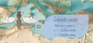
A total of 1791 Culicoides biting midges were captured in the study, of which 1596 were females (i.e. 1551 parous, 39 unfed and 6 engorged) and 195 were males. All the specimens were morphologically identified as Culicoides paolae (Fig. 2A) and only 18 parous females as Culicoides obsoletus complex (Fig. 2B). The sequencing analysis of COI gene amplicons, conducted in 20 and 5 specimens classified as C. paolae and C. obsoletus complex, respectively, confirmed the morphological identification. Four parous females (i.e. 0.26%) of the 1596 collected, tested positive for Dirofilaria spp. DNA presence. In particular, two specimens of C. paolae were positive for D. immitis (i.e. 0.13%) and two C. paolae were positive for D. repens (i.e. 0.13%) by qPCR. The D. immitis positive C. paolae were also positive for humans (i.e. n = 1; 99.7% nucleotide identity with the GenBank sequence, accession number MK617272) and dog DNA (i.e. n = 1; 100% nucleotide identity with the GenBank sequence, accession number, MW435612). The positive specimens for D. repens were collected in July in site 5 and were positive for human DNA (n = 2; 99.3 and 98.9% nucleotide identity, accession numbers MN503463 and LS997794).
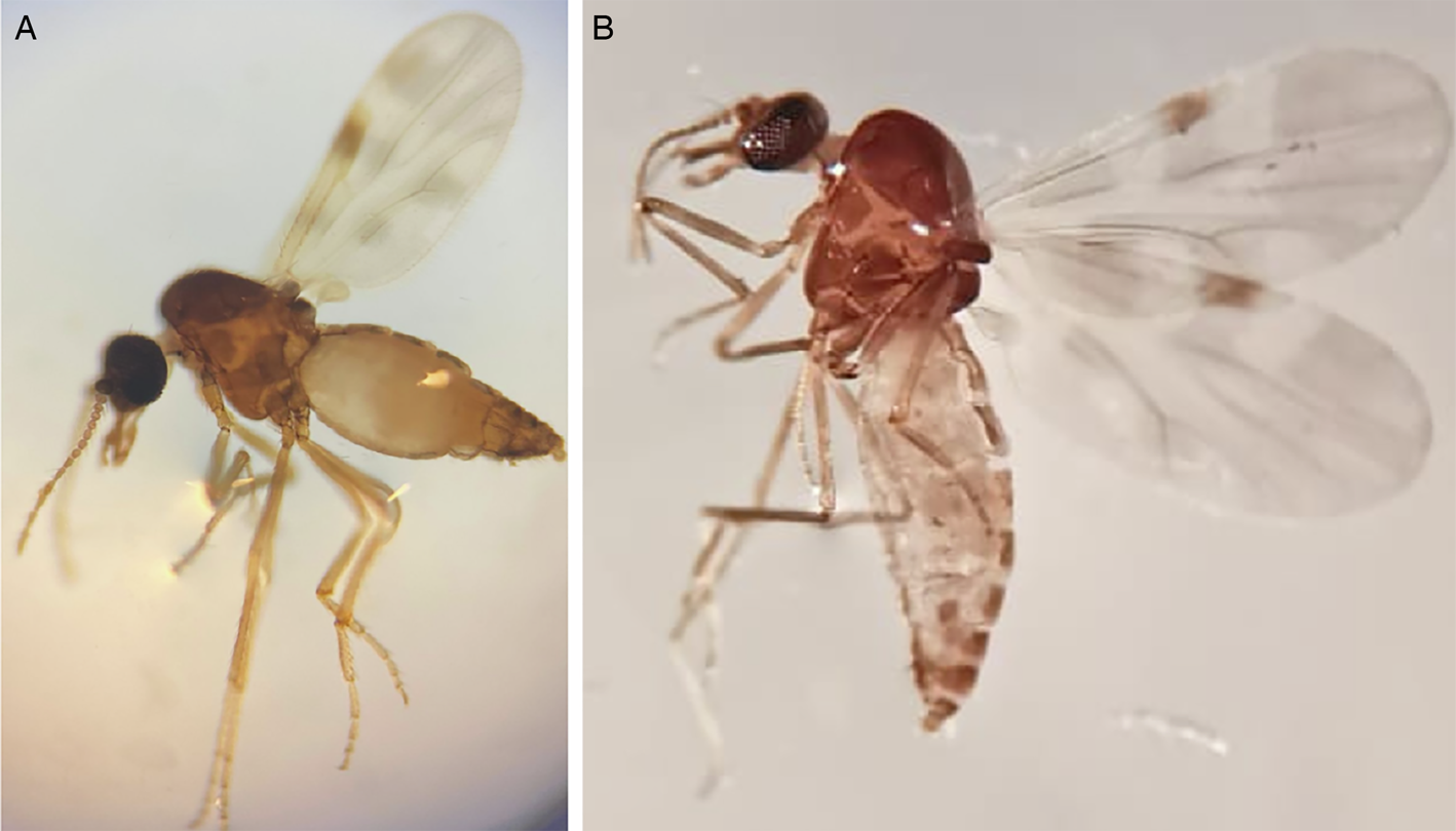
Fig. 2. Parous female specimen of Culicoides paolae (A) and Culicoides obsoletus complex (B).
Light traps were more effective at capturing biting midges compared to those baited with BG-lure; in fact, all the specimens, but one, have been collected with CDC light traps. During the same sampling period, the traps collected also 262 mosquitoes (i.e. 107 males and 155 females) belonging to six species (i.e. Aedes mariae, Culex pipiens, Aedes albopictus, Culex laticinctus, Culiseta annulate and Aedes caspius) as reported in Brianti et al. (Reference Brianti, Panarese, Napoli, De Benedetto, Gaglio, Bezerra-Santos, Mendoza-Roldan and Otranto2021).
Discussion
Dirofilaria immitis and D. repens have been herein molecularly identified for the first time in C. paolae biting midges collected from the island of Linosa, with a focus on HW infection. To date, Culicoides spp. have been mainly studied for their vectorial role in the transmission of bluetongue disease virus and African horse sickness virus (Borkent, Reference Borkent and Marquardt2004; Veiga et al., Reference Veiga, Martínez-de la Puente, Václav, Figuerola and Valera2018). However, they are, along with simuliids, the vectors of mansonellosis (Khanzadeh et al., Reference Khanzadeh, Khaghaninia, Maleki-Ravasan, Koosha and Oshaghi2020), and are also considered as competent vectors of onchocercidae worms (Mellor et al., Reference Mellor, Boorman and Baylis2000; Shelley and Coscaro'n, Reference Shelley and Coscaro'n2001; Ta-Tang et al., Reference Ta-Tang, Crainey, Post, Luz and Rubio2018; Khanzadeh et al., Reference Khanzadeh, Khaghaninia, Maleki-Ravasan, Koosha and Oshaghi2020), such as M. perstans and M. streptocerca (Shelley and Coscaro'n, Reference Shelley and Coscaro'n2001; Ta-Tang et al., Reference Ta-Tang, Crainey, Post, Luz and Rubio2018). Therefore, the presence of Dirofilaria spp. DNA in C. paolae biting midges may represent an additional clue in understanding the filarial vector competence, although the molecular detection alone is not sufficient to demonstrate this. On the other hand, the detection of filarial DNA in parous females indicates a potential survival of the nematode to the digestion process and, eventually, its development to further stages.
The species diversity of Culicoides spp. observed in Linosa is significantly lower compared to those observed either in the closest Sicily (i.e. 23 species; Blanda et al., Reference Blanda, Blanda, La Russa, Scimeca, Scimeca, D'Agostino, Auteri and Torina2018) or in North Africa (i.e. 14 species in Algeria, Hakima et al., Reference Hakima, Hwang and Lee2020; 54 species in Morocco, Bourquia et al., Reference Bourquia, Garros, Rakotoarivony, Gardès, Huber, Boukhari, Delécolle, Baldet, Mignotte, Lhor, Khallaayoune and Balenghien2019). The low species diversity observed in Linosa could be related to the dimension of the island, the paucity of ecotypes and microhabitats present as well as the limited presence of animals. Indeed, several factors may influence the presence, the abundance and composition of the Culicoides fauna, including both biotic (e.g. vegetation, human presence, other animal presence) and abiotic (light, soil, water, air, climatic factors, etc.) factors. The two species of Culicoides, C. paolae, the most representative species, and C. obsoletus complex, herein described, are widespread in the Mediterranean basin (Blanda et al., Reference Blanda, Blanda, La Russa, Scimeca, Scimeca, D'Agostino, Auteri and Torina2018; Bourquia et al., Reference Bourquia, Garros, Rakotoarivony, Gardès, Huber, Boukhari, Delécolle, Baldet, Mignotte, Lhor, Khallaayoune and Balenghien2019; Hakima et al., Reference Hakima, Hwang and Lee2020).
The molecular prevalence of Dirofilaria spp. in the Culicoides specimens collected in Linosa is lower compared to that of the mosquito population collected in the same island (0.6%; Brianti et al., Reference Brianti, Panarese, Napoli, De Benedetto, Gaglio, Bezerra-Santos, Mendoza-Roldan and Otranto2021). Interestingly, in the same period none of the mosquitoes analysed tested positive for D. repens, as well as any of the sampled dogs in the island scored positive for D. repens (Brianti et al., Reference Brianti, Panarese, Napoli, De Benedetto, Gaglio, Bezerra-Santos, Mendoza-Roldan and Otranto2021). Noteworthily, the two C. paolae positive for D. repens were positive for human DNA and, considering the low seroprevalence in humans in the island (i.e. 3.96%) (Mendoza-Roldan et al., Reference Mendoza-Roldan, Gabrielli, Cascio, Manoj, Bezerra-Santos, Benelli, Brianti, Latrofa and Otranto2021), the positive C. paolae may have acquired the D. repens microfilariae elsewhere, for instance in the island of Lampedusa where D. repens infects both dogs and humans (Brianti et al., Reference Brianti, Panarese, Napoli, De Benedetto, Gaglio, Bezerra-Santos, Mendoza-Roldan and Otranto2021; Mendoza-Roldan et al., Reference Mendoza-Roldan, Gabrielli, Cascio, Manoj, Bezerra-Santos, Benelli, Brianti, Latrofa and Otranto2021). Considering that mosquitoes are competent and recognized vectors of Dirofilaria spp., the epidemiological importance of Culicoides species should be carefully assessed in future experimental studies and by detecting further developmental stages in these insects. During the past few decades, other blood-sucking arthropods have been investigated as potential vectors of HW infection. For instance, fleas of the genus Ctenocephalides could develop the filarial nematode in their Malpighian tubules with the infective stage larvae living in the haemocoelic cavity of the arthropod (Summers, Reference Summers1940). Also, blackflies, belonging to the Simulium turgaicum complex, have been regarded as potential vectors of Dirofilaria spp. (Khanzadeh et al., Reference Khanzadeh, Khaghaninia, Maleki-Ravasan, Koosha and Oshaghi2020) even if they exist a definitive proof of their involvement in the life cycle of Dirofilaria spp. is yet to be assessed.
This study confirmed the general feeding habits of C. paolae, which can adapt to any type of hosts (e.g. humans and dogs). In the present survey, the majority of positive specimens fed on humans and/or dogs. This may be related to host availability in the island since only 10 cows and less than 20 sheep are present in Linosa (Brianti et al., Reference Brianti, Panarese, Napoli, De Benedetto, Gaglio, Bezerra-Santos, Mendoza-Roldan and Otranto2021). Considering this lack of hosts the probability that the infection could be transmitted from dogs to humans through Culicoides spp. bites will increase. Accordingly, the seroprevalences of D. immitis and D. repens in humans were, respectively, 7.7 and 3.96% in Linosa and 7.9 and 19.93% in Lampedusa (Mendoza-Roldan et al., Reference Mendoza-Roldan, Gabrielli, Cascio, Manoj, Bezerra-Santos, Benelli, Brianti, Latrofa and Otranto2021), being slightly higher compared to those recorded in other similar surveys conducted in the human populations (e.g. 10.8 and 0.2% of D. immitis and D. repens prevalence, respectively, in Romania-Moldova, Ciuca et al., Reference Ciuca, Simòn, Rinaldi, Kramer, Genchi, Cringoli, Acatrinei, Miron and Morchon2018; 6.4% of D. immitis prevalence in the Canary Islands, Cabrera et al., Reference Cabrera, Carretòn, Morchòn, Falcòn-Cordòn, Falcòn-Cordòn, Simòn and Montoya-Alonso2018). This high seropositivity for D. immitis in humans was most likely associated with the hyperendemicity of D. immitis in dogs.
This study represents the first evidence, even if circumstantial, of the role of Culicoides spp. in the life cycle of Dirofilaria spp. The evaluation of the epidemiological role of other blood-sucking arthropods, different from mosquitoes, is still of importance and should be further investigated to better control the Dirofilaria spp. transmission to both human and animal hosts, mainly in geographically confined epidemiological contexts such as Linosa Island.
Acknowledgements
The authors thank Valeria Blanda and Antonella Caruana (Istituto Zooprofilattico Sperimentale della Sicilia ‘A. Mirri’, Palermo, Italy) for their support during the laboratory work.
Author contributions
E. N., R. P. and E. B. contributed to conceptualization and project administration. E. B., E. N., R. P., J. A. M. R., F. L. R. and D. O. contributed to methodology. E. B., E. N., R. P. and D. O. contributed to formal analysis. E. N., R. P., E. B. and D. O. contributed to data curation. E. N., R. P. and E. B. contributed to writing and original draft preparation. E. N., R. P., F. L. R., J. A. M. R., D. O. and E. B. contributed to editing.
Financial support
This research received no specific grant from any funding agency, commercial or not-for-profit sectors.
Conflict of interest
The authors did not disclose any conflict of interest.


