


Introduction
The genus Joyeuxiella (Cyclophyllidea: Dipylidiidae) comprises 4 species, namely Joyeuxiella pasqualei, Joyeuxiella echinorhyncoides, Joyeuxiella fuhrmanni and Joyeuxiella rossicum (Jones, Reference Jones1983; Schuster, Reference Schuster2020). Joyeuxiella pasqualei is the most frequent species diagnosed in Mediterranean Europe. Its prevalence ranges from 1.2% to 75.9% in cat populations, from Greece, Portugal and Spain (Calvete et al., Reference Calvete, Lucientes, Castillo, Estrada, Gracia, Peribáñez and Ferrer1998; Millán and Casanova, Reference Millán and Casanova2009; Waap et al., Reference Waap, Gomes and Nunes2014; Symeonidou et al., Reference Symeonidou, Gelasakis, Arsenopoulos, Angelou, Beugnet and Papadopoulos2018). Although commonly found in domestic cats, J. pasqualei has also been reported in wild carnivores, such as Iberian lynx (Lynx pardinus) and red foxes (Vulpes vulpes) (Dalimi et al., Reference Dalimi, Sattari and Motamedi2006; Millán and Casanova, Reference Millán and Casanova2007).
Joyeuxiella fuhrmanni has so far been reported only in cats from Southern Africa (Baer, Reference Baer1924; Ortlepp, Reference Ortlepp1933) and the Middle East (Schuster et al., Reference Schuster, Thomas, Sivakumar and O'Donovan2009, Reference Schuster, Mustafa, Baskar, Rosentel, Chester and Knaus2016; Alagaili et al., Reference Alagaili, Mohammed and Omer2011; El Azazi et al., Reference El-Azazy, Abdou, Khalil, AL-Batel, Henedi and Tahrani2016), while J. echinorhynchoides has been found in red foxes (V. vulpes) from Greece, Spain, Iran and Saudi Arabia (Papadopoulos et al., Reference Papadopoulos, Himonas, Papazahariadou and Antoniadou-Sotiriadou1997; Nabavi et al., Reference Nabavi, Manouchehri Naeini, Zebardast and Hashemi2014; Sanchis-Monsonís et al., Reference Sanchis-Monsonís, Fanelli, Martínez-Carrasco and Tizzani2020), and J. rossicum in domestic and wild cats, wolves and red foxes in Moldova, Ukraine, Uzbekistan, Kazakhstan and Russia (reviewed by Schuster, Reference Schuster2020).
In domestic dogs, Joyeuxiella spp. have been scantly recorded, with few reports of J. pasqualei in Greece (0.8% prevalence), J. echinorhynchoides in Iran (14.3% prevalence) and Joyeuxiella sp. in Germany, Jordan and South Africa, with prevalences of 0.1%, 3.2% and 5%, respectively (Haralabidis et al., Reference Haralabidis, Papazachariadou, Koutinas and Rallis1988; Minnaar et al., Reference Minnaar, Krecek and Fourie2002; Barutzki and Schaper, Reference Barutzki and Schaper2003; Nabavi et al., Reference Nabavi, Manouchehri Naeini, Zebardast and Hashemi2014). Animals infected by these cestodes are usually asymptomatic. However, the attachment of adult parasites to the intestinal mucosa may provoke villous necrosis and pleating of the small intestine (Papazoglou et al., Reference Papazoglou, Diakou, Patsikas, Anagnostou, Vagiatis, Papastefanou and Kosmas2006).
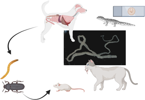
Information on the biology of Joyeuxiella spp. is scant, and gaps in the life cycle, such as the identification of putative intermediate hosts (e.g. coprophagous insects) are still to be clarified. Up to date, it is known that the adult cestodes inhabit the small intestine of carnivores (definitive hosts), and it is suggested that the cysticercoids develop in coprophagous insects, which may act as first intermediate hosts (Bowman et al., Reference Bowman, Hendrix, Lindsay and Barr2002; Papazoglou et al., Reference Papazoglou, Diakou, Patsikas, Anagnostou, Vagiatis, Papastefanou and Kosmas2006). Additionally, reptiles and small mammals (e.g. rodents, shrews) are regarded as secondary intermediate hosts (Bowman et al., Reference Bowman, Hendrix, Lindsay and Barr2002; Papazoglou et al., Reference Papazoglou, Diakou, Patsikas, Anagnostou, Vagiatis, Papastefanou and Kosmas2006; Galán-Puchades et al., Reference Galán-Puchades, Mas-Coma, Valero and Fuentes2021).
This study aims to report the occurrence of J. pasqualei in a dog living in a closed environment, which triggered us to delve into the biology of this cestode by further studying experimental models (i.e. laboratory mice and insects) and biological samples from a naturally infected cat and reptiles collected in the same garden.
Material and methods
Sampling procedures
A male 6-year-old German Shepherd dog (living in the garden of one of the authors; 41.081039° N, 16.991501° E, Bari, Apulia region, Southern Italy), physically healthy and with no clinical signs of gastrointestinal parasitosis, started releasing proglottids in the feces for several days. Fecal samples (n = 6) containing these proglottids were collected at different time points (2 samples per week) and sent to the parasitology laboratory of the Department of Veterinary Medicine at the University of Bari (Italy) for identification. Later, a female cat about 5 years of age was found road-killed near the garden, and subsequently necropsied at the pathology laboratory. At necropsy, adult cestodes were found in the small intestine, and adult nematodes were observed in the oesophagus, small and large intestines.
The proglottids were washed in saline solution (0.9%), counted and stored in 70% ethanol. Representative samples of the mature fresh proglottids from the dog and the cat were compressed and fixed in an alcohol–formalin–acetic acid (AFA), dehydrated in an ethanol series (30%, 50%, 70%) and stained with carmine acid mounted in Canada balsam (Schmidt, Reference Schmidt1986). A morphometrical examination of the mature proglottids and scolex size was performed using a compound microscope (Leica DM-LB2) and all measurements were obtained with Leica Las version 4.5.0 software (Leica Microsystems, Wetzlar, Germany). Cestodes were identified according to morphological keys by Jones (Reference Jones1983) and confirmed with molecular techniques as described below.
Italian wall lizards (Podarcis siculus; n = 28), and common wall geckos (Tarentola mauritanica; n = 13) were also collected in the garden and its surroundings to investigate natural infection by J. pasqualei. These animals were submitted to necropsy soon after euthanasia.
Experimental infection of rodents
A total of 10 laboratory mice (Mus musculus) were experimentally infected using an orogastric tube with 50 μL of the macerated content of gravid proglottids recovered from the dog's feces. Animals were kept in cages with food and water provided ad libitum. Mice were divided in 2 groups (A and B) of 5 individuals each, from which animals were euthanized after 3 weeks (group A) and 4 weeks (group B) post experimental infection. Euthanasia was performed by cervical dislocation by a trained veterinarian. Thereafter, animals were dissected using a protocol previously described (Petersen, Reference Petersen1986) and examined for the presence of cysticercoids in the body cavities and organs at 100× magnification using an optical microscope. All ethical standards and requirements regarding experimental work on rodent model host were fulfilled, and experiment was carried out in an accredited facility.
Experimental infection of invertebrate hosts
Darkling beetles (Tenebrio molitor; adults n = 50; larvae n = 50) were separated in groups of 10 specimens, and 12 h before the experimental feeding the insects were left without food in separate plastic cages (15 × 15 × 8 cm) at 25°C and 65% relative humidity. The experimental feeding was performed with 10 μL of the macerated content of gravid proglottids recovered from the dog's feces. The macerated content was checked under the microscope to confirm the presence of egg capsules, and then placed in small pieces of apple and offered to the insects, which were observed for 1 h to confirm their active feeding. The insects were kept at a constant temperature (25°C) and humidity (65%) in separate group cages for larvae and adults and provided with water and food (apple and ground nuts) ad libitum. Four weeks post infection, the adult arthropods were euthanized and dissected to check for the presence of cysticercoids. The larvae were kept alive until they become adults, which were also euthanized and dissected.
Molecular and phylogenetic analysis
Genomic DNA extraction of proglottids, cysts and insects was performed using a commercial kit (QIAamp DNA Micro Kit; Qiagen, Hilden, Germany) following the manufacturer's instructions. Thereafter, samples were screened by conventional PCR using the forward JB3 (5′-TTTTTTGGGCATCCTGAGGTTTAT-3′), and reverse primers JB4.5 (5′-TAAAGAAAGAACATAATGAAAATG-3′), which amplify a ~400 bp of the cox1 gene (Bowles et al., Reference Bowles, Blair and McManus1992). Amplicons were purified and sequenced in both directions, using the Taq Dye Deoxy Terminator Cycle Sequencing Kit v.2 (Applied Biosystems, Foster City, California, USA) in an automated sequencer (ABI-PRISM 377; Applied Biosystems, Foster City, California, USA). Sequences were edited and aligned using the Mega7 software and compared with those available in the GenBank database, using the Basic Local Alignment Search Tool (BLAST, http://blast.ncbi.nlm.nih.gov/Blast.cgi).
Phylogenetic analysis of the cox1 gene sequence of J. pasqualei was performed along with other cestode sequences available in the GenBank database. The phylogenetic tree was inferred using the maximum likelihood (ML) method based on the Hasegawa–Kishino–Yano model (Hasegawa et al., Reference Hasegawa, Kishino and Yano1985), which was selected via Bayesian information criterion (BCI) by the best-fitting substitution model using MEGA7 software (Kumar et al., Reference Kumar, Stecher and Tamura2016). A discrete Gamma distribution was used to model evolutionary rate differences among sites [5 categories (+G, parameter = 0.3264)]. The analysis was performed with 1000 bootstrap replications using MEGA7 software (Kumar et al., Reference Kumar, Stecher and Tamura2016). Ancylostoma ceylanicum cox1 sequence (KC247743.1) was used as outgroup.
Morphological and molecular confirmation
Voucher samples of the adult cestodes (collected from the cat), proglottids (collected on the dog's feces) and cysts (collected from geckoes), were morphologically identified by 2 independent experts from 2 different reference laboratories of the University of Sassari and University of Bari, Italy. Thereafter, these samples were submitted to molecular analysis and the obtained sequences were deposited in the GenBank database according to the blind species level confirmation by both reference laboratories.
Results
Proglottids recovered from the dog's feces and adult cestodes detected at necropsy of the cat were morphologically identified as J. pasqualei. Briefly, the white strobilae were flattened dorso-ventrally with each proglottid containing 2 lateral genital pores. Strobilae were 30.58 ± 7.30 cm long and 1.11 ± 0.31 mm wide with ± 170 proglottids on average. The scolex, 526.9 μm ± 91.7 in diameter, was provided with 4 round suckers and a conical rostellum, 141 μm long × 118 μm wide, was followed by a short unsegmented neck (Fig. 1). Immature segments were 1.30 ± 0.30 mm long and 0.82 ± 0.26 mm wide while the mature segments were 2.57 ± 0.90 mm long and 0.97 ± 0.43 mm wide and contained 2 sets of female reproductive organs. The testes were located anteriorly and posteriorly to vasa deferentia. Gravid segments were rice seed shaped, 4.26 ± 0.43 mm long and 1.56 ± 0.09 mm wide (Fig. 1) and egg capsules containing a single egg (Fig. 2) were distributed between and also laterally to the longitudinal excretory vessels (Fig. 1).

Fig. 1. (a) Single proglottids of J. pasqualei collected in the feces of a dog – scale bar 2000 μm. (b) Joyeuxiella pasqualei adult, presenting scolex and strobila – scale bar 1500 μm. (c) Conical rostellum, see arrow – scale bar 300 μm. (d) Gravid proglottid containing single eggs – scale bar 200 μm.

Fig. 2. Egg capsule of J. pasqualei recovered from gravid proglottid – scale bar 20 μm.
An average of 5 proglottids were recovered from each fecal sample of the dog, meanwhile a total of 17 adults were recovered from the small intestine of the cat. Two out of 13 (15.4%) wall geckoes had natural infections of J. pasqualei cysts in the liver and attached to the small intestine (Fig. 3). All 28 P. siculus lizards were negative for the parasite. At necropsy of the experimentally infected rodents, no cysts were observed after 3- and 4-weeks post infection. None of the dissected T. molitor beetles had cysticercoids of the parasite after 4 weeks post experimental infection.

Fig. 3. Joyeuxiella pasqualei cysts isolated from liver and intestine of Tarentola mauritanica geckos. (a) Cysts spread through the intestinal serosa of gecko – scale bar 1 mm. (b) Cyst isolated from the liver of gecko – scale bar 500 μm. (c) Cyst after removal of the first wall layer – scale bar 500 μm. (d) Presence of J. pasqualei larvae within the cyst – scale bar 500 μm. (e and f) Larvae outside the cysts – scale bar 250 μm. Notice the presence of a conical rostellum characteristic of J. pasqualei.
Nucleotide sequences were obtained from the adult cestodes, proglottids and cysts morphologically identified as J. pasqualei and the BLAST analysis of the sequences showed the highest nucleotide similarities with the cox1 of Versteria sp. (i.e. 83.84%; accession number: KT223035.1), Echinococcus sp. (i.e. 83.67%; accession number: JQ690286.1), Raillietina sonini (i.e. 83.59%; accession number: EU665479.1), Taenia polyacantha (i.e. 83.33%; accession number: EU544581.1), and with the complete genome of D. caninum (i.e. 81.61%; accession number: MN099047.1) available in GenBank. In addition, when blasting the only J. pasqualei cox 1 sequence (product size 100 bp) available in Genbank (KY310707.1) with our representative sequence (product size 396b), a nucleotide identity of 93% was found (cover query of 100%). Sequences obtained were deposited at the Genbank database as J. pasqualei under the accession number: ON981095.
At the phylogenetic analysis of the cox1 gene, J. pasqualei sequence from our study clustered in a sister clade with the species Progamotaenia macropodis, being also related to a bigger taxon that includes D. caninum (Fig. 4).

Fig. 4. Phylogram based on cox1 sequences inferred by using the ML method according to the Hasegawa–Kishino–Yano model. Numbers near the nodes are the support bootstrap values ordered as ML. Ancylostoma ceylanicum was used as outgroup. Joyeuxiella pasqualei representative sequence from this study is in bold.
Discussion
This study reports the occurrence of J. pasqualei in a domestic dog and cat, providing further information on the biology of this parasite. This cestode has been considered a frequent parasite affecting cats in Africa, Asia, Europe and the Middle East (Schuster et al., Reference Schuster, Mustafa, Baskar, Rosentel, Chester and Knaus2016); however, its role as a parasite of dogs has been scarcely studied with scant information on its epidemiology in this animal species. The above situation could be due to the morphological similarities of proglottids with the well-known D. caninum, leading to misdiagnosis by veterinary practitioners.
The biological cycle of Joyeuxiella spp. is still not clear and previous experimental studies investigating the development of this parasite in putative first intermediate hosts have failed (Witenberg, Reference Witenberg1932; Ortlepp, Reference Ortlepp1933; Schuster and Montag, Reference Schuster and Montag2000). For example, in an experiment with dung beetles of the genera Hister, Aphodius and Onthophagus fed with cow dung containing broken up and whole Joyeuxiella sp. proglotids, Ortlepp (Reference Ortlepp1933) did not find any larval forms of the cestode after the 10th and the 24th day post initial feeding. Similarly, in the present study, meal worms and their adults did not act as first intermediate hosts for J. pasqualei as no metacestodes were observed in the organs and muscles of the insects. Therefore, further studies focusing on the experimental infection of J. pasqualei in possible intermediate hosts are needed to better understand the biology of this cestode.
The finding of J. pasqualei cysts in the liver and intestines of T. mauritanica (15.4%) collected on the garden supports evidence of geckoes as second intermediate hosts in the life cycle of this parasite (Schuster, Reference Schuster2020). In particular, this gecko species has been regarded as one of the main species known to harbour J. pasqualei cysts (Schuster, Reference Schuster2020). Small mammals, such as rodents, are also suggested to act as secondary intermediate hosts, with the few reports available mostly for J. rossicum (Schuster, Reference Schuster2020). In the present study, cysts did not develop in laboratory mice after experimental feeding with eggs from gravid proglottids, further suggesting that in the life cycle of J. pasqualei, a first intermediate host (e.g. coprophagous insects with cysticercoids) is needed to establish infection in the second intermediate hosts.
Infection by J. pasqualei in pets (i.e. cats, dogs) has been associated with villous necrosis, and pleating of the small intestine (Papazoglou et al., Reference Papazoglou, Diakou, Patsikas, Anagnostou, Vagiatis, Papastefanou and Kosmas2006). Nevertheless, most reported cases describe asymptomatic infections in these hosts (Calvete et al., Reference Calvete, Lucientes, Castillo, Estrada, Gracia, Peribáñez and Ferrer1998; Waap et al., Reference Waap, Gomes and Nunes2014; Symeonidou et al., Reference Symeonidou, Gelasakis, Arsenopoulos, Angelou, Beugnet and Papadopoulos2018). Indeed, in the case described herein, the dog did not present any clinical sign, and was free of the infection after anthelmintic treatment. In the case of the cat, no intestinal macroscopic lesions were observed during the necroscopy, even though the animal presented a high parasitic load of J. pasqualei.
The molecular analysis of the present study provides more robust data for the diagnostic confirmation of J. pasqualei as we provided cox 1 sequences (396 bp) according to the morphological confirmation of adults and immature forms of the parasite. Although a J. pasqualei sequence from a previous study is already available in Genbank (Poon et al., Reference Poon, Tam, Lau, Cheng, Yuen, Schuster and Woo2017), the small product size (100 bp) provided does not bring sufficient information for further analyses. In addition, when comparing the previous sequence with ours, we found some differences in 6 nucleotide positions, suggesting a considerable genetic diversity within this species.
Data presented herein highlight the occurrence of J. pasqualei in a dog and provide further insights into the biology of this cestode. In addition, the inability of laboratory mice to become infected by direct ingestion of gravid proglottids suggests the need of a first intermediate host (possibly coprophagous insects) to the completion of this parasite's life cycle. Therefore, further studies are advocated to elucidate the transmission biology of this little-known tapeworm.
Acknowledgements
The authors would like to thank Lucia Macedo (Federal Rural University of Pernambuco) for the technical support on molecular analysis.
Data availability
The data that support the findings of this study are available from the corresponding author, upon reasonable request.
Author's contributions
M. A. B.-S. performed methodology, analysis, wrote the first draft of the manuscript and reviewed the last version of the manuscript. J. A. M.-R., R. P. L., G. A., R. S., A. V., G. S. and D. M. performed methodology, and reviewed the last version of the manuscript. D. O. conceived and supervised the study and reviewed the last version of the manuscript.
Financial support
This research received no specific grant from any funding agency, commercial or not-for-profit sectors.
Conflict of interest
The authors declare there are no conflicts of interest.
Ethical standards
Protocols for the collection of reptiles were approved by the ethical committee of the Department of Veterinary Medicine of the University of Bari, Italy (Prot. Uniba 12/20), and authorized by the Ministry for Environment, Land and Sea Protection of Italy (Approval Number 0073267/2019), the Societas Herpetologica Italica and the Istituto Superiore per la Protezione e la Ricerca Ambientale (Approval Number 71216). Also, the authorization for the experimental infection of mice was provided by the ethical committee (approval PP03-21).


