


INTRODUCTION
Steinernema carpocapsae is an insect entomopathogenic nematode that is symbiotically associated with the bacterium Xenorhabdus nematophila. This symbiotic complex is considered a promising biological control agent against a wide range of important agricultural insect pests (Ehlers, Reference Ehlers2001; Kaya et al. Reference Kaya, Aguillera, Alumai, Choo, de la Torre, Fodor, Ganguly, Hazir, Lakatos, Pye, Wilson, Yamanaka, Yang and Ehlers2006). Entomopathogenic nematodes are highly virulent and usually kill the insect host quickly, and considerable interpopulation variation has been demonstrated (Rosa et al. Reference Rosa, Cabral and Simoes2002). The rate of parasitism and of mortality caused by a nematode strain is both host- and developmental stage-specific (Hao et al. Reference Hao, Montiel, Lucena, Costa and Simões2012). Virulence factors from S. carpocapsae are crucial to the success of this biological control agent (Shapiro-Ilan et al. Reference Shapiro-Ilan, Lewis, Son and Tedders2003). The virulence of the symbiotic complex was attributed to the ability of the bacterium to excrete a large set of toxins and enzymes (Forst et al. Reference Forst, Dowds, Boemare and Stackebrandt1997). In S. carpocapsae, virulence factors that were lethal to insects were identified from its secreted compounds (Burman, Reference Burman1982; Laumond et al. Reference Laumond, Simões and Boemare1989).
Expressed sequence tag (EST) analysis facilitated the identification of genes encoding proteins that were released by S. carpocapsae and that were highly homologous to virulence factors released by other parasitic nematodes (Hao et al. Reference Hao, Montiel, Abubucker, Mitreva and Simões2010), including an aspartic protease. Aspartic proteases have catalytic aspartic acid residues in their active site clefts; these proteases include pepsins, renins, cathepsins D and E and chymosins (Dunn, Reference Dunn2002; Williamson et al. Reference Williamson, Brindley, Knox, Hotez and Loukas2003). Two well-known aspartic proteases identified in humans are pepsin and renin, but aspartic proteases have also been identified in various parasitic species (Antonov et al. Reference Antonov, Ginodman, Kapitannikov, Barshcvskaya, Gurova and Rumsh1978). In parasitic nematodes, aspartic proteases have been associated with the digestion of host haemoglobin in the trichostrongylid Haemonchus contortus and in the hookworms Ancylostoma caninum and Necator americanus (Williamson et al. Reference Williamson, Brindley, Abbenante, Prociv, Berry, Girdwood, Pritchard, Fairlie, Hotez, Dalton and Loukas2002). Additionally, aspartic proteases have been shown to degrade skin macromolecules and to aid skin penetration in hookworms, suggesting that the role of aspartic proteases in nematode parasitism is not limited to the digestion of haemoglobin (McKerrow et al. Reference McKerrow, Brindley, Brown, Gam, Staunton and Neva1990). These additional functions of aspartic proteases in hookworms suggest that aspartic proteases may play a role in the biology of parasitic nematodes that do not feed on blood, such as Strongyloides stercoralis, Onchocerca volvulus and Brugia malayi, in which aspartic proteases have previously been identified (McKerrow et al. Reference McKerrow, Brindley, Brown, Gam, Staunton and Neva1990; Jolodar and Miller, Reference Jolodar and Miller1998). In addition, aspartic proteases have been found in parasitic nematodes, including Schistosoma mansoni (Schulmeister et al. Reference Schulmeister, Heyers, Morales, Brindley, Lucius, Meusel and Kalinna2005), Schistosoma japonicum (Becker et al. Reference Becker, Harrop, Dalton, Kalinna, McManus and Brindley1995), and the free-living nematode, Caenorhabditis elegans (Geier et al. Reference Geier, Banaj, Heid, Bini, Pallini and Zwilling1999).
In S. carpocapsae, serine proteases are associated with the invasion of insects (Toubarro et al. Reference Toubarro, Lucena-Robles, Nascimento, Santos, Montiel, Veríssimo, Pires, Faro, Coelho and Simões2010) or with the evasion of insect defences (Balasubramanian et al. Reference Balasubramanian, Hao, Toubarro, Nascimento and Simões2009, Reference Balasubramanian, Toubarro and Simões2010); a metalloprotease (Jing et al. Reference Jing, Toubarro, Hao and Simões2010) and aspartic proteases have also been described in this species (Balasubramanian et al. Reference Balasubramanian, Toubarro, Nascimento, Ferreira and Simões2012a, Reference Balasubramanian, Nascimento, Ferreira, Martinez and Simõesb). Although S. carpocapsae infections in insects are increasingly frequent, our understanding of the host–parasite relationship is limited. The main pathogenic mechanisms remain unclear, particularly the role of the nematode compared with the role of the host. However, little is known about the molecular events taking place during nematode infection.
Steinernema carpocapsae does not feed on blood; thus, the role of aspartic proteases in S. carpocapsae parasitic processes remains unclear. However, aspartic proteases may be involved in the digestion of other host macromolecules. Therefore, the goal of this study is to identify the S. carpocapsae aspartic protease from expressed genes, to study aspartic protease gene expression in S. carpocapsae developmental stages and in vitro induced nematodes with an insect homogenate. This report provides a transcriptional analysis of the putative genes that are likely associated with nematode parasitism and/or development. Our results from S. carpocapsae demonstrate that transcriptomic and genomic sequence data can be combined to identify a gene's function and that doing so can provide fresh insight into the genetic mechanisms associated with parasitism. In the present work, we cloned a putative aspartic protease gene, sc-asp110, and examined its expression in different S. carpocapsae developmental stages, allowing a unique comparison among the parasite's life-cycle stages.
MATERIALS AND METHODS
Insect homogenate preparations for induction and nematode collection
Galleria mellonella were reared with pollen and wax (1 : 1) in plastic boxes in the dark at 27 °C with 65% relative humidity (RH). The larvae were collected, frozen in liquid nitrogen, ground, and homogenized with 1% Tyrode using a homogenizer (Glas-Col, USA). The homogenate was centrifuged at 2400 g for 15 min at 4 °C; then, the supernatant was collected, and a 1% antibiotic solution (penicillin, streptomycin and neomycin; Sigma, USA) was added to 5% of the supernatant that was used for the nematode induction. Infective juveniles (IJs) were produced in G. mellonella larvae, harvested in a white trap and stored at 10 °C for 1–3 months (Dutky, Reference Dutky1959). Ten millilitres (25 000 mL−1) of IJs was surface-disinfected with 0·5% sodium hypochlorite (bleach) for 10 min followed by washing 3 times with 0·8% NaCl at room temperature (23±2 °C) and used for the induction in different defined times.
Total RNA isolation and cDNA synthesis
Total RNA was isolated from the desired nematodes using TRIzol (Invitrogen, Germany) according to the manufacturer's instructions. cDNA was synthesized using the Super Script TM First-Strand Synthesis System for RT-PCR (Invitrogen, Germany) according to the manufacturer's instructions.
Full-length cDNA cloning
Sc-ASP110 5′ and 3′ RACE cDNA were obtained using the SMART™ RACE cDNA Amplification Kit (Clontech-Takara, UK). Based on the sequence in the EST library constructed in our laboratory, the specific primers Sc-ASP110 5′ (5′-GAGTTGATGACCCAGAGGTTGGAG-3′) and Sc-ASP110 3′ (5′-ATCCAGGTCGGCTCCTACAA-3′) were designed for 5′RACE and 3′RACE, respectively. The PCR conditions were as follows: 94 °C for 5 min followed by 25 cycles at 94 °C for 30 s, 56 °C for 30 s and 72 °C for 30 s, with a final extension at 72 °C for 7 min. The PCR product was re-amplified for another 25 cycles. The PCR products were cloned into the pCR4-TOPO vector (Invitrogen, Germany) and then transformed into TOP10 cells by heat shock. The DNA inserts isolated from positive clones were sequenced (Stabvida, Portugal), and full-length cDNA was obtained by joining the two fragments.
Genomic DNA extraction
Genomic DNA was extracted according to the method described by Ausbel (Reference Ausbel1989). Briefly, 1 mL of IJs was washed 3 times with distilled water and homogenized in liquid nitrogen. A 500 μL aliquot of lysis buffer (100 mm Tris–HCl (pH 8·0), 200 mm NaCl, 50 mm EDTA, 0·5% SDS and 0·2 mg mL−1 proteinase K) was added to the homogenate. The solution was heated to 65 °C for 2 h, and the solution was cooled at room temperature. A 500 μL aliquot of phenol was added to the solution; then, the solution was homogenized and centrifuged at 12 000 g for 10 min at 4 °C. The supernatant was collected, and 420 μL of chloroform and isoamyl alcohol (24 : 1) was then added. After gentle mixing, the solution was centrifuged at 12 000 g for 10 min at 4 °C. Two volumes of 95% cold ethanol were added to the supernatant; the solution was incubated at −20 °C for 1 h and was centrifuged at 16 000 g for 10 min to precipitate the DNA. The DNA pellet was then washed with 2 volumes of 70% ethanol. The DNA pellet was air dried, resuspended in 50 μL of TE with RNase (20 μg mL−1) and incubated at 37 °C for 1 h. The DNA quality and quantity were assessed by 0·8% (w/v) agarose gel electrophoresis and by UV spectrophotometry, respectively.
Genomic DNA cloning
To obtain the genomic sequence, DNA was amplified by PCR using the gene-specific primers Sc-ASP110-F (5′-ATGAAGCTGAAGGCCTCCGGC-3′) and Sc-ASP110-R (5′-TTAAAGGCTGTGGTGGGCCTTG-3′) designed from the full-length cDNA. The thermal cycling conditions were as follows: 94 °C for 5 min, followed by 30 cycles at 94 °C for 30 s, 55 °C for 30 s and 72 °C for 1 min, with a final extension at 72 °C for 7 min. The PCR product was confirmed by electrophoresis, cloned into the pCR4-TOPO vector (Invitrogen, Germany), and then transformed into TOP10 cells by heat shock. The DNA inserts in plasmids that were isolated from positive clones were then sequenced (Stabvida, Portugal).
Analysis of sc-asp110 gene expression by qRT-PCR
Total RNA was isolated from different nematode development stages (L1/L2, L3 IJs, L3 haemocelium, L3 gut, L4 and adult) in infected G. mellonella larvae at 0, 6, 12, 24, 48 and 72 h after induction. The RNA was reverse-transcribed into cDNA products and the relative gene expression of sc-asp110 was quantified by qRT-PCR using 18S rRNA as an endogenous control. The primers for 18S rRNA were 18S-F (5′-TGATGAGGAGCTAATCGGAAACG-3′) and 18S-R (5′-CACCATCCACCGAATCAAGAAAG-3′). The primers for Sc-ASP110-F were (5′-GCAAGTCAAACACCTTCAAGCC-3′) and Sc-ASP110-R (5′-GCGGTCCATCCATACGGTAAAG-3′). qRT-PCR was performed using SYBR green mix according to the manufacturer's instructions (Applied Biosystems, USA). The qRT-PCR conditions were as follows: 95 °C for 10 min, and 60 cycles at 95 °C for 15 s and 60 °C for 60 s. qRT-PCR data from three replicate samples were analysed using Relative Manager Software (Applied Biosystems, USA) to estimate the transcript levels of each sample using the 2−∆∆ Ct method (Livak and Schmittgen, Reference Livak and Schmittgen2001).
In situ hybridization
In situ hybridization was performed using nematodes in the parasitic stage as described by de Boer et al. (Reference de Boer, Yan, Davis, Smant and Baum1998), with the following modifications. Nematodes were fixed in 1× paraformaldehyde at 4 °C for 18 h, followed by 4 h incubation in 0·1× paraformaldehyde at 22 °C. Partial digestion with proteinase K (0·5 mg mL−1) was performed at 22 °C for 20 min. Sc-ASP110-F (5′-GCAAGTCAAACACCTTCAAGCC-3′) and Sc-ASP110-R (5′- GCGGTCCATCCATACGGTAAAG-3′) primers were used to amplify a 287-bp PCR fragment, which was then column-purified and used as a template in a symmetric PCR to amplify sense and antisense DNA with primers Sc-ASP110-F and Sc-ASP110-R, respectively. Single-stranded Sc-ASP110-specific DNA was labelled with digoxigenin (Roche, Germany) and was hybridized overnight at 55 °C under constant agitation at a dilution of 1 : 10. DIG-labelled probes were visualized by incubation with alkaline-phosphatase conjugated anti-DIG antibody (1 : 1000 dilutions) and with NBT/BCIP substrate colour reactions.
Bioinformatics analysis
Protein motifs were identified using the Simple Modular Architecture Research Tool (SMART) (http://smart.embl-heidelberg.de/) and the Conserved Domain search from NCBI-CD search with threshold score of 67.835 (http://www.ncbi.nlm.nih.gov/Structure/cdd/wrpsb.cgi). The signal peptide was analysed using the SignalP program (http://www.cbs.dtu.dk/services/SignalP/), prediction of non-classical protein secretion-SecretomeP with threshold score is 0.5 (http://www.cbs.dtu.dk/services/SecretomeP/), and the theoretical isoelectric point and the molecular weight were predicted using the Compute pI/MW program (http://expasy.org/tools/protparam.html). Sequence similarities were analysed using the BLAST program from NCBI (http://www.ncbi.nlm.nih.gov/BLAST/). A multiple sequence alignment was generated using the CLUSTAL Win BioEdit 7.0 program. Phylogenetic analysis was conducted using the MEGA5 program. Homology modelling data were obtained by the I-TASSER Zhang-Server with C-score of 1.56 (http://zhanglab.ccmb.med.umich.edu/), and homology modelling was performed with PyMOL (http://www.pymol.org/). Protein disulfide bond connectivity was analysed by DiANNA (http://clavius.bc.edu/~clotelab/DiANNA/).
RESULTS
Sequence analysis of Sc-ASP110
The full-length cDNA of Sc-ASP110 was deposited in the GenBank database under the accession number JX392861. Sc-ASP110 consists of 1112 nucleotides, with an open reading frame of 1026 bp, a 3-UTR region of 86 bp and a poly-A signal (AATAAA) (Fig. 1). The ORF encodes a protein of 341 amino acid residues (GenBank accession No. AFP21684) with a typical catalytic aspartic domain (aa 18–337). The putative mature protein contains 341 amino acid residues with a calculated theoretical pI of 4·7 and a molecular mass of 37·1 kDa.
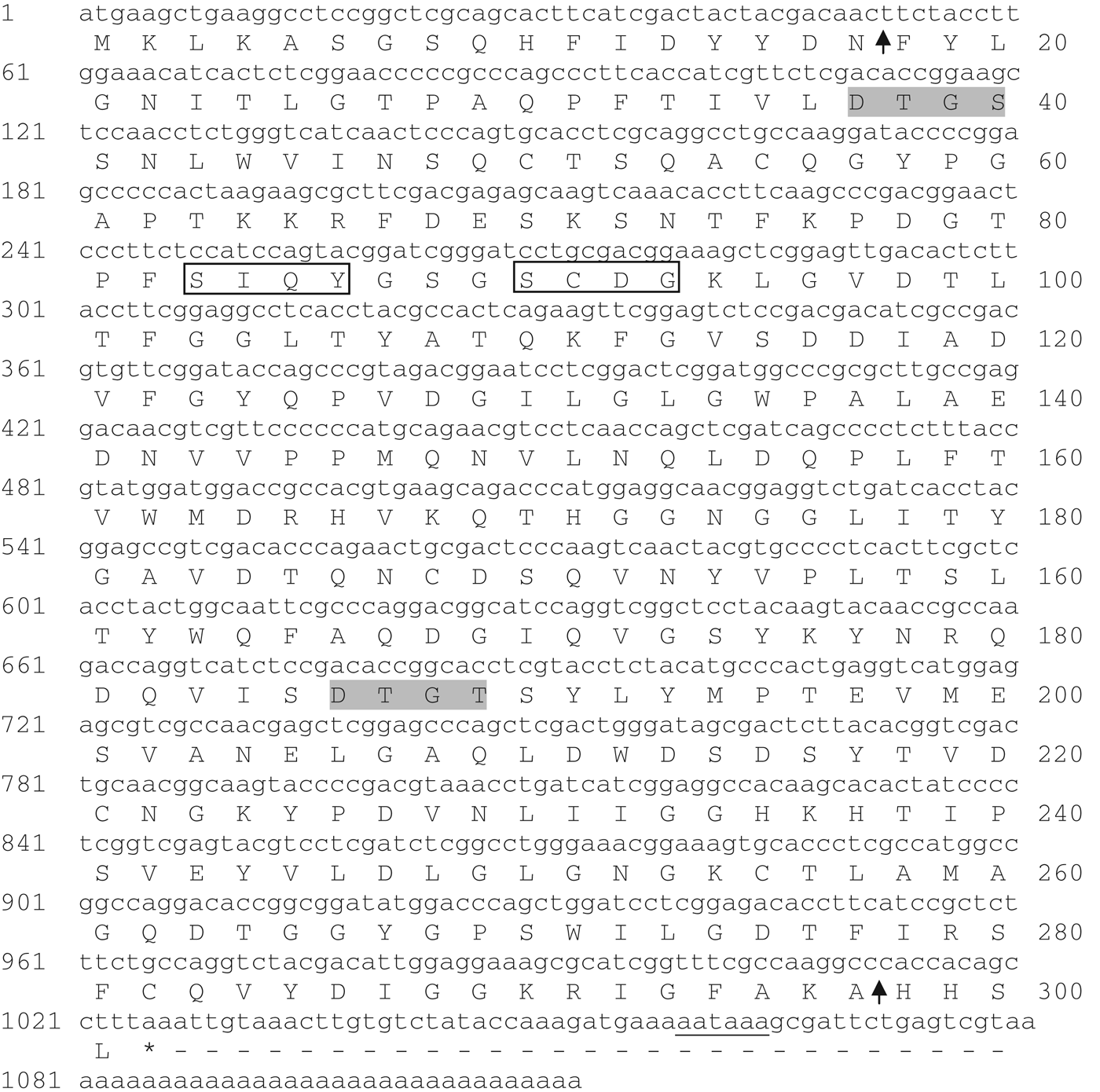
Fig. 1. Nucleotide sequence of the full-length cDNA and deduced amino acid sequence of Sc-ASP110. The number on the left is for the nucleotide sequence, whereas the number on the right is for the amino acid sequence. In the nucleotide sequence, the polyadenylation signal is underlined. In the amino acid sequence, the aspartyl domain is marked with an arrow. Catalytic motifs are shaded and active sites are boxed. The asterisk (*) at the end of the amino acid sequences shows the stop codon.
Sequence homology and phylogenetic analysis
A similarity analysis using the NCBI-BLASTp program showed that the deduced amino acid sequence of Sc-ASP110 has the following similarities: 77% to Steinernema feltiae (GenBank Accession No. ACS32298), 65% to Caenorhabditis briggsae CBR-ASP-1 (XP_002647755), 64% to Caenorhabditis remanei CRE-ASP-1 (XP_003093500), 64% to Caenorhabditis brenneri CBN-ASP-1 (EGT51351), 61% to C. elegans ASP-1 (NP_741677), 60% to S. stercoralis (AAD09345), 60% to Strongyloides ratti (ACR56785), 56% to Parastrongyloides trichosuri ASP-1 (AAT79343), 48% to C. elegans (AAB06576) and 45% to C. remanei CRE-ASP-6 (XP_003110463) (Fig. 2).

Fig. 2. Alignment of the amino acid sequence of aspartic protease from Steinernema carpocapsae (Accession No. JX392861) with aspartic proteases from Steinernema feltiae (ACS32298), Caenorhabditis briggsae (XP_002647755), Caenorhabditis remanei (XP_003093500), Caenorhabditis elegans (NP_741677), Strongyloides stercoralis (AAD09345), and Strongyloides ratti (ACR56785). Areas shaded in green followed by blue indicate a high degree of homology (>50% similarity), and unshaded areas are regions of variability among the proteins. This figure is reproduced in colour online.
To search for a phylogenetic relationship between Sc-ASP110 and other homologous aspartic proteases, a phylogenetic tree was reconstructed based on the deduced amino acid sequences from 18 species, including parasitic and free-living nematodes. This analysis showed that Sc-ASP110 was clustered with the aspartic protease of the closely related parasitic nematode S. feltiae (Fig. 3).

Fig. 3. A molecular phylogenetic tree of Sc-ASP110 was generated by the neighbour-joining (NJ) method using the MEGA 5 program. An unrooted phylogenetic tree was generated based on the alignment of the amino acid sequences from 18 animal species. The numbers indicate the frequency with which this node is recovered per 100 bootstrap replications in a total of 1000. The scale bar indicates an evolutionary distance of amino acid substitutions per position. The accession numbers of amino acid sequences were as follows: Caenorhabditis brenneri CBN-ASP-1 (EGT51351), Caenorhabditis brenneri ASP-1 (ACE00320), Caenorhabditis remanei CRE-ASP-1 (XP_003093500), Caenorhabditis briggsae CBR-ASP-1 (XP_002647755), Caenorhabditis elegans ASP-1 (NP_741677), Caenorhabditis elegans ASP-2 (AAB06576), Steinernema carpocapsae (AFP21684), Steinernema feltiae (ACS32298), Strongyloides stercoralis (AAD09345), Strongyloides ratti ASP-1 (ACR56785), Caenorhabditis elegans ASP-3 (CAA08899), Caenorhabditis briggsae CBR-ASP-5 (XP_002635086), Caenorhabditis remanei CRE-ASP-5 (XP_003110542), Caenorhabditis elegans ASP-5 (NP_505135), Caenorhabditis brenneri CBN-ASP-6 (EGT57061), Caenorhabditis briggsae CBR-ASP-6 (XP_002635088), Caenorhabditis elegans ASP-6 (NP_505133) and Caenorhabditis remanei CRE-ASP-6 (XP_003110463).
Gene structure of sc-asp110
The sc-asp110 gene was 1073 bp without the untranslated region (UTR) (GenBank Accession No. JX499022). The sc-asp110 gene structure consists of two exons and one intron (47 bp) that has similarity to the aspartic protease gene of S. mansoni (GenBank Accession No. AJ318869). A typical (GT-AG) intron splice motif was identified in the sc-asp110 gene, whereas in S. mansoni, a (GG–AG) intron splice motif has been described.
Analysis of sc-asp110 gene expression
The expression level of sc-asp110 was analysed throughout the S. carpocapsae life cycle in parasitized G. mellonella larvae by qRT-PCR. The results showed that this gene is upregulated at the L4 stage with a small decrease observed at the adult stage, followed by the L1/L2 stage. A low expression level was observed at the L3 stage in the gut and at the L3 stage in the haemocelium. No significant gene expression was detected in the non-induced infective juveniles (Fig. 4A).

Fig. 4. Determination of the homogenate-induced expression of Sc-ASP110 by PCR amplification. (A) qRT-PCR analysis of the expression in nematode life-cycle stages (IJ(R), Infective juvenile resistant; L3 (hem), L3 in haemocelium; L3 (gut), L3 in gut). (B) qRT-PCR expression in induced and non-induced nematodes. The expression ratios were calculated according to the 2−∆∆Ct method. Bars represent the s.d.s of three independent replicates. In both assays, the 18S gene was used as an endogenous control.
Quantitative analysis obtained by qRT-PCR showed that the maximum expression of the sc-asp110 gene appeared at 24 h after induction, and this time point is significantly different from any other time points (P<0·05). The second-highest expression levels appeared at 12 and 48 h after induction, which are significantly different (P<0·05) from the expression levels at 72 h after induction. Both quantitative and statistical analyses showed that the expression level at 24 h was 4·2-fold higher than at any other time point analysed; the expression levels at other time points were as follows: 1·44-fold at 6 h, 3·15-fold at 12 h, 2·73-fold at 48 h and 1·69-fold at 72 h after induction (Fig. 4B).
In-situ hybridization
Digoxigenin (DIG)-labelled antisense cDNA probes were used to localize the expression of sc-asp110. This antisense probe specifically hybridized to the sc-asp110 transcripts in the body walls of dorsal cells. The signal was specific and was reproducible, indicating a highly specific hybridization signal to the cDNA target in the dorsal cells (Fig. 5). The signal was not observed in a parallel hybridization with the sense cDNA probe.

Fig. 5. Hybridization of digoxygenin-labelled antisense cDNA probes (dark staining) to Sc-asp110 transcripts accumulating exclusively within the body walls of the dorsal cells of Steinernema carpocapsae. (A) Control; (B) Expressed full nematode; (C) Enlarged expressed nematode (arrows indicate the location of mRNA expression).
Homology modelling
The structural homology of Sc-ASP110 was obtained based on the aspartic protease structure of Homo sapiens (PDB ID: 2v0zC). The Sc-ASP110 protein is an α and β mixed class protein with 8 helices and 18 beta-strands. Asp37 and Asp 226 are predicted to be in the active site of the protease, and an active site in a cleft between subdomains (Fig. 6). The two Asps at the active site play key catalytic roles and are conserved among all aspartic family members. In Sc-ASP110, the DTGS and DTGT catalytic motifs are located in both N- and C-terminal lobes. Each motif contributes a catalytic residue of Asp. This model also shows that Sc-ASP110 consists of three disulfide bridges between residues C50 and C55, C91 and C322, and C261 and C295; these disulfide bridges connect short loops and may improve the protein's structural stability. The respective positions of the cysteine residue in disulfide bridges are highly conserved among the aspartic proteases.

Fig. 6. Modelling structure of Sc-ASP110. Sc-ASP110 α-helix and β-sheets arrangement, with predicted active site residues. Amino acid numbers of Sc-ASP110 are based on the full-length sequence (N, C, terminal). This figure is reproduced in colour online.
DISCUSSION
Using sequence data from an EST library, three S. carpocapsae aspartic proteases were identified. In this study, a 37·1 kDa putative aspartic protease gene (sc-asp110) expressed by S. carpocapsae was examined. To understand the role of aspartic proteases in the nematode parasitic process, the expression of sc-asp110 was analysed during the development and the parasitism of S. carpocapsae. The sc-asp110 full-length cDNA was obtained by the rapid amplification of the cDNA ends based on an EST fragment from S. carpocapsae. The EST library was constructed from parasitic stage nematodes (Hao et al. Reference Hao, Montiel, Abubucker, Mitreva and Simões2010). The sc-asp110 gene structure has two exons and one intron that are quite similar to the S. mansoni aspartic gene structure. The C. elegans genome has at least 6 aspartic proteases, with a range of exon structures (Tcherepanova et al. Reference Tcherepanova, Bhattacharyya, Rubin and Freedman2000; Morales et al. Reference Morales, Kalinna, Heyers, Mann, Schulmeister, Copeland, Loukas and Brindley2004). Introns were shown to regulate the expression of the Plasmodium falciparum var genes (Calderwood et al. Reference Calderwood, Gannoun-Zaki, Wellems and Deitsch2003) and to play a role in post-translational processing through alternative gene splicing (Volkman et al. Reference Volkman, Barry, Lyons, Nielsen, Thomas, Choi, Thakore, Day, Wirth and Hartl2001; Muhia et al. Reference Muhia, Swales, Eckstein-Ludwig, Saran, Polley, Kelly, Schaap, Krishna and Baker2003). The exon/intron structure of aspartic proteases in invertebrates is highly varied (Morales et al. Reference Morales, Kalinna, Heyers, Mann, Schulmeister, Copeland, Loukas and Brindley2004).
Sc-ASP110 was predicted to contain 341 amino acid residues with a molecular mass of 37·1 kDa and an isoelectric point of 4·7. In Sc-ASP110 signal peptide was not found and this might be non-classical secretory proteins. Non-classical secretory proteins are exported via an endoplasmic reticulum/Golgi-independent pathway to perform extracellular functions. The non-classical secretion pathway has close relation with cell multiplication, immune response and pathogenic infection was described (Nickel, Reference Nickel2003). The putative S. carpocapsae aspartic protease with signal peptide Sc-ASP113 has a molecular mass of 44·7 kDa with a theoretical pI of 5·1, whereas Sc-ASP155 has a molecular mass of 23·8 kDa with a theoretical pI of 5·0 (Balasubramanian et al. Reference Balasubramanian, Toubarro, Nascimento, Ferreira and Simões2012a, Reference Balasubramanian, Nascimento, Ferreira, Martinez and Simõesb). In addition, the Angiostrongylus cantonensis and the C. elegans aspartic proteases were predicted to have molecular masses of 46 kDa (Hwang et al. Reference Hwang, Chang and Wang2010) and 42·7 kDa, respectively (Tcherepanova et al. Reference Tcherepanova, Bhattacharyya, Rubin and Freedman2000).
We used a BLASTp multiple amino acid sequence alignment of the putative S. carpocapsae aspartic protease Sc-ASP110 and found that Sc-ASP110 shares high sequence identity with aspartic proteases of other nematodes, including: S. feltiae (77%); C. briggsae (65%); C. remanei (64%); C. brenneri (64%); S. stercoralis (60%); S. ratti (60%); C. elegans (60%); P. trichosuri (58%); C. briggsae (47%); and S. ratti (44%). These high homologies suggest that Sc-ASP110 has catalytic activity in the active site, thus confirming a common characteristic of aspartic proteases (Szecsi, Reference Szecsi1992). However, Sc-ASP110 has 39·2% identity to Sc-ASP113 and 12·6% identity to Sc-ASP155, thus suggesting that the Sc-ASP110 protein is quite different from the previous 2 S. carpocapsae aspartic proteases reported (Balasubramanian et al. Reference Balasubramanian, Toubarro, Nascimento, Ferreira and Simões2012a,Reference Balasubramanian, Nascimento, Ferreira, Martinez and Simõesb). A phylogenetic reconstruction analysis of other aspartic proteases, including those aspartic proteases belonging to the parasitic and non-parasitic nematodes, clustered Sc-ASP110 with the aspartic protease of a nematode of the same genus, S. feltiae.
Many nematodes secrete aspartic proteases, although the secretion functions of the aspartic protease during nematode development remain unclear. The function of aspartic proteases in the nematode may be complex because proteases are not only involved in larval development but also play a role in the invasion process (Yang et al. Reference Yang, Wei, Qin and Zheng2009). We studied the expression profile of sc-asp110 during different stages of the nematode life cycle and the infection time course. The qRT-PCR data showed that sc-asp110 transcripts had high expression levels in the L4 stage, followed by the adult stage, thus supporting the conclusion that sc-asp110 may function in the development of parasitism, particularly in nutrition by hydrolysing host tissues. In S. mansoni, the aspartic protease is over expressed in the gut of the adult female, which suggests that the primary function of this enzyme is in the digestion of haemoglobin (Brindley et al. Reference Brindley, Kalinna, Wong, Bogitsh, King, Smyth, Verity, Abbenante, Brinkworth, Fairlie, Smythe, Milburn, Bielefeldt-Ohmann, Zheng and McManus2001). Similarly, the H. contortus putative aspartic protease is almost exclusively expressed in L4 larvae and in adult worms (Longbottom et al. Reference Longbottom, Redmond, Russell, Liddell, Smith and Knox1997). So far, we found three distinct aspartic protease genes transcribed by S. carpocapsae, which we termed sc-asp113, sc-asp155 and sc-asp110; sc-asp113 and sc-asp155 are highly expressed in the L3 stage inside the gut (Balasubramanian et al. Reference Balasubramanian, Toubarro, Nascimento, Ferreira and Simões2012a, Reference Balasubramanian, Nascimento, Ferreira, Martinez and Simõesb). The qRT-PCR data revealed dramatic differences in the expression profile of these genes. The sc-asp110 expression levels in the nematode increased significantly at 24 h after induction, followed by 12 and 48 h after induction, thus suggesting that sc-asp110 is an inducible gene with a time-dependent expression. In contrast, the sc-asp113 and the sc-asp155 aspartic protease genes were upregulated in nematodes at 6 h after induction (Balasubramanian et al. Reference Balasubramanian, Toubarro, Nascimento, Ferreira and Simões2012a,Reference Balasubramanian, Nascimento, Ferreira, Martinez and Simõesb). Furthermore, sc-asp110 was induced by insect tissues, thus supporting the previous hypothesis that this protease participates in the late phase of parasitism, when the nematode is established in the host cavity.
In situ hybridization showed that digoxigenin-labelled anti-sense probes generated from S. carpocapsae cDNA specifically hybridized with transcripts in the body walls of dorsal cells, but the gland cells that are the source of the signal in this nematode could not be determined. No hybridization was observed with the control sense cDNA probes. The presence of signal peptides from the deduced amino acid sequences of Sc-ASP113 and Sc-ASP155 confirmed aspartic protease mRNA expression in the oesophageal glands of subventral cells. Similarly, the presence of signal peptides from the deduced amino acid sequences of BX-VAP-1, -2 and -3, and the detection of their corresponding mRNA expression in the oesophageal glands suggested that protease is secreted from the stylet of the nematode Bursaphelenchus xylophilus (Lin et al. Reference Lin, Jian, Zhao, Yang and Liu2011). Notably, the remarkable conservation of similar mechanisms of infection in plant and animal parasitic nematodes has been described previously (Bellafiore et al. Reference Bellafiore, Shen, Rosso, Abad, Shih and Briggs2008). The proteins and metabolites secreted from the oesophageal glands (subventral and dorsal glands) of parasitic nematodes are thought to be responsible for compatibility (Hussey, Reference Hussey1989), while the secretory proteins from its oesophageal glands are considered to play essential roles in both infection and pathogenicity (Kikuchi, Reference Kikuchi, Zhao, Futai, Sutherland and Takeuchi2008). In the deduced amino acid sequence of Sc-ASP110, no signal peptide was found by either the predictions of classical or non-classical protein secretion analysis. Although the molecular properties of the S. carpocapsae aspartic protease have been studied in detail, the physiological roles of aspartic protease inside the parasite body have not yet been examined; therefore, its physiological roles remain unclear. However, Sc-ASP110 may be involved in physiologic processes such as tissue invasion, feeding, embryogenesis, host immune evasion and nematode survival in the host. The data presented here are the initial step to understanding the role of the Sc-ASP110 aspartic protease in the parasitism of S. carpocapsae. Further analysis should help us to understand the biological functions of this interesting aspartic protease.
ACKNOWLEDGEMENTS
Dr N. Balasubramanian would like to thank FRCT for the post-doctoral grant (No. M3.1.7/F/009A/2009) and would like to acknowledge Fundação para a Ciência e Tecnologia (FCT, P) (No. PTDC/AGR-AAM/104487/2008).
FINANCIAL SUPPORT
This work was supported by Fundo Regional Science and Technology (FRCT), Azores, Portugal.


