


INTRODUCTION
Flatworms occupy a pivotal position in animal evolution when cephalization and an organized nervous system first appear. The flatworm central nervous system (CNS) has an archaic brain located in the head region and paired longitudinal nerve cords, which are cross-linked at regular intervals by transverse commissures. In turn, the CNS is linked to a simple peripheral nervous system (PNS), consisting of smaller nerve cords and nerve plexuses that supply all the major body structures, in particular the tegumental region, the somatic musculature, the alimentary tract (where present) and reproductive organs. The result is a sophisticated organization of central and peripheral neuronal elements that effectively links distant parts of the body. The importance of the nervous system in flatworms cannot be overstated. In the absence of a body cavity and a circulating body fluid, flatworms lack the capacity for classical endocrine cellular communication. Thus the coordination of behaviour, movement, metabolism and reproduction, which are so critical for survival of the organism, must be performed by the nervous system, possibly through a combination of synaptic and paracrine signalling (reviewed by Halton & Gustafsson, 1996).
As a field of study, flatworm neurobiology emerged nearly 40 years ago with the first identification of acetylcholine, serotonin and catecholamines (dopamine and noradrenaline) in crude extracts of a variety of worms (Barker, Bueding & Timms, 1966; Chou, Bennett & Bueding, 1972). In the years that followed, other putative neurotransmitters were identified, including glutamate and its derivative, γ-aminobutyric acid (GABA), neuropeptides and bioactive gases, such as nitric oxide. Beyond this initial discovery, studies of flatworm transmitters have progressed slowly, in part because of the limitations of the system itself. Technical problems arising from the worm's acoelomate structure have impeded the application of standard electrophysiology techniques, with the result that very little is known about individual neuronal circuits and their links to behaviour. Even the simplest tasks, such as the visualization or dissection of neuronal elements, can present a challenge in these animals due to the diffuse nature of the nervous system. Nevertheless, a few important advances over the last 15 years have renewed interest in flatworms and are beginning to accelerate the pace of this research. One of the most important developments came through the groundbreaking work of Halton and colleagues who have combined the power of confocal microscopy with immunofluorescence to provide a first detailed map of individual transmitter systems in flatworms (see Halton & Gustafsson, 1996; Halton, 2004; Halton & Maule, 2004). This work has greatly improved our understanding of flatworm neuroanatomy. It has also helped to visualize potentially important sites of neurotransmitter activity, which is an important first step towards the elucidation of transmitter function. A second development of note was the introduction of isolated muscle fibres as an experimental system to test putative neuromuscular transmitters, an approach first pioneered by Blair et al. (1991) in studies of Schistosoma mansoni. Whereas earlier functional studies had relied on the application of test transmitters onto whole worms or muscle strip preparations, this new approach enabled researchers to test substances directly on isolated muscle, thereby providing a better understanding of how transmitters work at the cellular level. An important outcome of this work has been the identification of acetylcholine, serotonin and neuropeptides as major regulators of neuromuscular function in all flatworm taxa, parasitic as well as free-living. Finally, the third and, in some ways, most promising recent development has been the application of molecular strategies to clone flatworm genes of neuronal relevance. Although very much in its infancy, this approach has already provided important new information about the structural and functional characteristics of pivotal neuronal proteins, notably transmitter receptors (Hamdan et al. 2002a; Bentley et al. 2004). These studies will benefit greatly from the continuing growth of flatworm DNA sequencing projects and, particularly, the recent release of two comprehensive schistosome expressed sequence tag (EST) databases, each covering >90% of the transcriptome (Hu et al. 2003; Verjovski-Almeida et al. 2003). As discussed later, even a cursory search through these databases is sufficient to identify numerous ESTs related to neurotransmitter function, including several putative receptors which, in principle, can now be cloned and characterized. With the impending completion of the S. mansoni genome project, our major challenge for future research is how best to use this new wealth of genetic data to explore molecular and functional properties of the flatworm nervous system.
This chapter is organized into two sections. The first is a review of the major transmitter systems in flatworms, with particular emphasis on the functional and biochemical properties of individual neurotransmitters and, where known, the cell surface receptors that mediate their effects. In the second section we direct our attention specifically to issues of molecular biology and review the few examples of flatworm transmitter receptors that have been cloned to date. Our intention in this section is to highlight important structural features of transmitter receptors as inferred from the flatworm sequences, and also to discuss strategies for heterologous expression and functional studies of worm receptors. The neuropeptide system is reviewed elsewhere in this volume and will not be discussed further. Instead, we will focus our attention on the classical transmitters, in particular the biogenic amines and acetylcholine, as well as neuroactive amino acids.
OVERVIEW OF MAJOR TRANSMITTER SYSTEMS IN FLATWORMS
In its strictest definition, a neurotransmitter is a chemical messenger that is synthesised by a neuron, released at a synaptic junction and is capable of eliciting a response by interaction with specific receptors located on the post-synaptic membrane. In addition, a neurotransmitter must be rapidly inactivated and removed from the synaptic cleft shortly after a response is elicited. As will become apparent throughout this discussion, the details of where neuroactive substances are synthesized, where they are released and how they are inactivated are generally lacking in studies of flatworms. Therefore none of these substances can be categorically classified as neurotransmitters. We will use the term here only superficially to describe a chemical messenger that is present in neurons and is biologically active. Across phylogeny, neurotransmitters and modulators are often classified into two major categories, the so-called classical transmitters and the neuropeptides. The classical transmitters comprise a diverse array of small chemical substances that can be further subdivided into at least four structural classes, (1) the choline esters, represented by acetylcholine, (2) the biogenic amines, (3) the excitatory acidic amino acids, primarily glutamate, and (4) the inhibitory amino acids, glycine and GABA. More recently, two additional classes of neuroactive agents have been identified, including gases (nitric oxide), and the purines, adenosine and adenosine triphosphate (ATP). The biogenic amines comprise the largest subset of classical transmitters and as such deserve special consideration (Fig. 1). These are small organic molecules that share a common protonated amine as the key functional group. Serotonin (5-hydrotryptamine: 5HT) is an indoleamine produced from metabolism of tryptophan whereas catecholamines (dopamine and noradrenaline) and phenolamines (tyramine and octopamine) are both produced from tyrosine. Histamine differs from the rest in that it is an imidazolamine derived from decarboxylation of histidine. As discussed in the next section, there is compelling evidence that most, if not all of these substances are present in the flatworm nervous system and are biologically active. A summary of the major classes of flatworm transmitters and their more commonly known activities are described in Table 1.

Fig. 1. Structural formula of major biogenic amines. Serotonin (5-hydroxytryptamine: 5HT) and histamine are derived from metabolism of tryptophan and histidine, respectively. The catecholamines (dopamine, noradrenaline) and phenolamines (octopamine, tyramine) are derivatives of tyrosine.
Serotonin (5-hydroxytryptamine: 5HT)
5HT is one of the most ubiquitous neuroactive agents in the animal kingdom. It has been found in every phylum examined thus far and it has a very broad functional spectrum, ranging from classical neurotransmission to neuromodulation and hormonal effects, both in vertebrates and invertebrates (reviewed by Walker, Brooks & Holden-Dye, 1996). Among the flatworms, 5HT has been identified in all major classes, including trematodes, cestodes and free-living turbellarians (reviewed by Davis & Stretton, 1995; Halton & Maule, 2004). In situ localization studies using an anti-5HT antibody in a variety of worms reported strong immunoreactivity throughout the nervous system, particularly the cerebral ganglia and commissure of the brain region, longitudinal and transverse nerve cords, the nerve plexuses that supply the sub-tegumental musculature, the muscles of the suckers and, where present, the rostellum and reproductive structures (Webb & Mizukawa, 1985; Fairweather et al. 1987; Gustafsson, 1987; Gustafsson et al. 1987; Maule et al. 1990; Reuter et al. 1995).
5HT biosynthesis, transport and inactivation
A major criterion in the identification of a neurotransmitter is that the substance must be produced endogenously from suitable precursors. 5HT is derived from tryptophan in a two-step biosynthetic pathway. The first reaction is also rate-limiting and is catalyzed by the enzyme tryptophan hydroxylase (TPH), which converts tryptophan to 5-hydroxytryptophan (5HTP). 5HTP is subsequently decarboxylated to form serotonin (see Kaufman & Ribeiro, 1996). The issue of whether 5HT is synthesized by flatworms has produced conflicting results. Whereas some studies reported de novo biosynthesis (Ribeiro & Webb, 1983a, 1984) others detected synthesis only from the immediate precursor, 5-hydroxytryptophan (5HTP), but not tryptophan itself (Bennett & Bueding, 1973; Cyr, Gruner & Mettrick, 1983), leading to speculation that the first enzyme, TPH, was lacking in some parasites. More recently, a full length cDNA for TPH was cloned from S. mansoni and was shown to encode an active enzyme with the same functional characteristics of the mammalian orthologue (Hamdan & Ribeiro, 1999). In addition TPH, was found to be expressed at least at the RNA level in all major developmental stages of S. mansoni, parasitic as well as free-living (Hamdan & Ribeiro, 1999; Boyle, Hillyer & Yoshino, 2003). Thus the entire enzymatic pathway appears to be conserved in the flatworms. It is unclear, however, how efficient this pathway is, especially in the parasites. There is some evidence that the level of TPH expression may be down-regulated in parasitic stages, for example in the adults compared to cercaria (Hamdan & Ribeiro, 1999) or in sporocysts relative to miracidia (Boyle et al. 2003). In addition, the level of TPH-like activity in crude extracts of adult S. mansoni is low (Hamdan & Ribeiro, 1999), which may explain why earlier studies based on less sensitive methods were unable to detect the enzyme.
If the parasites are unable to produce enough 5HT on their own, they may supplement endogenous stores with 5HT obtained from the host. Many parasitic flatworms live in environments that are rich in 5HT and there is ample evidence that the parasites can take up the amine from the medium in a dose-dependent manner (Bennett & Bueding, 1973; Catto & Ottensen, 1979; Cyr et al. 1983; Webb, 1985; Wood & Mansour, 1986; Boyle et al. 2003). In some instances, notably S. mansoni sporocysts, the intake of exogenous 5HT may be high enough to cause a decline in serotoninergic levels within the host (Manger, Christensen & Yoshino, 1996; Boyle et al. 2003). Most of this transport is saturable and Na+ dependent, consistent with a carrier-mediated event, although low levels of serotonin may also permeate the tegument by simple diffusion (Abd El-Razek & Webb, 1997). The carrier responsible for the saturable transport has not yet been identified but there is strong indication it may be similar to the serotoninergic Na+-cotransporter (SERT) of other species. A number of studies have shown that 5HT transport (Webb, 1985; Wood & Mansour, 1986; Boyle et al. 2003) and a putative transporter site (Ribeiro & Webb, 1987) in flatworms are sensitive to inhibition by known SERT blockers, notably fluoxetine (Prozac) and the tricyclic imipramines. More recently, a SERT-like EST was reported in the S. mansoni and S. japonicum EST databases (Hu et al. 2003; Verjovski-Almeida et al. 2003), thus providing the strongest evidence yet that a transporter of this type is present in the flatworms.
Aside from the recruitment of host 5HT, the flatworm SERT is likely to play an important role in the recycling and inactivation of the amine. Autoradiographic studies of 3H-5HT transport in Hymenolepis diminuta identified a concentrative, sodium-dependent transport system located within the CNS and musculature, as well as non-neuronal sites, including male reproductive structures and the tissues surrounding the genital pouch (Osloobi & Webb, 1999). This internal transport system is believed to mediate the re-uptake of neuronally released 5HT and may also sequester excess host-derived amine that would otherwise accumulate to toxic levels. In addition there is biochemical evidence that serotonin is metabolized to a 5-hydroxyindole acetic acid (5-HIAA)-like substance in H. diminuta (Ribeiro & Webb, 1984) and S. mansoni (Nimmo-Smith & Raison, 1968), presumably through the activity of a monoamine oxidase (MAO). Thus, flatworms appear to have mechanisms in place for the internalization and degradation of 5HT, not unlike those seen in other organisms.
Neuromuscular and metabolic effects of 5HT
5HT plays a number of different roles in flatworms, the best established of which is the regulation of neuromuscular function. There is ample evidence that 5HT stimulates the motility of intact flatworms in vitro (Hillman & Senft, 1973; Mellin et al. 1983; Holmes & Fairweather, 1984; Sukhdeo et al. 1984; Maule et al. 1989; Boyle, Zaide & Yoshino, 2000) and also promotes contraction of muscle strips and cut worm preparations (Pax et al. 1981, 1984; Thompson & Mettrick, 1989). 5HT induced muscle contraction when applied directly onto isolated muscle fibres of the planarian Procerodes littoralis (Moneypenny et al. 2001) suggesting a direct interaction with muscle receptors. In contrast, 5HT did not initiate contraction in preparations of S. mansoni muscle fibres, though it significantly potentiated depolarization-induced contraction of the muscle (Day, Bennett & Pax, 1994). When tested together with other known myoactive substances, including neuropeptides and glutamate, 5HT potentiated their effects in muscle fibres of S. mansoni but not P. littoralis (Day et al. 1994; Moneypenny et al. 2001). These conflicting results may be due to variation in the type of muscle preparations used in the two studies, or they may reflect fundamental differences between the free-living and parasitic worms, a notion that will re-emerge when we discuss cholinergic transmission. Nevertheless, the data clearly suggest that 5HT has excitatory neuromuscular activity in these animals, either as a classic transmitter acting directly on the musculature or as a positive modulator of muscle activity. That 5HT was able to potentiate peptide- and glutamate-induced muscle contraction in S. mansoni suggests a possible role in the modulation of transmitter release and/or activity. In addition, 5HT is known to stimulate glycogenolysis and phosphofructokinase activity in a variety of flatworm systems (see Mansour, 1979, 1984; Rahman, Mettrick & Podesta, 1983, 1985; see Pax et al. 1996) and thus may also facilitate contraction indirectly by increasing energy production in the muscle.
Presumably, this diversity of effects is made possible by the existence of multiple 5HT receptors. Studies of other organisms have identified as many as seven different types of receptors (5HT1–5HT7) of which six are members of the G protein-coupled receptor (GPCR) superfamily and the remaining type (5HT3; MOD-1) is a ligand-gated ion channel (LGIC) (Boess & Martin, 1994). Many of these receptors have been cloned from invertebrates, in particular 5HT1-, 5HT2- and 5HT7-like receptors, which are thought to represent the three archetypal pathways of GPCR-mediated signalling (Gi/o, Gq/11 and Gs, respectively). To date, the only 5HT receptors cloned from flatworms are planarian (see below) and none has been categorically identified by functional expression assays. There is, however, ample biochemical and functional evidence for the existence of serotoninergic receptors in flatworms. Specific 5HT binding sites have been identified in membrane preparations of Fasciola hepatica (McNall & Mansour, 1984) and H. diminuta (Ribeiro & Webb, 1986, 1987). The putative trematode receptor and one of the H. diminuta sites were tentatively linked to the production of cAMP (McNall & Mansour, 1984; Mansour & Mansour, 1986, 1989; Ribeiro & Webb, 1987) and cAMP-dependent protein phosphorylation (Ribeiro & Webb, 1991), suggesting that adenylate cyclase and, possibly, a Gs coupled GPCR were involved in serotoninergic signalling in these parasites. Attempts to characterize flatworm receptors by using various classic serotoninergic agonists and antagonists produced unusual pharmacological profiles that did not conform to mammalian prototypes (McNall & Mansour, 1984; Ribeiro & Webb, 1986, 1987; Tembe et al. 1993; Day et al. 1994; Pax et al. 1996; Boyle et al. 2000) and therefore the parasite receptors may be quite different from those of the host. Both in trematodes and H. diminuta the putative adenylate cyclase-linked receptor exhibited highest affinity for methiothepin and ergot alkaloids, including ergoline derivatives such as metergoline or d-lysergic acid diethylamide (LSD), whereas a structurally dissimilar drug, ketanserin, generally bound with lower affinity (McNall & Mansour, 1984; Ribeiro & Webb, 1986, 1987). This drug profile compares well with that obtained from whole worm motility or muscle assays (Tembe et al. 1993; Day et al. 1994; Pax et al. 1996; Boyle et al. 2000), suggesting the same 5HT receptor that stimulates adenylate cyclase may also mediate the excitatory effects of serotonin on motor activity. More research is needed to unravel the properties of this complex system and to identify other types of 5HT receptors in flatworms.
Effects of 5HT on tissue regeneration
Although most of this research has focused on neuromuscular effects, there is evidence that 5HT has other types of activities, for example as a possible modulator of flatworm reproduction (see Halton & Gustafsson, 1996) and, among turbellarians, as a modulator of tissue differentiation and regeneration. The case for serotoninergic control of planarian regeneration is particularly strong. A number of studies have shown that 5HT levels increase transiently in regenerating planarians (Martelly & Franquinet, 1984) and that a 5HT-sensitive, [3H]-LSD binding site is involved in the regeneration process; classic serotoninergic antagonists that inhibit [3H]-LSD binding also inhibit head regeneration in decapitated animals (Saitoh, Yuruzume & Nakata, 1996). More recently, two putative 5HT receptor cDNAs were cloned from planaria and their expression levels were examined by northern blot analyses (Saitoh et al. 1997). One of the two sequences, 5HTLpla4, showed up to 3-fold higher expression levels in regenerating animals compared to controls, suggesting that 5HT and its receptor were both upregulated during tissue regeneration. Structurally, 5HTLpla4 resembles a 5HT7 receptor, which is positively linked to adenylate cyclase (Boess & Martin, 1994) and 5HT is known to stimulate adenylate cyclase in crude planarian extracts (Franquinet, Moigne & Hanoune, 1978). Thus cAMP may serve as one of the early intracellular mediators in this response. It remains to be determined how this signal is carried to the nucleus to influence cell division and differentiation.
Catecholamines
Next to serotonin, catecholamines are the most widely distributed biogenic amines throughout the animal kingdom. Dopamine (DA), in particular, is active in virtually every phylum examined to date, including nematodes. Surprisingly, however, the evidence for a catecholamine system in the flatworms is sketchy. DA and/or noradrenaline (NA) have been identified in cestodes (H. diminuta and Diphyllobothrium dendriticum), trematodes (F. hepatica, S. mansoni, S. japonicum) and several species of planarians (Chou et al. 1972; Bennett & Gianutsos, 1977; Gianutsos & Bennett, 1977; Ribeiro & Webb, 1983b; Gustafsson & Eriksson, 1991; Joffe & Kotikova, 1991). In addition, there is compelling evidence that flatworms can synthesize catecholamines endogenously. The conversion of tyrosine to DA was first demonstrated in crude extracts of H. diminuta (Ribeiro & Webb, 1983b). More recently, a full-length cDNA for the rate-limiting biosynthetic enzyme, tyrosine hydroxylase (SmTH), was cloned from S. mansoni. Functional expression studies demonstrated that SmTH has high activity and shares many of the characteristics of the mammalian orthologue, including an absolute requirement for a tetrahydrobiopterin cofactor (Hamdan & Ribeiro, 1998). Thus a catecholaminergic system appears to be in place within the flatworms. What is less clear is how it functions, particularly in the parasites. There is some indication that catecholamines, like 5HT, contribute to the regulation of neuromuscular activity. However, the precise nature of their effects has not been clarified and may vary from species to species. Application of exogenous DA or NA to intact schistosomes produced a lengthening of the animal without an apparent change in motility (Hillman & Senft, 1973; Tomosky, Bennett & Bueding, 1974; Mellin et al. 1983), an effect that was attributed to inhibition (relaxation) of the circular musculature (Pax et al. 1984). In contrast, DA and NA were reported to stimulate muscle contraction in Diclidophora merlangi (Maule et al. 1989), whereas in F. hepatica there is evidence for catecholamines causing both stimulation and inhibition of muscle function (Holmes & Fairweather, 1984; see Davis & Stretton, 1995). Very little is known about the receptors responsible for these various effects. The putative DA receptor of schistosome circular muscle appears to be sensitive to classical dopamine antagonists such as haloperidol and spiroperidol (Mellin et al. 1983; Pax et al. 1984). On the other hand, the excitatory effects of catecholamines in D. merlangi could not be inhibited by a variety of classical dopamine or adrenergic blockers and therefore these receptors are probably different from their mammalian counterparts (Maule et al. 1989).
Perhaps the strongest evidence for catecholamines as flatworm transmitters comes from studies of free-living worms. Dopamine has pronounced effects on the locomotory activity of planarians (Venturini et al. 1989; Palladini et al. 1996). These effects are mediated by two types of dopamine receptors, which have been tentatively identified as D1- and D2-like based on their selectivities for mammalian dopaminergic antagonists. Interestingly, the two receptors can be linked to different locomotory behaviours in the worm; D1 stimulation produces a type of motor response that includes screw-like hyperkinesias (SLH response), whereas D2 stimulation triggers a distinctive C-like posture, described as a CLP response. Both responses can be inhibited by the respective D1 and D2 blockers (Venturini et al. 1989; Palladini et al. 1996; Raffa, Holland & Schulingkamp, 2001). Further evidence that dopamine is required for locomotion comes from studies of 6-hydroxydopamine (6-OHDA), a neurotoxin that selectively targets dopaminergic neurons. Treatment of planarians with 6-hydroxydopamine (6-OHDA) decreased the level of dopamine immunoreactivity in the CNS and also produced significant loss of motility, ranging from hypokinesia to complete paralysis over prolonged incubation (Caronti et al. 1999). Other studies have shown that planarians are sensitive to treatment with cocaine and nomifensin, two well-known dopamine re-uptake inhibitors, and therefore a mammalian-like re-uptake system is likely to be present in these animals. With such well defined behaviours, planarians provide a potentially useful experimental model for studies of dopamine in flatworms.
Phenolamines
The phenolamines, octopamine and its immediate precursor, tyramine, have broad-spectrum activities as neuromuscular transmitters, sensory transmitters and metabolic neurohormones in most invertebrate phyla, in particular arthopods, molluscs and nematodes (see Walker et al. 1996). In C. elegans there is strong evidence for tyramine and octopamine signalling pathways operating through separate receptors and mediating specific behaviours, such as feeding, locomotion and reproduction (see Komuniecki et al. 2004). Given the importance of this system in the invertebrates, it is surprising how little is known about phenolamines in flatworms, free-living or parasitic. There is evidence that octopamine is present in H. diminuta and is enriched in the region of the scolex (Ribeiro & Webb, 1983b). It has also been shown that exogenously supplied tyrosine is converted to tyramine and octopamine in crude extracts of H. diminuta, an indication that the complete biosynthetic pathway may be present (Ribeiro & Webb, 1983b). Unfortunately, this early evidence has not been followed up and it remains unclear if phenolamines are present in other flatworms or if they are biologically active.
Histamine
Histamine has been identified in crude extracts of several parasitic flatworms, both cestodes and trematodes (Mettrick & Telford, 1963; Ercoli, Payares & Nunez, 1985; Wikgren et al. 1990; Eriksson et al. 1996) and some trematodes have exceptionally high levels of endogenous histamine. Amphibian parasites, in particular, such as the frog lung parasite, Haplometra cylindracea, have among the highest concentrations of histamine ever reported in the animal kingdom (Eriksson et al. 1996). In situ localization studies of H. cylindracea have shown widespread distribution through the peripheral and central nervous system of the parasite. Discrete histamine-containing neurons have also been identified in larval stages of D. dendriticum (Wikgren et al. 1990). How the parasites obtain this histamine remains unclear. Most studies to date suggest that parasite histamine is obtained from the host. H. diminuta appears unable to synthesize histamine endogenously and takes it up from the host through tegumental transport, possibly by simple diffusion (Yonge & Webb, 1992). Similarly, histamine is probably derived from the host in the cestode Ameiva dorsalis and F. hepatica (Mettrick & Telford, 1963). In contrast, H. cylindracea and another amphibian parasite, Mesocoelium monody can synthesize histamine endogenously at high rate (Mettrick & Telford, 1963; Eriksson et al. 1996). It is unclear why some parasites synthesize histamine while others apparently do not. It has been suggested that the capacity for histamine biosynthesis may be an adaptation to the amphibian host environment, which has very low levels of circulating histamine (Eriksson et al. 1996).
The biological role of histamine in flatworms remains unclear but there is some evidence to suggest a neuromuscular function. Exogenously applied histamine affects the motility of H. diminuta, specifically in the posterior region of the strobila. The effect is dose-dependent, low concentrations of histamine causing an increase in motility whereas high concentrations have an inhibitory effect (Sukhdeo et al. 1984). In schistosomes, histamine has been implicated as a positive modulator of motility. Histamine stimulated the motility of S. mansoni cercaria in a dose-dependent manner. In addition, antihistaminic drugs caused severe paralysis of schistosomes, both larval and adults. The drug effects were reversible by addition of excess histamine, consistent with a receptor-mediated effect, and were caused only by antagonists of the mammalian H1 receptor, notably promethazine, diphenhydramine and bromopheramine. Other receptor blockers, including H2 and H3/H4 blockers, had no detectable effect on schistosome motility (Ercoli et al. 1985; Mousa, 2002). In 2002(a), Hamdan et al. cloned a G protein-coupled receptor from S. mansoni (SmGPCR) and showed, though functional expression studies, that the receptor was selectively activated by histamine. It is unknown, at present, if SmGPCR is the receptor responsible for the effects of histamine on schistosome motility.
An important question that must be asked when considering the effects of histamine in schistosomes is whether the parasite histaminergic system is part of an endogenous signalling pathway, or whether it is directed towards host histamine, which is elevated as a result of infection. It has long been known that S. mansoni cercariae can stimulate the release of histamine from rat peritoneal mast cells in vitro, the amount of histamine released being proportional to the number of cercariae present. The elevation in histamine at the site of cercarial penetration is thought to be the principal cause of the ‘swimmer's itch’ associated with primary schistosome exposure in human infections (Catto, Lewis & Ottensen, 1980). Schistosome-induced release of histamine has also been shown in cultures of human basophils treated with S. mansoni egg antigens (Falcone et al. 1996). Recently the biochemical factor responsible for this release was identified in an elegant study by Rao et al. (2002). These researchers cloned a schistosome homologue of the mammalian transitionally controlled tumour protein (SmTCTP) and showed that the recombinant protein was able to stimulate histamine release from cultured mast cells and basophils. It was suggested that the parasite stimulates histamine release in order to promote vasodilation at the site of entry, which in turn facilitates migration into the bloodstream (Rao et al. 2002). Accordingly, the schistosome histaminergic system may respond to the rise in host histamine levels by stimulating parasite motility during this process. This is an interesting possibility that points to SmTCTP and the histamine receptor, SmGPCR (Hamdan et al. 2002a) as potentially critical molecular components of the schistosome infection strategy.
Acetylcholine (ACh)
ACh is the quintessential neuromuscular transmitter of vertebrates and invertebrates. It is a small choline ester synthesized through the activity of choline acetyltransferase and it is degraded to choline and acetic acid by acetylcholinesterase (AChE). First implicated in fast excitatory responses at the vertebrate neuromuscular junction, ACh is known to play other roles as well, for example as an interneuronal transmitter in the vertebrate CNS, a modulator of smooth muscle contraction and cardiac function and, outside the neuromuscular system, as a modulator of glandular secretion. Among the invertebrates, ACh has been implicated as a neuromuscular transmitter, primarily excitatory (reviewed by Walker et al. 1996), though it may also have inhibitory effects (Kehoe & McIntosh, 1998), as a sensory transmitter in arthropods and a modulator of salivary gland secretion in insects (Walker et al. 1996). ACh exerts its numerous effects by interacting with two types of receptors, the muscarinic receptors, which belong to the G protein-coupled receptor superfamily and the nicotinic (acetylcholine) ligand-gated channels. New molecular evidence suggests that the two major types of cholinergic receptors are also present in flatworms (see Table 2).

Neuromuscular effects of ACh in parasitic flatworms
The neuroanatomy of the flatworm cholinergic system has been elucidated through numerous histochemical studies of the degradative enzyme, AChE (Halton, 1967; Wilson & Schiller, 1969; Maule et al. 1990, 1993) and immunolocalization of the transmitter itself, using an antibody against ACh (Samii & Webb, 1990). The results indicate that cholinergic neurons are enriched in the CNS, particularly in the longitudinal nerve cords, and contribute to the innervation of all major bodies of muscle, including attachment organs, the pharynx (where present), somatic and sub-tegumental muscle layers and muscularized reproductive structures. This distribution closely resembles that of the peptidergic neuronal system, with many neurons showing co-localization of AChE and peptide staining (see Halton & Gustafsson, 1996). Recently, AChE was cloned from S. haematobium and the recombinant protein was shown to have specific esterase activity towards ACh (Jones et al. 2002). In situ immunolocalization, using a specific antibody against the recombinant enzyme, confirmed the presence of AChE in the schistosome CNS and musculature but also identified significant immunoreactivity in the outer tegumental region (Jones et al. 2002). The functional relevance of the tegumental AChE will be discussed later in this section.
Exogenous application of ACh produces marked reduction of rhythmical movement and ultimately flaccid paralysis in most parasitic flatworms tested to date, including trematodes and cestodes. When applied to cut worms or muscle strips, ACh inhibited both the frequency and amplitude of muscle contraction in a dose-dependent manner. The same inhibitory effect was observed when the worms or muscle strips were treated with classical cholinergic agonists or AChE inhibitors, whereas antagonists generally increased motor activity (Pax et al. 1984; Sukhdeo et al. 1984; Sukhdeo, Sangster & Mettrick, 1986; Thompson, Sangster & Mettrick, 1986; Maule et al. 1989; McKay et al. 1989). These observations led to the identification of ACh as an inhibitory neuromuscular transmitter of flatworms. To explore the mode of action of ACh further, researchers tested the same cholinergic agents directly on preparations of isolated muscle fibres of S. mansoni. Non-selective cholinergic agonists such as carbachol, arecoline and ACh itself, all produced the same type of inhibitory response seen in the more complex preparations. On the other hand, type-selective agonists, including muscarine and nicotine, and most muscarinic and nicotinic antagonists tested had no effect. Among the antagonists, only α-bungarotoxin, a classic nicotinic channel blocker, was effective against ACh (Day et al. 1996). Based on these results, it was suggested that the cholinergic receptor of flatworm muscle was probably a nicotinic-like receptor, given its sensitivity to α-bungarotoxin, but the overall pharmacological profile did not conform to the mammalian prototype. The presence of a nicotinic receptor (nAChR) in flatworm muscle has now been confirmed by molecular data. Bentley et al. (2004) have recently cloned two schistosome nAChR subunits, one of which was found to be enriched in schistosome muscle, in particular the large subtegumental muscle layers and the muscles surrounding the caecum. How a nicotinic receptor, typically an excitatory cation channel, mediates the inhibitory effects of ACh on muscle cannot be explained at present. It is possible that ACh is acting indirectly by modulating activity of other neuromuscular transmitters, or that the nAChR of flatworm muscle is a gated anion (chloride) channel, similar to that recently identified in C. elegans and Aplysia (Kehoe & McIntosh, 1998; Jones & Sattelle, 2003). This latter possibility can be tested in future functional expression studies of the cloned schistosome nAChR subunits.
Neuromuscular effects of ACh in planarians
The cholinergic system of free-living planarians deserves special consideration because the effects that have been reported appear to be quite different from those in the parasites. Exogenous application of ACh or cholinomimetics to muscle fibres of the triclad, Bdelloura candida produced a stimulation, rather than inhibition of muscle function (Blair & Anderson, 1994) and the same observation was made in a subsequent study of P. littoralis muscle fibres (Moneypenny et al. 2001). When tested in intact animals, ACh was reported to cause hypokinesia with tonic contraction and a shortening of body length (Buttarelli et al. 2000), a very different response from the flaccid paralysis observed in the parasites. On the other hand, treatment with cholinergic antagonists produced hyperkinesia with screw-like movements, a type of behavioural response typically caused by positive modulators of motility such as dopamine. Further investigation suggested there may be an interaction between the cholinergic and dopaminergic systems in planarians and ACh may act by preventing dopamine-induced hyperkinesias (Venturini et al. 1989; Palladini et al. 1996; Buttarelli et al. 2000) though the precise mode of action has not been elucidated. Recently, two new Dugesia ESTs that resemble mammalian nAChR subunits were deposited in the database (Table 2). Their tissue expression, as determined by in situ hybridization, was found to be localized throughout the central nervous system (Cebria et al. 2002), suggesting ACh may have broad spectrum activities as a central neurotransmitter. It will be of interest to determine if these receptors contribute to modulation of dopaminergic function and locomotory activity in these animals.
Effects of ACh on glucose transport
As is the case in other organisms, the effects of acetylcholine in flatworms are not confined to the neuromuscular system. Recent studies of schistosomes have identified a cholinergic system located on the parasite surface that serves to regulate glucose uptake from the host (Camacho & Agnew, 1995; Camacho et al. 1995; Jones et al. 2002). Treatment with acetylcholine increased glucose uptake by intact schistosomes in vitro. The effect occurred at much lower concentrations of ACh (nM range) than those required to elicit a neuromuscular response (μM range), suggesting the two activities were unrelated. In S. haematobium, the rate of glucose transport was stimulated by ACh in a dose-dependent manner up to a concentration of 1 nM, above which the rate of transport returned to basal level and was eventually inhibited. Further studies suggested that the effect on transport was mediated by a tegumental nicotinic-like receptor. The cholinergic stimulation of glucose transport in S. haematobium was blocked most effectively by nicotinic antagonists such as α-bungarotoxin, curare and d-tubocurarine (Camacho & Agnew, 1995) and specific α-bungarotoxin binding sites, presumed to be nicotinic receptors were identified on the parasite surface (Camacho et al. 1995). The strongest evidence for a nicotinic receptor was obtained recently, following the cloning of the two previously mentioned schistosome nAChR subunits. Immunolocalization studies determined that whereas one of the subunits (ShAR1β) is enriched in muscle, the second (ShAR1α) is localized exclusively in the tegumental region (Bentley et al. 2004) and is presumed to be a component of the tegumental nicotinic receptor of schistosomes. In addition, an antibody directed towards recombinant schistosome AChE identified strong immunoreactivity in the tegument (Jones et al. 2002) and therefore the tegumental cholinergic system also includes AChE. The role of this AChE may be to prevent accumulation of ACh in the tegument, which might otherwise cause desensitization of the receptor. It has been suggested that the tegumental nicotinic receptor of schistosomes is activated by host-derived ACh and acts by modulating the activity of GLUT 1-like glucose transporters, which are also present on the parasite surface (Jones et al. 2002).
Glutamate
The acidic amino acids, in particular glutamate, are major excitatory neurotransmitters in both vertebrate and invertebrates. The fast excitatory effects of glutamate are mediated by direct gating of cation channels of which three major classes have been identified both in vertebrates and invertebrates. The three classes vary on the basis of primary sequence and are classified according to their agonist specificities as NMDA (N-methyl-D-aspartate), AMPA (α-amino-3-hydroxy-5-methyl-4-isoxazolepropionic acid) and kainate receptors. In addition, glutamate exerts some of its effects by interaction with as many as 8 different types of metabotropic G protein-coupled receptors (mGlu1-mGlu8). Among invertebrates, nematodes in particular, glutamate may serve a dual role both as an excitatory and inhibitory neurotransmitter. The inhibitory effects are mediated by direct gating of a chloride channel (GluCl), which appears to exist only in invertebrates. GluCls have attracted a great deal of attention recently because they are the molecular targets for the insecticidal/nematocidal drug, ivermectin, and the receptors have been implicated in the mechanism of ivermectin resistance in nematodes (reviewed by Raymond & Sattelle, 2002).
Glutamate has not been as widely investigated in flatworms as ACh or the biogenic amines but there is growing evidence that it may be just as important a neurotransmitter, particularly in the control of motor activity. The most compelling evidence for a glutamatergic system has come from studies of cestodes. Strong glutamate immunoreactivity has been reported in the CNS and PNS of several cestodes, including H. diminuta (Webb & Eklove, 1989), M. corti (Brownlee & Fairweather, 1996) and the fish tapeworm, Gyrocotyle fimbriata (Keenan & Koopowitz, 1982). Glutamate produced immediate, dose-dependent contraction of longitudinal body wall strips of H. diminuta (Thompson & Mettrick, 1989) and also increased spike activity in exposed longitudinal nerve cords of G. fimbriata (Keenan & Koopowitz, 1982). When tested in tissue preparations of H. diminuta, glutamate induced production of inositol triphosphates (IP3) (Webb, 1997), suggesting that some of its effects may be mediated by a metabotropic GPCR linked to phospholipase C-β. Glutamate is released from H. diminuta tissue slices by K+- or electrically-induced depolarization (Webb, 1988, 1995). In addition there is evidence for a non-tegumental glutamate transport system in H. diminuta, which may be involved in inactivation of neuronally released transmitter (Webb, 1986). Together, these findings point to glutamate as an important excitatory transmitter in the cestodes.
Trematodes also contain significant levels of glutamate in their nervous system. F. hepatica shows strong glutamate immunoreactivity in the cerebral ganglia and nerve commissure as well as the nerve plexuses that supply the subtegumental muscles and the oral and ventral suckers (Brownlee & Fairweather, 1996). Cercaria of S. mansoni and the duck parasite, Trichobilharzia ocellata have identifiable glutamate immunoreactivity in their cerebral ganglia, nerve ring and longitudinal nerve cords (Solis-Soto & Brink, 1994). Although the neurotransmitter is clearly present, the search for an effect of glutamate in trematodes has generated conflicting results. Initially, it was reported that glutamate had no effect on basal motor activity of intact S. mansoni in culture (Mellin et al. 1983). Subsequent studies demonstrated that glutamate was able to produce a dose-dependent muscle contraction when applied directly to S. mansoni isolated muscle fibres but the effect was mediated by a glutamate transporter, rather than specific glutamate receptors located on the muscle (Miller et al. 1996). The glutamate-induced contraction of S. mansoni muscle was inhibited by blockers of glutamate transport, or by removing Na+ from the medium, whereas classic glutamate agonists and antagonists were generally without effect. The researchers proposed that an electrogenic Na+-dependent glutamate transporter present in the muscle caused depolarization and ultimately contraction of the fibre (Miller et al. 1996). More recently a study by Mendonça-Silva, Pessoa & Noel (2002) identified the first putative glutamate receptor in crude membrane fractions of S. mansoni. A detailed binding analysis using [3H]-kainate as the ligand determined the receptor had highest affinity for glutamate and classic ligands of the AMPA/kainate receptor family, including kainate itself, quisqualate, AMPA and the kainate antagonist, 6,7-dinitroquinoline-2,3-dione (DNQX). In addition, the researchers showed that treatment of whole S. mansoni with kainate had a dramatic effect on worm behaviour, causing the animals to adopt a coiled corkscrew-like posture with apparent tonic muscle contraction (Mendonça-Silva et al. 2002). The effect was fully reversible upon removal of the drug and could be blocked by addition of DNQX, suggesting it was receptor mediated. This provided the first indication that a kainate-like glutamate receptor was involved in the control of neuromuscular function in trematodes.
Attempts to identify other glutamate receptors through biochemical means have not been as successful (Mendonça-Silva et al. 2002). There is, however, growing evidence that flatworms have a rich diversity of glutamate receptors, both gated channels and GPCRs. A superficial analysis of the S. mansoni EST database identified as many as 5 different putative glutamate receptors, including kainate and NMDA channel subunits and also a GPCR that shows high homology with mammalian metabotropic receptors (Table 2). Glutamate receptor ESTs have also been reported in Planaria and one of these sequences was found to be ubiquitously distributed throughout the planarian CNS (Cebria et al. 2002). Thus glutamate may be more important as a neuroactive agent in flatworms than is presently believed. The availability of these sequences offers great promise for future studies of glutamate activity and its mode of action in these animals.
Other putative neuroactive agents
The flatworm nervous system contains a number of potentially neuroactive substances whose functions are unknown at present. Most significantly, GABA has been identified within the CNS of turbellarians (Polycelis nigra, Dugesia tigrina), a cestode (M. expansa) and trematodes (F. hepatica, S. mansoni), using a combination of immunocytochemistry and biochemical analyses (Eriksson & Panula, 1994; Eriksson, Panula & Reuter, 1995a; Eriksson et al. 1995b; Mendonça-Silva et al. 2004). GABA is a major inhibitory neurotransmitter of the vertebrate CNS and has inhibitory activities in several invertebrate phyla as well, including nematodes. To date, the only evidence of GABA activity in flatworms has come from an early study of the marine polyclad, Notoplana acticola. Application of low levels of GABA decreased electrical activity in the ventral nerve cords of N. acticola, an effect that was reversible by co-application of picrotoxin (Keenan, Koopowitz & Bernardo, 1979). More recently, picrotoxin was shown to hinder the locomotory behaviour of S. mansoni (Mendonça-Silva et al. 2004), suggesting that a transmitter-gated anion channel, possibly a GABA-A-like receptor, may be involved in schistosome locomotion. In addition, there is evidence for GABA biosynthesis in both cestodes and trematodes (Eriksson et al. 1995b; Mendonça-Silva et al. 2004) and new molecular data suggest there may be several GABAergic receptors present. At least three putative GABA-A-like receptor subunits have been reported in the schistosome EST databases (Table 2). This suggests that GABA, like its precursor glutamate, may be an important neurotransmitter of flatworms.
Another putative neuroactive substance of note is the diffusible gaseous transmitter, nitric oxide (NO). The evidence for NO as a flatworm transmitter is indirect and comes primarily from the demonstration of the biosynthetic enzyme, nitric oxide synthase (NOS) in H. diminuta, F. hepatica and S. mansoni. The presence of NOS-containing neuronal fibres was first determined by using histochemical stains against NADPH diaphorase (Gustafsson et al. 1996, 2001), an enzyme that has since been identified as an isoform of NOS. Subsequent studies confirmed the presence of NOS by using a specific antibody raised against the non-inducible (neuronal) form of the enzyme (Kohn et al. 2001). The results identified significant immunoreactivity throughout the central and peripheral nervous systems of the parasite, suggesting that NO is likely to be produced within neuronal structures and used as a neurotransmitter. However the function of NO in flatworms remains elusive.
MOLECULAR BIOLOGY OF TRANSMITTER RECEPTORS IN FLATWORMS
Reading through the literature, it is easy to draw a parallel between the current state of receptor studies in flatworms and those of the mammalian CNS about 15 years ago, before the first cDNAs were cloned. As in the mammalian field at the time, the predominant experimental approach in flatworm studies has been physiological with comparatively little biochemistry or molecular biology. This approach has been useful to identify activities of individual transmitters in whole animals or tissues but it provided little information about the receptors involved. In the mammalian literature, the earlier physiological approach has been largely superceded by molecular strategies as more and more receptors were cloned and researchers began to test their activities in vitro under controlled conditions. The last decade, in particular, has witnessed an explosion of molecular oriented research, not only in the mammalian CNS but many invertebrate models as well, notably C. elegans. A combination of receptor cloning, heterologous expression studies, mutagenesis and molecular genetics have provided an unprecedented amount of information on structural and functional properties of all major types of transmitter receptors. More recently, the field has evolved again as researchers are returning to intact animal models to explore receptor function in vivo, this time by using reverse genetics approaches including gene knockouts and RNA interference (RNAi). By comparison the study of flatworm transmitter receptors is only just beginning. Problems arising primarily from limited DNA sequence information and the lack of functional expression systems have hindered progress in all aspects of flatworm molecular biology, including receptor studies. Nevertheless, as pointed out earlier, there have been important advances in the last few years, as a few receptors have now been cloned and more sequences are becoming available through EST and genomic sequencing projects. In this next section, we will review the current status of this research, with particular emphasis on classical transmitter receptors that have been cloned from flatworms, mainly biogenic amine and cholinergic receptors. Broader issues of interest to the field, including strategies for functional expression and ‘de-orphanizing’ of neurotransmitter receptors will be discussed.
Biogenic amine receptors
The difficulty in cloning flatworm receptors is evident by how few of these sequences are available in the database. At the time of writing, only 3 full-length biogenic amine receptors have been cloned, including the aforementioned planarian 5HT-like receptors (5HTLpla1, 5HTLpla4) (Saitoh et al. 1997) and the S. mansoni histamine receptor, SmGPCR (Hamdan et al. 2002a). To clone these sequences, researchers used multiple combinations of degenerate PCR primers targeting conserved regions of amine receptors, particularly within transmembrane domains 3 and 6, which are the most conserved across phylogeny. In the planarian studies, partial receptor sequences first amplified by degenerate PCR were subsequently labelled and used as probes to screen cDNA libraries for the full length clones. In the case of SmGPCR, the remaining 5′ and 3′ ends were identified by rapid amplification of cDNA ends (RACE) procedures and the entire cDNA was amplified directly by reverse transcription (RT)-PCR.
The planarian and schistosome sequences show a clear affiliation with the GPCR superfamily, in particular class A GPCRs, which are structurally related to the photoreceptor, rhodopsin (reviewed by Roth & Kristiansen, 2004). Hydropathy analyses predict a typical heptahelical organization consisting of seven transmembrane (TM) domains, separated by 3 intracellular and 3 extracellular loops. The N-terminal segment is short (<50 residues) and is predicted to be extracellular, whereas the C terminal segment is intracellular and of variable length (Roth & Kristiansen, 2004). The sequences also show close resemblance to biogenic amine receptors from other species, with levels of homology ranging from about 40% to 55% over the transmembrane regions. Although not yet tested for activity, the two planarian receptors were readily identified as serotoninergic based on sequence homology. A dendogram analysis determined that 5HTLpla1 and 5HTLpla4 were most closely related to the mammalian 5HT7 receptor and the 5HT7-like Drosophila 5HTdro1, both of which are Gs coupled receptors (Saitoh et al. 1997). In the case of SmGPCR the sequence analyses were less clear. SmGPCR shares approximately the same level of homology with all different types of amine receptors and therefore could not be identified on the basis of sequence. To identify the ligand SmGPCR was transfected into mammalian cells and was tested for activity with a battery of potential agonists, using assays that measured changes either in cAMP or Ca2+ (Hamdan et al. 2002a), the principal second messengers of GPCR-mediated signalling. Initial attempts to detect receptor activity were unsuccessful due to exceptionally low levels of receptor expression in the heterologous environment. However, the researchers were able to increase protein expression several fold by rewriting the SmGPCR sequence according to mammalian preferred codons (Hamdan, Mousa & Ribeiro, 2002b), which presumably improved translation efficiency in the foreign cell environment. Subsequent activity assays determined that SmGPCR was selectively activated by histamine in a dose-dependent manner. SmGPCR produced a robust stimulation of intracellular Ca2+ but not cAMP upon stimulation by histamine, suggesting the receptor is coupled to the phospholipase C-β and the inositol triphosphate (IP3) pathway of signal transduction, at least in the heterologous cell environment (Hamdan et al. 2002a).
The availability of these flatworm sequences gives us a first glimpse of how an amine GPCR may be structured at the molecular level. In the absence of detailed crystal structures, which are still lacking for all amine GPCRs, it is possible to obtain theoretical information about the organization of the helices by comparison with the crystal structure of the prototype, rhodopsin (Palczewski et al. 2000). A computer-generated model of SmGPCR suggests the seven TM helices are arranged in a counterclockwise orientation forming a helical bundle within the membrane (Fig. 2). The C-terminal end of TM7 extends into a short intracellular helix, which runs parallel to the plane of the membrane. Highly conserved GPCR signature peptides, including the DRY motif at the cytoplasmic end of TM3, the aromatic amino acid cluster motif of TM6 and the TM7 NPxxY motif are present both in SmGPCR and the planarian sequences. Recent evidence suggests these residues contribute to universal functional domains that mediate the conformational activation of all GPCRs (Visiers, Ballesteros & Weinstein, 2002). The binding site is located within the helical bundle near the extracellular interface. Studies primarily of mammalian 5HT, adrenergic and also histamine receptors have mapped the agonist binding site of amine GPCRs to a solvent-accessible crevice formed primarily by residues of TM3, TM5, TM6 and, to a lesser extent, TM4 and TM7. A number of principal ligand binding residues, in particular a TM3 aspartate (Asp 3.32) and a TM6 phenylalanine (F6.52) are conserved in most biogenic amine receptors (Visiers et al. 2002; Roth & Kristiansen, 2004) and are present in the planarian sequences as well. Interestingly, when we analyzed the schistosome receptor SmGPCR, we observed that one of the core ligand binding residues, the highly conserved TM3 aspartate was replaced with an asparagine (Asn3.32) (Fig. 2). The TM3 aspartate acts as a counterion for the protonated amino group of the biogenic amine and is thought to be an essential contributor to the agonist binding site (Visiers et al. 2002). However, SmGPCR was found to be active in the absence of this aspartate; moreover, a Asn→Asp mutation had no apparent effect on the activity of SmGPCR (Hamdan et al. 2002a) whereas the reverse Asp→Asn mutation in mammalian histamine receptors leads to a decrease in activity (Leurs, Smit & Timmerman, 1995). These findings suggest that a different SmGPCR residue may be involved in the anchoring of the biogenic amine. The results also reinforce the notion that schistosome transmitter receptors are different from those of the mammalian host. With cDNAs now available, it should be possible to elucidate the precise nature of these differences through structural analyses, mutagenesis and modelling.

Fig. 2. Schistosoma mansoni histamine receptor (SmGPCR). (A) Theoretical three-dimensional model of SmGPCR. The model was produced with the homology modelling program Composer of the Biopolymer module of Sybyl 6.9 (Tripos Inc. St. Louis), using the coordinates of bovine rhodopsin (1f88) (Palczewski et al. 2000) as a template. Details of the structural alignment and model refinement were as described previously (Xie, Dernovici & Ribeiro, 2005). The positions of the 7 transmembrane helices are shown. The extracellular N-terminus, intracellular C-terminus and third intracellular loop could not be modelled due to lack of structural information and were omitted. The receptor's predicted binding pocket (marked by the arrow) is located within a solvent accessible crevice formed by residues near the extracellular boundaries of helices 3, 4, 5, 6 and 7. (B) A protein sequence alignment of the TM3 region for selected biogenic amine receptors. The arrow marks the position of a highly conserved TM3 aspartate (Asp 3.32), which is replaced with an asparagine in the schistosome SmGPCR receptor. The invariant DRY motif of amine GPCRs is marked by asterisks.
Nicotinic (acetylcholine) ionotropic receptors
Nicotinic ACh receptors (nAChR) are members of the Cys-loop ligand gated channels (LGIC), so called because they share a conserved pair of disulfide linked cysteines separated by 13 residues in the extracellular N-terminal end. Cys-loop channels have a typical pentameric organization consisting of multiple arrangements of α, β, δ, ε and γ subunits, many of which are further subdivided into multiple structural subtypes. As many as 17 different nAChR subunits, (α1–α10, β1–β4, δ, ε, γ) have been reported among vertebrates and an even greater number may exist in invertebrates. C. elegans, for example, may have a total of 27 nAChR subunits, of which 20 are α and 7 are non-α (Jones & Sattelle, 2003). Alpha subunits are identified in part by the presence of the ligand binding motif, YxCC, in the long extracellular N-terminal segment. Some α subunits, notably α7–α9 are capable of forming functional homopentameric channels. However, the majority of known nAChR channels are heteromeric assemblies of α and non-α subunits.
The schistosome ShAR1 cDNAs cloned by Agnew and colleagues were the first cholinergic receptors and also the first members of the Cys-loop LGIC superfamily to be identified in any flatworm (Bentley et al. 2004). The researchers employed a degenerate PCR approach involving combinations of as many as 23 degenerate primers to amplify two partial sequences that resembled nAChR subunits. The remaining 5′ and 3′ ends were subsequently obtained directly from a cDNA library by anchored PCR using sequence-specific and vector-derived primers. Initially cloned from S. haematobium, virtually identical nAChR sequences were subsequently obtained from S. mansoni and S. bovis (Bentley et al. 2004). A sequence alignment of the schistosome sequences with other LGICs identified the conserved 13 residue Cys-loop motif and a typical topology of the Cys-loop family. Each subunit has a long extracellular N-terminus, followed by 4 transmembrane helices (M1–M4), which are separated by loops (Fig. 3). The extracellular N-terminal domain comprises the agonist binding site (Jones & Sattelle, 2003; Karlin, 2002) and many of the predicted ACh binding residues are present in the schistosome sequences. In addition, one of the two ShAR1 subunits contains the distinctive N-terminal YxCC peptide and therefore was classified as an alpha subunit (ShAR1α), whereas the second sequence was designated non-alpha (ShAR1β) (Bentley et al. 2004). In the pentameric structure the channel pore is formed by a bundle of five M2 helices, each contributed by one the subunits. Residues within or in the vicinity of M2 have been implicated in ion selectivity. In particular, the presence of negatively charged residues (Asp/Glu) near the cytosolic and extracellular TM boundaries of M2 (positions −1 and +20) confer selectivity to cations, whereas positively charged, polar or neutral residues at these sites favour anion permeability (Karlin, 2002) (Fig. 3). The cognate residues in the two schistosome ShAR1 subunits are acidic and thus are consistent with those of a cation channel (Bentley et al. 2004). Based on sequence similarity, ShAR1α and ShAR1β are most closely related to the homomeric channel-forming α subunits, including α7–α9 and the C. elegans orthologue, ACR-16 (Jones & Sattelle, 2003) but the overall level of homology is relatively low (<60%), suggesting the schistosome sequences are quite divergent.
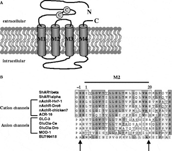
Fig. 3. Nicotinic (acetylcholine) receptors (nAChR). (A) Schematic representation of an individual nAChR subunit showing four transmembrane helices (M1–M4) and a long extracellular N-terminal segment carrying the signature Cys-loop motif. In the pentameric channel structure the pore is formed by five M2 helices contributed by each of the subunits. (B) A protein sequence alignment of the M2 regions of selected Cys-loop LGICs, including representative subunits of both cation-selective and anion (Cl−) channels. Rings of charge that are known to confer selectivity for anions or cations are marked by arrows. The predicted M2 regions of the S. haematobium ShAR1α, ShAR1β subunits (AY392150; AY392151) and a S. japonicum nAChR-like EST (BU769415) are shown. Other sequences were downloaded from public databases, using the following accession numbers: H. virescens Hv7-1, AAD32697; Drosophila Droα6, AF321445; GluClα-Dro, AAC47266; chicken-α7, JN0113; C. elegans, ACR-16, S68588; MOD-1, AAG36975; GluClα-Ce, S50864; GLC-3, CAB51708 (modified from Raymond & Sattelle, 2002).
Subsequent attempts to produce a functional nicotinic channel by expressing ShAR1α alone or jointly with ShAR1β in Xenopus oocytes have not been successful. Co-expression of ShAR1α with a vertebrate β subunit also failed to produce a response as did the expression of a chimeric form of ShAR1α that contained the M3–M4 region (second intracellular loop) of vertebrate α7. In contrast, expression of the wildtype α7 orthologue produced a fully functional homomeric channel (Bentley et al. 2004). It is unknown if the lack of ShAR1 activity was due to problems related to heterologous expression in the oocytes, or because the required subunit partners for ShAR1α and ShAR1β have yet to be discovered. As discussed in the next section, a number of new schistosome nAChR ESTs, both α-like and non-α subunits have been recently released into the database (Table 2). We noted that at least one of these sequences (BU769415) lacks the negatively charged amino acids of the M2 boundaries that determine cation selectivity (Fig. 3) and thus may be capable of forming anion channels. It will of interest to determine if any of these new putative subunits can partner with ShAR1α or ShAR1β and, in particular, if there are anion-selective nAChRs in schistosomes that might explain the inhibitory effects of ACh in parasitic worms.
Discovery of new transmitter receptors
Clearly, our next major challenge is to clone more receptors, a task that is bound to be facilitated by the new wealth of genomic sequences and ESTs available in the database. Already, we can identify numerous putative LGIC subunits, one full length neuropeptide GPCR and least 12 schistosome and planarian ESTs that strongly resemble neurotransmitter GPCRs (Table 2). Of note are muscarinic and 5HT-like receptor ESTs obtained from S. mansoni, the first to be identified in any of the parasitic flatworms. As more and more ESTs become available, it is expected the repertoire of full-length receptors will expand quickly in the near future.
Beyond cloning, the next step is to establish suitable in vitro systems for functional expression studies of the recombinant receptors. This is necessary not only to demonstrate activity but also to characterize the receptor's pharmacological profile and signalling mechanism. When combined with mutagenesis and protein modelling, these studies have the potential to identify important residues for ligand binding and conformational activation, which can help to elucidate the relationship between receptor structure and function. At present there are no cultured flatworm cells available for transfection of cloned cDNAs and therefore a heterologous environment must be used. The SmGPCR and ShAR1 studies described earlier (Hamdan et al. 2002a; Bentley et al. 2004) and many recent reports of cloned nematode receptors (see Raymond & Sattelle, 2002; Jones & Sattelle, 2003; Komuniecki et al. 2004) illustrate some of the more common strategies used for functional expression analyses of recombinant GPCRs and LGICs. For the most part, GPCRs are expressed in cultured mammalian cells and then tested by means of a radioligand binding assay or by measuring changes in intracellular second messengers. Other commonly used assays measure the incorporation of radiolabeled GTPγS into receptor-activated G proteins, or employ indirect methods based on reporter gene assays (Durocher et al. 2000) and yeast screening systems (Klein et al. 1998). Functional expression studies of LGICs are more challenging due to the pentameric nature of the channel and the great diversity of potential subunit arrangements. Researchers typically co-express multiple subunits either in Xenopus oocytes or mammalian cells with the expectation of finding the correct heteromeric composition (reviewed by Sine, 2002; Jones & Sattelle, 2003). The recombinant channels are subsequently tested for activity by electrophysiology or, recently, plate-based fluorometry (Fitch et al. 2003; Jensen & Kristiansen, 2004). An important consideration in studies of this nature is whether flatworm receptors can be expressed at sufficiently high levels to produce detectable activity, and whether they are properly targeted to the plasma membrane in the foreign cell environment. The choice of heterologous system can markedly influence the level of receptor expression and therefore it may be necessary to test more than one type of cell line when optimizing the system. The insect cell line, Sf9, has been used in place of mammalian cells for high-level expression of a nematode 5HT receptor (Smith et al. 2003). Cell growth and transfection conditions may also require optimization. Although most heterologous expression is done under standard cell culture conditions, expression of a C. elegans neuropeptide receptor was markedly increased when cell culture temperatures were shifted from 37 °C to 28 °C following transfection (Kubiak et al. 2003). Other strategies to improve translational efficiency of cloned cDNAs include codon-rewriting (Hamdan et al. 2002a,b) and the introduction of appropriate translational and membrane targeting sequences. In particular, the addition of a mammalian Kozak sequence upstream of the start codon (GCCACC) and a single adenine immediately after the stop codon have been shown to significantly increase expression of cloned helminth receptors in mammalian cells (Hamdan et al. 1999, 2002a,b).
Orphan receptors
The term ‘orphan’ has been used to describe a new receptor for which the ligand cannot be identified, either because the receptor sequence is novel or the expressed receptor fails to be activated by known transmitters. Nearly half of the endogenous GPCRs in mammals are orphans with no apparent ligand (Vassilatis et al. 2003) as are many of the GPCRs in the C. elegans genome. The number of orphan receptors among LGICs is also high; of the 90 neurotransmitter-gated channels in the C. elegans genome, there are at least 20 new subunits that cannot be paired with ligands (Jones & Sattelle, 2003). As new flatworm sequences become available it is likely that many novel receptors will be discovered as well. Orphan sequences present their own set of challenges as the receptors must be assayed with a battery of test substances in order to identify a potential ligand. Deciding which ligands to test can be a difficult task, particularly if the receptor is completely novel. In some instances it is possible to make educated guesses about the nature of the ligand; for example, a receptor that shows general characteristics of an amine GPCR may be tested first against a collection of biogenic amines and structurally related substances in order to identify an agonist. However, a different strategy must be used when the receptor fails to be activated by all the predicted ligands, or the structure is so unusual that a ligand cannot be predicted at all. The ‘orphan receptor strategy’ (reviewed by Lin & Civelli, 2004) is an alternative approach, which is designed to identify new transmitter substances by using orphan receptors as targets. The method involves expressing the receptor in a suitable surrogate system and then testing for activity by application of increasingly fractionated tissue extracts until an agonist can be identified. This strategy has been successful in the ‘de-orphanization’ of novel mammalian peptide receptors (see Lin & Civelli, 2004) and has potential for discovery of other types of receptors as well. Usually a high-throughput (HTS) activity assay is required in these studies, one that can be adapted to a 96-well (or larger) plate format and is suitable for screening multiple compounds at various concentrations. In the case of GPCRs, the most popular HTS assays are designed to measure mobilization of intracellular Ca2+ in intact transfected cells, using either an automated fluorometric imaging plate reader (FLIPR) or an aqueorin-based luminescence assay (Coward et al. 1999; Ungrin et al. 1999). GPCRs that do not normally signal through Ca2+ can be directed to this pathway by using permissive G proteins (Gα16) or G protein chimeras (Coward et al. 1999) and therefore, the Ca2+ assay can be used with a wide range of receptors, irrespective of their preferred signalling pathway. HTS technologies are also becoming increasingly popular in studies of ligand-gated channels. Plate-based fluorometric assays of LGICs measure changes in intracellular Ca2+ or membrane potential, using appropriate cell-permeable fluorescent dyes (Fitch et al. 2003; Jensen & Kristiansen, 2004). With these technologies now available, and the rapidly accumulating number of new sequences in the database, investigators are well poised to significantly advance our understanding of transmitter receptors in flatworms.
Flatworm receptors as drug targets
The availability of cloned receptors opens many new doors for future research, none perhaps as important as in the areas of receptor structure-function and pharmacology. By virtue of their pivotal biological roles and divergent structures, flatworm transmitter receptors are strong candidates for drug targeting. Heterologously expressed receptors may be used in mechanism-based screens of chemical libraries to identify selective antagonists. HTS plate-based fluorometric methods of the type described above are particularly attractive for drug discovery because of their exceptional screening capacity and adaptability to both GPCR- and LGIC-based assays. Alternatively, it is possible to use a rational drug design approach (reviewed by Thompson Klein & Geary, 1996) by which a drug may be designed to fit the target's binding site, provided the target structure is known. Detailed crystal structures of eukaryotic membrane proteins continue to be elusive and it is unlikely they will become available for any of the flatworm receptors in the near future. In their absence, however, a great deal of valuable information about the receptor's binding site can be obtained from computer generated homology models. Several highly refined three-dimensional models of biogenic amine receptors have been made available from structural alignments with rhodopsin (Visiers et al. 2002) and the same strategies can be used to model flatworm GPCRs. In the case of the ACh (nicotinic) channel, high-quality models are being generated in a modular fashion. The crystal structure of the snail ACh binding protein (AChBP) (Sixma & Smit, 2003) is used as a template for the receptor's extracellular binding domain, whereas the pore forming M2 bundle can be modelled from available NMR structure (reviewed by Capener et al. 2002). Combined with ligand docking and molecular dynamics simulations, these models are powerful tools for identification of putative agonist and antagonist binding sites, which can be subsequently tested by mutagenesis. The combination of basic molecular biology, modelling and drug screens has great potential to identify new selective drugs for control of parasitic infections.
CONCLUSIONS
Forty years since neurotransmitters were first discovered in worms, there is still much to be learned about their molecular properties and mode of action. This may change in the near future as a result of more DNA sequences being made available and new opportunities for molecular research. The area of neurotransmitter receptors, in particular, is poised to undergo a major shift from the predominantly physiological approaches of the past to a more biochemical exploration of structure and function. If recent advances in mammalian neurochemistry are any indication, we predict that a great deal of new information about the molecular architecture of the flatworm nervous system will emerge from this research. Ultimately, however, the lessons learned from in vitro molecular studies must be validated in animal models. As we move beyond the cloning era, it will be necessary to identify the biological roles of individual receptors and their relationship to worm behaviour in vivo. Unfortunately, many of the strategies used to manipulate gene expression in other systems, including transgenics and gene knockouts, are not yet possible in flatworms. However, there has been significant progress in ways of transfecting flatworms (Davis et al. 1999; Wippersteg et al. 2002) and, recently, RNA interference (RNAi) both in parasites (see Boyle & Yoshino, 2003) and planarians (Orii, Mochii & Watanabe, 2003). These important developments offer new prospects for functional genomics approaches and hold great promise for future advances in flatworm neurochemistry.
ACKNOWLEDGEMENTS
We thank Dr. G. Stirling for a critical reading of the manuscript and many helpful suggestions. Work in the Ribeiro laboratory is supported by funding from the Natural Sciences and Engineering Research Council of Canada (NSERC) and the Canadian Institutes of Health Research (CIHR).


