


Introduction
Rhodonite is a well-known pyroxenoid and typical rock-forming constituent of many manganese-rich rocks. The first mention of rhodonite appeared in the paper by Germar (Reference Germar1819), on the basis of data from Jasche who described it two years earlier (Jasche, Reference Jasche1817). This name was given initially to a rock formed by pinkish-red Mn-rich silicates (at present known as rhodonite and pyroxmangite), quartz, tephroite and Mn oxides. Somewhat later, the name rhodonite was transferred to the major manganese silicate of this rock which typically has a pink or red colour (ρόδον means rose in Greek). For a long time, several minerals with different chemical and crystal chemical features were all combined under the name rhodonite. A brief historical review is given by Shchipalkina et al. (Reference Shchipalkina, Pekov, Chukanov, Biagioni and Pasero2019b).
Some confusion with the term ‘rhodonite’ remained up to recent times, even after detailed elaboration of the crystal chemistry of manganese pyroxenoids. This is due to the absence of a clear definition of rhodonite as a mineral species with significant chemical variations, mainly in the Mn, Ca and Fe contents, in samples belonging to the rhodonite structure type; the situation was additionally complicated by the ordering of some cations in the rhodonite-type structure. In accordance with modern criteria in mineral nomenclature, such a situation cannot be described in the framework of a single mineral species, thus a mineral group needed to be established and defined. In 2019, we proposed a nomenclature for the rhodonite group and this proposal was accepted by the Commission on New Minerals, Nomenclature and Classification of the International Mineralogical Association (IMA–CNMNC) as Proposal 18-I (Miyawaki et al., Reference Miyawaki, Hatert, Pasero and Mills2019).
According to the IMA-accepted nomenclature, the rhodonite group includes isostructural pyroxenoids with the general crystal chemical formula M (5)AM (1–3)B 3M (4)C[Si5O15] in which the species-defining components are: A = Ca or Mn2+, B = Mn2+ and C = Mn2+ or Fe2+ (Shchipalkina et al., Reference Shchipalkina, Pekov, Chukanov, Biagioni and Pasero2019b). The structural formula of rhodonite-group minerals can be written as VIM(1)VIM(2)VIM(3)VIM(4)VIIM(5)[Si5O15] (Roman numerals mean coordination numbers of metal cations M) and simplified to VIIM(5)VIM(1–4)[Si5O15]. In the simplified formula, two types of M cations with different coordination numbers are distinguished. At present, three valid mineral species belonging to this group are known, namely rhodonite defined as CaMn4[Si5O15], ferrorhodonite with the idealised formula CaMn3Fe[Si5O15] (Shchipalkina et al., Reference Shchipalkina, Chukanov, Pekov, Aksenov, McCammon, Belakovskiy, Britvin, Koshlykova, Schafer, Scholz and Rastsvetaeva2017) and vittinkiite described in the present paper. A proposal on vittinkiite was submitted by us to the IMA–CNMNC in 2017, however, it could only be accepted as a mineral species after the approval of the nomenclature of the rhodonite group in August 2019. Vittinkiite (IMA 2017–082a, Shchipalkina et al., Reference Shchipalkina, Pekov, Chukanov, Zubkova, Belakovskiy, Britvin and Koshlyakova2019a) is defined as an analogue of rhodonite with the prevailing of Mn2+ over Ca at the A [= M(5)] site and, thus, with the idealised, end-member formula MnMn4[Si5O15]. The minerals now known as rhodonite and vittinkiite were earlier joined under the general name ‘rhodonite’ and typically described with the simplified formula MnSiO3, considering only the gross dominant metal cation Mn2+ and ignoring cation distribution between different M sites.
Vittinkiite can be named figuratively ‘an old new mineral’. This rhodonite-type mineral with a low Ca content has been known for a long time, however, it is much rarer than rhodonite sensu stricto, the idealised end-member formula of which is CaMn4[Si5O15]. Such a Ca-poor rhodonite-group mineral has been reported undeniably from two localities in Finland, namely Vittinge (Sundius, Reference Sundius1931) and Simsiö (Hietanen, Reference Hietanen1938), the Taguchi manganese deposit in Aichi Prefecture, Japan (Momoi, Reference Momoi1964), the Xanthi area in Greece (Sapountzis and Christofides, Reference Sapountzis and Christofides1982), and the Malosedel'nikovskoe, Kurganovskoe, Kozhaevskoe and Faizulinskoe manganese and rhodonite deposits in the Urals, Russia (Brusnitsyn, Reference Brusnitsyn2013). It is noteworthy that, based on the chemical data on Uralian rhodonite, Brusnitsyn and Zaitsev (Reference Brusnitsyn and Zaitsev2000) suggested distinguishing two separate mineral species with the idealised formulae CaMn4[Si5O15] and MnMn4[Si5O15] under the conventional names ‘rhodonite-I’ and ‘rhodonite-II’, respectively.
Earlier published representative chemical analyses of ‘rhodonite’ corresponding to vittinkiite (i.e. with Ca < 0.5 atoms per formula unit = apfu) are given in Table 1. The data on crystal structures of several Ca-poor rhodonite-type minerals (now defined as vittinkiite) and synthetic MnMn4[Si5O15] (Ohashi and Finger, Reference Ohashi and Finger1975; Peacor et al., Reference Peacor, Essene, Brown and Winter1978; Pertlik and Zahiri, Reference Pertlik and Zahiri1999; Narita et al., Reference Narita, Koto and Morimoto1977) are summarised and discussed by Shchipalkina et al. (Reference Shchipalkina, Pekov, Chukanov, Biagioni and Pasero2019b).
Table 1. Chemical composition of vittinkiite and representative earlier published analyses.

“–” indicates the content is below the detection limit.
Vittinkiite was named by us after the Vittinki (an old Swedish name is Vittinge) iron mines, Isokyrö, Western and Inner Finland Region, Finland. The choice of this name has a double reason: (1) the first published analysis of ‘rhodonite’ with Ca < 0.5 apfu (#2 in Table 1: Sundius, Reference Sundius1931) was from Vittinki; and (2) the Fersman Museum specimen cat#15061 that became the holotype is from the same locality.
Rhodonite from Vittinki (Vittinge) was first described in 1863 by Adolf Erik Nordenskiöld (Nordenskiöld, Reference Nordenskiöld1863). The specimen studied by us, which became the holotype of vittinkiite, is deposited in the systematic collection of the Fersman Mineralogical Museum of the Russian Academy of Sciences, Moscow under the catalogue No. 15061. It was acquired before 1926 from the collection of Adolf Erik Nordenskiöld (1832–1901) and catalogued as rhodonite in 1926. We believe that it is identical or close to the material described in Nordenskiöld (Reference Nordenskiöld1863).
Besides data on the holotype vittinkiite, this present work provides data on several vittinkiite specimens from other localities studied by us and discusses the history of identification of two natural MnSiO3 polymorphs: pyroxmangite Mn7[Si7O21] and vittinkiite MnMn4[Si5O15].
Experimental
Samples
Rhodonite-group minerals from the collection of the Fersman Mineralogical Museum were studied by electron microprobe analysis and infrared (IR) spectroscopy and/or powder X-ray diffraction (PXRD) was used to confirm they belonged to the rhodonite structural type. Among 60 samples studied from 30 worldwide localities, we found vittinkiite in nine specimens from six localities. These are specimens from Vittinki iron mines, Isokyrö, Finland (No. 15061, now the holotype of vittinkiite); New England Range, New South Wales, Australia (No. 61533); Ridder Mine (now Leninogorskiy Mine), NW Altai, Kazakhstan (No. 80484); Sultanuizdag, Uzbekistan (Nos. 78835, 78836 and 77378); Nozhiy Lake, Aginsk district, Zabaikal'sky Krai, Russia (No. 25677); and Tsang-Ping, Hebei, China (No. 58691) All these specimens are visually similar to each other. They consist of massive aggregates of pinkish vittinkiite associated with quartz, tephroite, black unspecified Mn oxides and sometimes pyroxmangite.
The samples of rhodonite, pyroxmangite and pyroxferroite used to obtain reference IR spectra originate from the collection of one of the coauthors (N.V.C.). Rhodonite from Långban, Sweden forms pink fine-grained aggregate in a skarn. Pyroxmangite samples from the Southern Faizulinskoe Mn deposit, South Urals, Russia and from the Razoare Mn deposit, Maramures, Romania occur as crimson granular aggregates. Mn-rich pyroxferroite from a metamorphosed xenolith of gneiss hosted by alkaline basalt of the Bellerberg paleovolcano, Eifel, Germany forms imperfect brownish-red crystals.
In the holotype specimen, vittinkiite is light pink with a white streak. It forms massive aggregates composed by tabular single-crystal grains up to 2 mm across associated intimately with quartz and pyroxmangite (Fig. 1). The lustre is vitreous. The mineral is translucent in aggregates and transparent in thin fragments. Cleavage is perfect on {201} and good on {021} and {210}. The density measured by flotation in heavy liquids (Clerici solutions) is 3.68(2) g cm−3. The density calculated using the empirical formula is 3.737 g cm−3.

Fig. 1. Rock consisting of vittinkiite (Vit), quartz (Q) and pyroxmangite (Pxm) from Vittinki (Vittinge) iron mines, Isokyrö, Finland (the holotype vittinkiite).
In plane polarised light vittinkiite is pale pink to colourless, depending on grain thickness, and non-pleochroic. Vittinkiite is optically biaxial (+), with α = 1.725(4), β = 1.733(4) and γ = 1.745(5) (589 nm). 2V estimated by the curvature of the interference isogyre in conoscopic mode on the sections perpendicular to the optical axes is 75(10)°; 2Vcalc = 79°. Orientation: Y ∧ b = 22°. The dispersion of optical axes is weak, r < v.
Composition
Chemical data for vittinkiite were obtained using a Jeol JSM-6480LV scanning electron microscope equipped with an INCA-Wave 500 wavelength-dispersive spectrometer (WDS mode). The acceleration voltage was 20 kV, the beam current was 10 nA and the electron beam was defocused to 4 μm × 4 μm. The contents of other elements with atomic numbers higher than that of carbon are below detection limits. The standards used for quantitative analysis were diopside for Mg, wollastonite for Ca, ferrosilite for Fe and Si, pure Zn for Zn, and pure Mn for Mn. Analytical data are given in Tables 1 and 2.
Table 2. Chemical composition of vittinkiite from the collection of the Fersman Mineralogical Museum RAS, our data.

“–” indicates the content is below the detection limit. *For four analyses.
Single-crystal X-ray diffraction
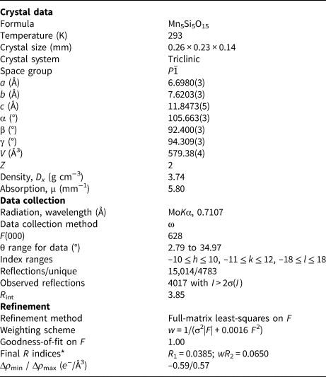
Single-crystal X-ray diffraction was performed using an Xcalibur S CCD diffractometer with MoKα radiation (λ = 0.71073 Å). Data reduction was performed using CrysAlisPro Version 1.171.37.35 (Agilent Technologies, 2014). A total of 15,014 reflections within the sphere limited by θ = 34.97° were obtained. The experimental details of data collection and refinement values are shown in Table 3. After averaging of equivalent reflections, the experimental set contained 4017 reflections with I > 2σ(I). The data on rhodonite obtained by Peacor and Niizeki (Reference Peacor and Niizeki1963) was used for the initial model for the structure refinement. The structure was refined using the JANA2006 suite of programs (Petříček et al., Reference Petříček, Dušek and Palatinus2006). Atomic scattering factors for neutral atoms together with anomalous dispersion corrections were taken from International Tables for X-ray Crystallography (Ibers and Hamilton, Reference Ibers and Hamilton1974). The final refinement cycles finished with R 1 = 3.85%, wR 2 = 6.50% and GooF = 1.00 for all data. Fractional atomic coordinates, refined site-scattering values, equivalent atomic displacement parameters (U eq), and anisotropic atomic displacement parameters U ij are given in Table 4. The selected interatomic distances are presented in Table 5. The crystallographic information file has been deposited with the Principal Editor of Mineralogical Magazine and is available as Supplementary material (see below).
Table 3. Crystal data and refinement details for vittinkiite.
*R 1 = Σ||F obs|–|F calc||/Σ|F obs|; wR 2 = {Σ[w(|F obs|–|F calc|2]/Σ[w|F obs|2]}½; GoF = {Σw(|F obs|–|F calc|)2/(n–p)}½ where n is the number of reflections and p is the number of refined parameters.
Table 4. Atom coordinates, site multiplicities (Q) and Wyckoff letter, displacement parameters (U eq/Å2) and anisotropic displacement parameters (Å2) for vittinkiite

*U eq is defined as one third of the trace of the orthogonalised Uij tensor.
Table 5. Selected interatomic distances for cation sites in the crystal structure of vittinkiite.

Powder X-ray diffraction data
Powder X-ray diffraction data (PXRD) were collected using a Rigaku R-AXIS Rapid II diffractometer (image plate), CoKα (λ = 1.79021 Å), 40 kV and 15 mA, rotating anode with the microfocus optics, Debye-Scherrer geometry, d = 127.4 mm and exposure = 15 min. The data were integrated using the software package Osc2Tab (Britvin et al., Reference Britvin, Dolivo-Dobrovolsky and Krzhizhanovskaya2017). Calculated intensities were obtained by means of STOE WinXPOW v. 2.08 program suite based on the atomic coordinates and unit-cell parameters obtained from the single-crystal data. Data are listed in Table 6.
Table 6. Powder X-ray diffraction data (d in Å for CoKα radiation) for vittinkiite*.

* For the calculated pattern, only reflections with intensities ≥1 are given.
The strongest lines are given in bold
Infrared spectroscopy
Samples for infrared spectroscopy were powdered, mixed with anhydrous KBr and pelletised (the ratio sample:KBr was 1:150 in mass proportion). The infrared absorption spectra of a transparent, homogeneous crystal of vittinkiite and reference samples of rhodonite, pyroxmangite and pyroxferroite were obtained using an ALPHA FTIR spectrometer (Bruker Optics) at a resolution of 4 cm−1. A total of 16 scans were collected for each spectrum. The IR spectrum of an analogous pellet of pure KBr was used as a reference.
Results and discussion
Infrared spectroscopy
Bands in the IR spectrum of vittinkiite (Fig. 2a) and their assignments are (cm−1, s – strong band, sh – shoulder): 1112, 1053s and 1020s, (Si–O stretching vibrations of the Si–O–Si bridges), 950s, 930sh, 915sh, 891s and 874 (Si–O stretching vibrations of apical Si–O bonds), 718, 693, 664, 578 and 559 (O–Si–O bending vibrations), 515, 492, 458s and 391 (lattice modes involving Si–O–Si bending and M⋅⋅⋅O stretching vibrations where M = Mn, Fe and Ca). The IR spectrum of vittinkiite is close to that of rhodonite (Fig. 2; see also Chukanov, Reference Chukanov2014). Note that the intensity of the band in the range from 390 to 395 cm−1 in IR spectra of rhodonite-type minerals increases with the lowering of Ca content at the M5 site. Based on this observation, we assign this band to M 5Mn⋅⋅⋅O stretching vibrations.

Fig. 2. Infrared absorption spectra of (a) the holotype vittinkiite and (b) rhodonite with the composition Ca0.9Mn3.7Mg0.4(Si5O15) from Långban, Sweden.
The other distinctive features of vittinkiite that distinguish it from rhodonite are a high intensity of the band at 950 cm−1 and shifts of all strong bands of Si–O stretching vibrations towards lower wavenumbers as compared to analogous bands of rhodonite (cm−1): 1060 → 1053, 1026 → 1020, 950 → 949 and 898 → 891. These features are characteristic of all vittinkiite samples independent of their origin.
Crystal structure of vittinkiite
The crystal structure of rhodonite was first solved by Liebau et al. (Reference Liebau, Hilmer and Lindemann1959). The rhodonite structure contains the chains of tetrahedra with a repeat unit [Si5O15] and ribbons formed by edge-sharing metal-centred polyhedra М1, M2, M3, M4 and M5 (Fig. 3). The polyhedra М1, M2 and M3 are slightly distorted octahedra with mean cation–oxygen distances 2.21–2.23 Å, occupied predominantly by Mn. The polyhedron M4 is a strongly distorted octahedron with the shortest and the longest cation–oxygen distances in the ranges 1.95 to 1.99 Å and 2.77 to 2.91 Å, respectively. This site concentrates Fe, Mg and Zn (Peacor and Niizeki, Reference Peacor and Niizeki1963; Ohashi and Finger, Reference Ohashi and Finger1975; Peacor et al., Reference Peacor, Essene, Brown and Winter1978; Nelson and Griffen, Reference Nelson and Griffen2005; Leverett et al., Reference Leverett, Williams and Hibbs2008; Shchipalkina et al., Reference Shchipalkina, Chukanov, Pekov, Aksenov, McCammon, Belakovskiy, Britvin, Koshlykova, Schafer, Scholz and Rastsvetaeva2017). The shortest M4–O distances are observed in samples with the highest contents of cations smaller than Mn2+, namely Mg2+ and Fe2+. The site M5 has seven-fold coordination and mean a cation–oxygen distance in the range 2.40–2.42 Å and in most cases is occupied predominantly by Ca (usually, together with subordinate Mn). Mutual alignment of the polyhedral ribbons and chains of tetrahedra is shown in Fig. 3.

Fig. 3. Сrystal structure of vittinkiite (a and b) and the fragment of polyhedral ribbon with marked sites (c). The Mn-centred (M) polyhedra are purple and Si-centred tetrahedra are yellow. Dashed lines contour the polyhedral slab (a) and polyhedral ribbon (b).
Based on e ref values, coordination numbers and interatomic distances, the occupancies of cationic sites in holotype vittinkiite are as follows (assumed cation assignment is given in square brackets taking into account the electron microprobe data): M1, M2, and M3–Mn2+, M4–Mn2+ with minor Mg2+ [Mn0.94Mg0.06], and M5–Mn2+ with minor Ca2+ [Mn0.92Ca0.08].
The main, species-defining difference between vittinkiite M (5)MnM (1–3)Mn3 M (4)Mn[Si5O15] and rhodonite M (5)CaM (1–3)Mn3M (4)Mn[Si5O15] is the predominant cation at the M5 site, i.e. Mn2+ or Ca, respectively.
Polymorphism of MnSiO3
Four polymorphs of a manganese metasilicate with the simplified end-member formula MnSiO3 are known. They belong to the rhodonite, pyroxmangite (Narita et al., Reference Narita, Koto and Morimoto1977), garnet (Fujino et al., Reference Fujino, Momoi, Sawamoto and Kumazawa1986) and clinopyroxene (Tokohami et al., Reference Tokohami, Horiuchi, Nakano, Akimoto and Morimoto1979) structure types. These four compounds demonstrate the following polymorphic transformations accompanied by a regular increase in the density of crystal structures and change in coordination numbers (given in Roman numerals) of Mn and Si with expansion of SiO4 tetrahedra and compression of Mn-centred octahedra (Fig. 4): rhodonite VII(M5)MnVI(M1–4)Mn4[IVSi5O15] (space group P  $\bar{1}$) → pyroxmangite VII(M1–2)MnVI(M3–7)Mn5[IVSi7O21] (P
  $\bar{1}$) → synthetic clinopyroxene VII(M2)MnVI(M1)Mn[IVSi2O6] (P21/c) → synthetic tetragonal garnet VIIIMn3VIMnVISi[IVSiO4]3 (I41/a) (Akimoto and Syono, Reference Akimoto and Syono1972; Momoi, Reference Momoi1974; Fujino et al., Reference Fujino, Momoi, Sawamoto and Kumazawa1986). The comparative data for these polymorphs are given in Table 7.

Fig. 4. Projections of crystal structures of MnSiO3 polymorphs: rhodonite type (vittinkiite, this work) (a), pyroxmangite (Narita et al., Reference Narita, Koto and Morimoto1977) (b), clinopyroxene (Tokohami et al., Reference Tokohami, Horiuchi, Nakano, Akimoto and Morimoto1979) (c) and tetragonal garnet (Fujino et al., Reference Fujino, Momoi, Sawamoto and Kumazawa1986) (d). The unit cells are outlined.
Table 7. Comparative data for vittinkiite, pyroxmangite and four synthetic polymorphs of MnSiO3.

*Including the data on vittinkiite and its synthetic analogue
**Including the data on pyroxmangite and its synthetic analogue
Roman numerals in the idealised formula mean the coordination numbers
The repeat unit in pyroxenoids increases with the decrease in size of octahedrally coordinated cations, e.g. as a consequence of the main components Ca – Mn – Mg in the pyroxenoid series wollastonite – bustamite – rhodonite – pyroxmangite (Liebau, Reference Liebau1962; Takeuchi, Reference Takeuchi1977, and references therein). The composition of these MnSiO3 polymorphs remains stable, but the transformation from the rhodonite-type to clinopyroxene-type crystal structure occurs through a compound with pyroxmangite structure type. This can be defined by the controlling factor of pressure that acts due to the effect of a smaller cation (for example, Mg in the MnSiO3–MgSiO3 system). The relationship between pyroxenoids (wollastonite, bustamite, rhodonite and pyroxmangite structural types) and pyroxenes has been the focus of many researchers (Burnham, Reference Burnham1971; Morimoto, Reference Morimoto, Koto and Shinohara1966; Narita, Reference Narita1973; Koto et al., Reference Koto, Morimoto and Narita1976; Pinckney and Burnham, Reference Pinckney and Burnham1988; Henry, Reference Henry1998) who report several approaches to the interpretation of causes and triggers of their reversible transitions.
Only two of the above-discussed polymorphs of MnSiO3 are found in nature, namely pyroxenoids with different periodicities of the silicate chain SinO3n, vittinkiite, ideally MnMn4[Si5O15], and pyroxmangite, ideally Mn7[Si7O21]. The rhodonite-type pyroxenoid, i.e. vittinkiite, is considered as a relatively low-pressure polymorph of MnSiO3 whereas pyroxmangite is a high-pressure modification. For pure MnSiO3, the transformation from the rhodonite-type form (vittinkiite) to the pyroxmangite-type form occurs at a pressure of 3 kbar and temperature ~350–400°C, though pyroxmangite can be metastable at low temperature and atmospheric pressures (Maresch and Mottana, Reference Maresch and Mottana1976). This has been confirmed indirectly by pyroxmangite found originating from cavities in effusive rocks of the Eifel volcanic region, Germany (Shchipalkina et al., Reference Shchipalkina, Aksenov, Chukanov, Pekov, Rastsvetaeva, Schafer, Ternes and Schuller2016). The heating experiments undertaken by Aikawa (Reference Aikawa1979) show that MnSiO3 with the pyroxmangite structure can exist metastably in the temperature range 400–1000°C, due to the high-activation energy of the pyroxmangite → ‘rhodonite’ (i.e. vittinkiite) transformation. According to high-pressure and high-temperature experiments by Akimoto and Syono (Reference Akimoto and Syono1972), the MnSiO3 polymorphs with clinopyroxene (P21/c) and garnet structures (I41/a) appeared at pressures higher than 60 and 125 kbar, respectively. A complete phase diagram for the four MnSiO3 polymorphs was reported by Akimoto and Syono (Reference Akimoto and Syono1972).
The effects of isomorphous substitutions on the stability of rhodonite- and pyroxmangite-type crystal structures and on PT conditions of the transformations rhodonite ↔ pyroxmangite were examined experimentally by Ito (Reference Ito1972) for the MnSiO3–MgSiO3 system. The influence of different impurities on the stability of these two structural types was also discussed by Pinckney and Burnham (Reference Pinckney and Burnham1988), though that work concentrates mainly on the Mn–Mg substitutions. The general approximation of effects of substitutions of Mn for Ca, Mg and Fe in MnSiO3 was reported by Maresch and Mottana (Reference Maresch and Mottana1976). According to their data, Fe2+ and Mg cations, smaller than Mn2+, stabilise the pyroxmangite-type structure whereas Ca stabilises the rhodonite-type structure. The influence of these impurities can be an objective cause of paragenesis of pyroxmangite- and rhodonite-group minerals despite the different fields of stability.
The MgSiO3–MnSiO3 system includes two pyroxenes known in nature: clinopyroxene kanoite, MnMg[Si2O6] (Kobayashi, Reference Kobayashi1977) and orthopyroxene donpeacorite, (Mn,Mg)MgSi2O6 (Petersen et al., Reference Petersen, Anovitz and Essene1984). Gnos et al. (Reference Gnos, Armbruster and Nyfeler1996) showed that rhodonite-group minerals, pyroxmangite, kanoite and donpeacorite can occur together, occasionally forming complex intergrowth. The coexistence of different pyroxenes and pyroxenoids with species-defining Mn, Ca and Mg may indirectly confirm the affinity of their structural types to the species-defining cations and impurities, in which the content and ratios determine the pyroxene or pyroxenoid structural type.
Identification of pyroxenoids with the idealised formula MnSiO3
The occurrence of rhodonite-group minerals (rhodonite, vittinkiite or ferrorhodonite) and pyroxmangite, a pyroxenoid with the generalised formula (Mn,Fe,Mg,Ca)7[Si7O21], in paragenesis, despite the difference in their fields of stability, leads to confusion in the identification of these minerals. All of them are important constituents of silicate manganese assemblages, which form at greenschist-facies metamorphism (Roy, Reference Roy1981; Dasgupta et al., Reference Dasgupta, Banerjee, Fukuoka and Bhattacharya Roy1990; Brusnitsyn Reference Brusnitsyn2000, Reference Brusnitsyn2013, Reference Brusnitsyn2015; and references therein).
It appears that the end-member polymorphs, vittinkiite and pyroxmangite, were confused during earlier studies in the ‘pre-structural’ period. Ford and Bradley (Reference Ford and Bradley1913) noticed that the holotype pyroxmangite was considered initially to be a highly ferrous rhodonite, because most physical properties and chemical features of rhodonite and pyroxmangite are very similar. In the ‘pre-structural’ period, these minerals were distinguished by optical methods, using 2V and the angle between cleavage planes. The 2V values are different: 63–87° for rhodonite and 37–46° for pyroxmangite (Deer et al., Reference Deer, Howie and Zussman1978). Ноwever, specimens of both rhodonite and pyroxmangite can show a wide variation in 2V ranging from 40 to 72° (Aikawa, Reference Aikawa1984, and references therein). Such intermediate values have been explained as due to the submicroscopic lamellar structure of pyroxmangite–rhodonite intergrowths: the thickness of lamellae is smaller than the wavelength used for the measurement of 2V (Aikawa, Reference Aikawa1984), which further complicates the identification of these minerals.
The chemical composition of the pyroxenoids discussed cannot be the decisive characteristic for their identification in most common cases because the compositional fields of rhodonite- and pyroxmangite-type pyroxenoids are partially overlapped (Shchipalkina et al., Reference Shchipalkina, Pekov, Chukanov, Biagioni and Pasero2019b and references therein). As shown in Fig. 5a, there is a continuous isomorphous series between vittinkiite and rhodonite. The formal border between vittinkiite and rhodonite can be fixed at 0.5 Ca apfu: this is a chemical criterion of subdivision of these mineral species. However, based on literature and our data, we conclude that vittinkiite is not as common in nature as rhodonite (Fig. 5a). Moreover, vittinkiite containing <0.1 Ca apfu is yet unknown. Members of the rhodonite group with 0.4–1.2 Ca apfu are the most widespread in nature. The correlation between contents of Ca and (Mg+Fe) in rhodonite-group minerals is discernible, but indistinctly (Fig. 5a), whereas for pyroxmangite and pyroxferroite this correlation is considerable (Fig. 5b). Generally, there are some regularities in the distribution of Ca, Mg and Fe inherent to rhodonite-group minerals and pyroxmangite occurring together. The content of (Mg+Fe) is higher in pyroxmangite whereas coexisting rhodonite is typically more Ca-rich than pyroxmangite. In general, rhodonite occurs mainly in Mn-bearing rocks enriched in Ca whereas pyroxmangite is more common for Ca-depleted manganolites (Aikawa, Reference Aikawa1979; Brusnitsyn, Reference Brusnitsyn2000, Reference Brusnitsyn2013; Reference Brusnitsyn2015 and references therein). However, pyroxmangite and members of the rhodonite–vittinkiite series can occur in intimate intergrowths, typically with lamellar textures (Maresch and Mottana, Reference Maresch and Mottana1976; Aikawa, Reference Aikawa1979, Reference Aikawa1984; Brusnitsyn, Reference Brusnitsyn2000, Reference Brusnitsyn2013, Reference Brusnitsyn2015, and references therein).

Fig. 5. Ratios of the major metal cations in minerals of the rhodonite group (a) and pyroxmangite–pyroxferroite series (b), based on literature (Sundius, Reference Sundius1931; Hietanen, Reference Hietanen1938; Peacor and Niizeki, Reference Peacor and Niizeki1963; Momoi, Reference Momoi1964; Burnham, Reference Burnham1971; Ohashi and Finger, Reference Ohashi and Finger1975; Peacor et al., Reference Peacor, Essene, Brown and Winter1978; Sapountzis and Christofides, Reference Sapountzis and Christofides1982; Pinckney and Burnham, Reference Pinckney and Burnham1988; Nelson and Griffen, Reference Nelson and Griffen2005; Shchipalkina et al., Reference Shchipalkina, Chukanov, Pekov, Aksenov, McCammon, Belakovskiy, Britvin, Koshlykova, Schafer, Scholz and Rastsvetaeva2017; Brusnitsyn, Reference Brusnitsyn2000, Reference Brusnitsyn2013; Reference Brusnitsyn2015; Shchipalkina et al., Reference Shchipalkina, Aksenov, Chukanov, Pekov, Rastsvetaeva, Schafer, Ternes and Schuller2016; and reference therein) and our data. In (a) samples with determined crystal structures are marked by filled circles: blue – rhodonite; light purple – vittinkiite; deep purple – synthetic analogue of the end-member vittinkiite; red – ferrorhodonite; green – Zn-rich variety of rhodonite (‘fowlerite’); and grey – Mg-enriched variety of rhodonite. Other vittinkiite samples are shown by empty purple circles whereas rhodonite and ferrorhodonite samples are empty blue circles. In (b) samples with a determined crystal structure are marked by black (filled) squares.
The most efficient and accurate methods for discrimination of these manganese pyroxenoids are PXRD and IR spectroscopy. The main reflections in the PXRD patterns of rhodonite-group minerals and pyroxmangite appear in the range d = 3.60–2.50 Å. However, identification of individual phases in a mixture can be hindered during routine PXRD because of the presence and overlapping of numerous strong reflections in the same region (Brusnitsyn, Reference Brusnitsyn2013).
As our data show, vittinkiite (and rhodonite-group minerals in general) and pyroxmangite are distinguished clearly by the IR spectra in the region of 600–700 cm−1. The triplet (631–641) + (655–662) + (674–684) cm−1 with the strongest band in the range of 655–662 cm−1 is a characteristic feature of pyroxmangite (Fig. 6). In addition, pyroxmangite differs from rhodonite-type minerals in relative intensities of the bands of Si–O stretching vibrations. These features of the IR spectra of pyroxmangite remain for a wide range of the Mn:Fe ratio, up to the point where Fe prevails over Mn, which corresponds to pyroxferroite (Fig. 6). Our study of rhodonite and pyroxmangite specimens from the collection of the Fersman Mineralogical Museum has showed that IR spectroscopy is useful for the identification of individual phases but is not suitable for their mixtures.

Fig. 6. Infrared absorption spectra of (a) Fe-free pyroxmangite with the empirical formula (Mn6.74Mg0.16Ca0.09)(Si7O21) from the Southern Faizulinskoe Mn deposit, South Urals, Russia; (b) Fe-bearing pyroxmangite with the empirical formula (Mn4.2Fe2.2Mg0.4Ca0.2)(Si7O21) from the Razoare Mn deposit, Maramures, Romania; and (c) Mn-rich pyroxferroite with the empirical formula (Fe3.0Mn2.8Mg1.1Ca0.1)(Si7O21) from a metamorphosed xenolith of gneiss hosted by alkaline basalt of the Bellerberg palaeovolcano, Eifel, Germany.
Acknowledgements
We thank three anonymous referees for their valuable comments. This work was supported by the Russian Foundation for Basic Research, grant 18-05-00332 (in part for the crystal structure and crystal chemistry of vittinkiite). The infrared spectroscopy investigation was performed in accordance with the state task, state registration No. ААAА-А19-119092390076-7. The technical support by the SPbSU X-Ray Diffraction Resource Center in the powder XRD study is acknowledged.
Supplementary material
To view supplementary material for this article, please visit https://doi.org/10.1180/mgm.2020.75


