


Introduction
The genus Synarthonia Müll. Arg. is characterized by solitary ascomata, becoming mono- to pluri-carpocentral synascomata embedded in a slightly elevated to immersed pseudostroma, with a thin white thalline margin and Arthonia-type asci with transversely septate (macrocephalic) or muriform ascospores. The genus, represented by only two species (Müller Argoviensis Reference Müller Argoviensis1891, Reference Müller Argoviensis1895) viz. S. bicolor Müll. Arg. and S. stigmatidialis Müll. Arg., remained unfamiliar to most lichenologists until the discovery of an allied new genus Synarthothelium (Sparrius Reference Sparrius2009). Recently, Menezes et al. (Reference Menezes, Lima, Xavier-Leite, Maia, Aptroot and Cáceres2013) added another species, S. sarcographoides Aptroot et al. from Brazil. Out of the three species, only S. bicolor is known from India (Jagadeesh Ram & Sinha Reference Jagadeesh Ram and Sinha2009).
During the course of studies on specimens of the family Arthoniaceae, types of S. bicolor and S. stigmatidialis were examined. Study of the types and protologue of S. sarcographoides Aptroot et al. revealed some additional information about the genus. These studies also led to the discovery of two new species, S. psoromica and S. sikkimensis from India. The taxonomic significance of these findings is discussed here.
Material and Methods
The specimens examined are housed in BSA and G herbaria. Morphological details were examined using a Nikon SMZ 1500 stereomicroscope. Anatomical details were studied using a Nikon Eclipse 50i compound microscope. Hand-cut sections of thalli and ascomata mounted in distilled water, KOH solution (K), and lactophenol cotton blue (LPCB) were studied. The amyloid reactions were tested in Lugol's iodine solution without (I) or with pretreatment with KOH (KI). All measurements were made on material mounted in distilled water and drawings were made with the help of an Ernst Leitz Wetzlar (Germany) microscope (in ×10) with sections mounted in distilled water. The length, breadth, and length/breadth ratio (l/b) of ascospores are given as: (min–)(
 $$--><$>\bar{x} $$$
–SD)– (
$$--><$>\bar{x} $$$
–SD)– (
 $$--><$>\bar{x} $$$
+ SD) (–max), where ‘min’ and ‘max’ are the extreme values,
$$--><$>\bar{x} $$$
+ SD) (–max), where ‘min’ and ‘max’ are the extreme values,
 $$--><$>\bar{x} $$$
the arithmetic mean, and SD the corresponding standard deviation, followed by the number of measurements (n). The chemistry was studied by spot tests and thin-layer chromatography following Orange et al. (Reference Orange, James and White2001).
$$--><$>\bar{x} $$$
the arithmetic mean, and SD the corresponding standard deviation, followed by the number of measurements (n). The chemistry was studied by spot tests and thin-layer chromatography following Orange et al. (Reference Orange, James and White2001).
Taxonomic Treatment
Synarthonia Müll. Arg.
Bull. Soc. R. Bot. Belg. 30: 85 (1891).
Type species: Synarthonia bicolor Müll. Arg.
Thallus corticolous, crustose, endophloeodal to epiphloeodal, smooth, cracked to rimose-like, smooth to verruculose, sorediate or esorediate, ecorticate. Prothallus white fibrous-like or rhizomorph-like, pale brownish to dark brown when in contact with other lichen species. Photobiont trentepohlioid, in short chains or single-celled, cells globose to ellipsoid.
Ascomata solitary when young, later becoming mono- to pluri-carpocentral synascomata embedded in a slightly elevated to immersed pseudostroma, with a thin white thalline margin, without algal cells; disc thinly white or greyish pruinose to epruinose, pale brown to dark brown when pruina removed. Excipulum hyaline to pale brownish or straw-coloured, composed of brown pigmented or hyaline conglutinated hyphae, non-carbonized. Epithecium pale brownish, formed by densely branched and anastomosing tips of paraphysoids, with some greyish gelatinous material and hyaline crystals. Hymenium hyaline, not inspersed, rarely inspersed with some hyaline granular crystals. Paraphysoids branched and anastomosing, apices thickened, with or without brown pigment. Hypothecium hyaline to pale brownish. Asci 8-spored, Arthonia-type, without KI+ blue ring structures. Ascospores hyaline or brownish, transversely septate (macrocephalic) or muriform, with or without thin epispore.
Pycnidia not seen.
Chemistry
Including psoromic acid and lichexanthone.
Notes
The ascomata of Synarthonia are solitary when young, with a well-developed white thalline margin especially in S. bicolor, or poorly developed to inconspicuous white thalline margin in all other species examined. Ascomata are later grouped to form mono-carpocentral synascomata, as observed in S. bicolor and the newly described S. sikkimensis. Finally, they become pluri-carpocentral synascomata, appearing as ascomata developed in a pseudostroma. The type of S. bicolor (c. 1 cm2), with only a few ascomata, has mono-carpocentral synascomata. Other specimens of S. bicolor examined have distinct solitary to mono-carpocentral ascomata in the peripheral part of the thallus, turning into pluri-carpocentral synascomata in the central part.
Synarthothelium is another genus having mono- to pluri-carpocentral synascomata with a thalline margin similar to Synarthonia, but with Arthothelium-type asci and ascospores more than 40 μm long. Synarthonia superficially resembles some species of Syncesia, due to its synascomata and thinly white pruinose discs, but can be easily distinguished by its non-carbonized hypothecium, Arthonia-type asci and macrocephalic, transversely septate or muriform ascospores.
Study of the protologue of the recently amended genus Reichlingia Diederich & Scheid (Frisch et al. Reference Frisch, Thor and Sheil2014), where ascomata are divided and individual hymenia separated by deep or incomplete fissures, and ascomata are densely covered by coarse white pruina, shows its morphological similarity with Synarthonia. In Synarthonia there are also some deep fissures separating individual ascomata in synascomata, with the fissures sometimes crossing the level of the hypothecium (Fig. 1B & F). The drawing by Müller Argoviensis of a cross-section of the ascomata of S. stigmatidialis associated with the holotype shows a fissure separating the individual ascomata (Fig. 3B). We have not seen any type specimens of Reichlingia species. Further studies may show that Reichlingia may be a synonym of Synarthonia.

Fig. 1 A & B, habits of Synarthonia bicolor (Jagadeesh 1016A); C & D, habits of S. psoromica (holotype); E–G, S. sikkimensis (holotype); E, rhizomorph-like prothallus; F, thallus with marginal soredia; G, centre of thallus. H, habit of S. stigmatidialis (holotype). Scales: A–H = 2 mm. In colour online.
Synarthonia bicolor Müll. Arg.
Bull. Soc. R. Bot. Belg. 30: 86 (1891); type: Costa Rica, San José, 1890, Pittier 5292 (G00110644—holotype!).
(Figs 1A & B, 2A & B; 3A, C & D)
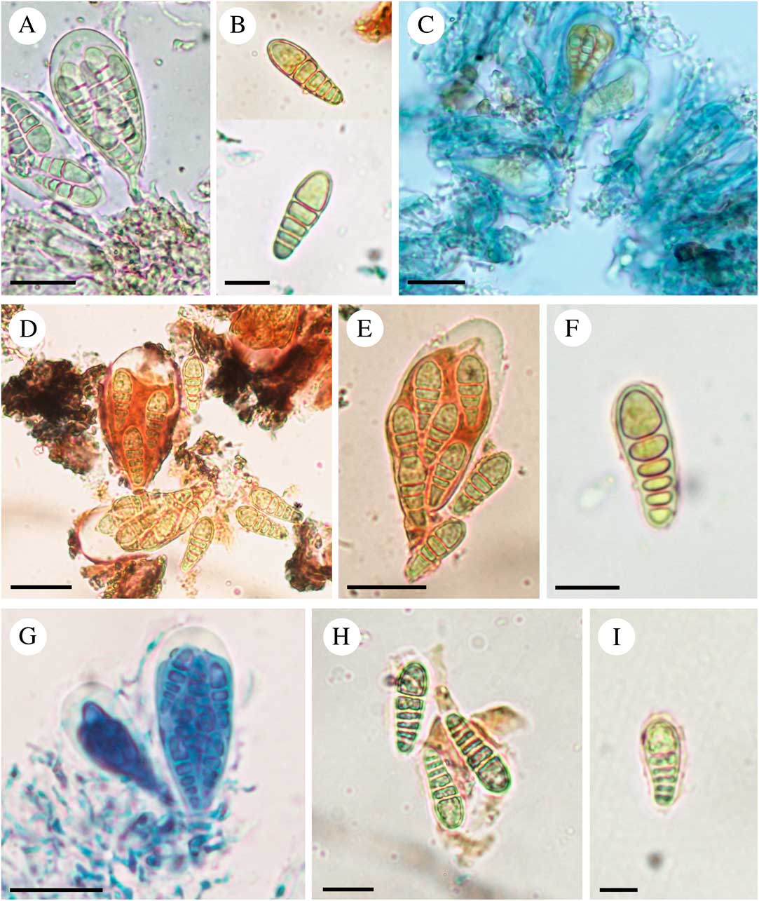
Fig. 2 A & B, Synarthonia bicolor; A, ascus (Jagadeesh 754B); B, ascospores (holotype). C, asci and paraphysoids of S. psoromica (holotype); D–F, S. sikkimensis (holotype); D, asci; E, ascus with 3-septate ascospores; F, an ascospore with thin epispore. G–I, S. stigmatidialis (holotype); G, asci; H, ascospores; I, an ascospore with thin epispore. A, in water; B, D–F, H & I, in Lugol's iodine solution; C, in KI; G, in LPCB. Scales: A, C, D, E & G = 20 μm; B, F, H & I = 10 μm. In colour online.

Fig. 3 A & B, drawings of cross-section of synascomata by Müller Argoviensis associated with holotype; A, Synarthonia bicolor; B, S. stigmatidialis. C & D, S. bicolor (Jagadeesh 1016A); C, section showing monocarpocentral synascomata; D, section showing pluricarpocentral synascomata. E, synascomata cross-section of S. psoromica (holotype); F, synascomata cross-section of S. sikkimensis (holotype), arrow showing deep fissures separating individual ascomata. Scales: C–F = 100 μm.
Thallus corticolous, crustose, white to whitish grey, some areas yellowish to brownish (in type), smooth, 50–80 μm thick; calcium oxalate crystals absent. Border line dark brown, c. 0·2 mm wide. Algal cells 8–15 × 6–10 μm.
Ascomata solitary when young, with a well-developed white thalline margin lacking algal cells, turning into mono-carpocentral synascomata and finally becoming pluri-carpocentral synascomata forming a pseudostromatic structure, with a hardly visible white thalline margin, individual ascomata irregular to lirellate or rounded; disc pale brownish to brownish, epruinose or rarely white pruinose. Excipulum hyaline to pale brownish, 10–15 μm thick, K–. Epithecium brownish to somewhat greyish, with some gelatinous material and hyaline crystals, 10–25 μm thick, K+ olivaceous. Hymenium hyaline, clear, rarely with some minute crystals, 35–80 μm high, I+ directly red, KI+ blue. Paraphysoids richly branched and anastomosing up to the tip, 1·0–1·5 μm wide; apices slightly thickened, 2·0–2·5 μm wide, brown pigmented. Hypothecium hyaline to pale yellowish, 18–44 μm thick, K–, I+ red, KI+ blue. Asci 8-spored, Arthonia-type, 43–72 × 18–27 μm. Ascospores hyaline, pale brownish at maturity, old spores±warty and with shrivelled surface, (3–)4–5-septate, macrocephalic, (17·2–)20·0–24·0(–26·8) × (5·0–)7·0–8·5(–9·2) μm (n = 50), l/b =(2·4–)2·6–3·0(–3·5) μm, epispore not seen.
Chemistry
Thallus K–, C–, P–, UV+ yellow. TLC: lichexanthone present.
Notes
The ascomata of S. bicolor are mainly epruinose, especially in the type specimen, and rarely with some sparse minute hyaline crystals in the hymenium. The width of ascospores in the protologue is 8·5–9·5 μm, according to Sparrius (Reference Sparrius2009) it is 4–5 μm, whilst the present study shows the width to be (5·0–)7·0–8·5(–9·2) μm. It is the only species with a UV+ yellow thallus and lichexanthone as a secondary metabolite.
Specimens examined. India: West Bengal: Sundarbans Biosphere Reserve, on Excoecaria agallocha L., 2004, Jagadeesh 754B (BSA); Haldibari, on Excoecaria agallocha L., 2004, Jagadeesh 957 (BSA); Maya dwip, Sundarbans National Park, on Excoecaria agallocha L., 2004, Jagadeesh 1016A (BSA).
Synarthonia psoromica S. Joseph & G. P. Sinha sp. nov.
MycoBank No.: MB811393
Synarthonia psoromica is characterized by immersed synascomata, 3-septate ascospores and a thallus with psoromic acid as a secondary metabolite.
Type: India, Tamil Nadu, Nilgiris, Armbi Forest, on bark of Rapanea wightiana (Wall. ex A. DC.) Mez, 11°25′09·5"N, 76°41′13·2"E, 2410 m, 6 December 2012, S. Joseph 8140 (BSA—holotype & isotype).
(Figs 1C & D, 2C & 3E)
Thallus corticolous, rimose in appearance, whitish, c. 100 μm thick; calcium oxalate crystals absent. Prothallus pale brownish, fibrous-like. Algal cells 11–16 × 9–14 μm.
Ascomata grouped in a slightly immersed pseudostroma, pluri-carpocentral synascomata, usually separated from the thallus by a narrow slit, with an inconspicuous to thin thalline margin, individual ascomata rounded to slightly elongated; disc thinly white pruinose. Excipulum hyaline to pale brownish, 10–20 μm thick, K– or K+ slightly olivaceous. Epithecium pale brownish, 13–20 μm thick, with some hyaline to pale brownish crystals, K+ olivaceous. Hymenium hyaline, not inspersed, 40–70 μm high, I+ red, KI+ pale blue. Paraphysoids richly branched and anastomosing up to apices, 1·0–1·8 μm wide, apices slightly thickened, 2·0–2·5 μm wide, not pigmented. Hypothecium hyaline to pale yellowish, with some brownish hyphal inclusions, 15–60 μm thick, I+ blue, KI+ deep blue. Asci 8-spored, Arthonia-type, 34–50 × 15–20 μm. Ascospores hyaline, brownish at maturity, (2–)3-septate, (12·5–)13·0–14·7(–15·8) × (4·0–)4·4–5·2(–5·5) μm (n = 30), l/b = (2·4–)2·6–3·0(–3·4) μm, macrocephalic, epispore not seen.
Chemistry
Thallus K–, C–, P+ yellow, UV–. TLC: psoromic acid present.
Notes
Synarthonia psoromica is easily distinguished from other known species by its synascomata embedded in a slightly immersed pseudostroma, an I+ red hymenium, smaller and less septate (2–3-septate) ascospores and the presence of psoromic acid.
Synarthonia sarcographoides Aptroot et al.
In Menezes et al., Lichenologist 45: 616 (2013); type: not seen.
The species recently described from north-east Brazil (Menezes et al. Reference Menezes, Lima, Xavier-Leite, Maia, Aptroot and Cáceres2013) can be distinguished from other Synarthonia species by its brown, broadly ellipsoid, 7 × 1–2-septate, muriform ascospores, 20–22 × 11·0–12·5 μm.
Synarthonia sikkimensis S. Joseph & G. P. Sinha sp. nov.
MycoBank No.: MB811394
Similar to S. stigmatidialis but differs by the sorediate thallus, rhizomorph-like prothallus and larger ascospores [(17·0–)19·0–23·0(–25·7) × (5·5–)6·5–7·5(–8·6) μm].
Type: India, Sikkim, Lachung-Dombang, near Yakche, bridge area, 27°43′35·5"N, 88°45′17·9"E, 3040 m, 20 March 2012, G. P. Sinha & S. Joseph 7192 (BSA—holotype).
(Figs 1E–G, 2D–F, 3F)
Thallus corticolous, crustose, rimose-like, smooth to verrucose, 100–180 μm thick, centrally with some pustule-like structures, becoming sorediate at margins; calcium oxalate crystals absent. Prothallus rhizomorph-like, pale brownish when in contact with other lichens. Algal cells 10–17 × 7–14 μm.
Ascomata solitary along thallus margin, elongated to lirellate, centrally grouped in slightly elevated pseudostroma, pluri-carpocentral synascomata, with a thin white margin, individual ascomata rounded to slightly elongated; disc thinly white pruinose. Epithecium pale brownish, 10–20 μm thick, K+ slightly greenish. Hymenium hyaline, not inspersed, 50–80 μm high, I+ blue turning red or I+ red with blue streaks, KI+ blue. Paraphysoids richly branched and anastomosing, 0·8–1·5 μm wide, with more branching network at the epithecium, apices thickened, 2·0–2·8 μm wide, brown pigmented. Hypothecium hyaline to pale brownish, with some brownish hyphal inclusions, 20–40 μm thick, K+ greenish, I+ blue turning red or I+ red with blue streaks, KI+ blue. Asci 8-spored, Arthonia-type, 45–55 × 16–25 μm. Ascospores hyaline, brown granular and warty at maturity, 3–5-septate, macrocephalic, (17·0–)19·0–23·0(–25·7) × (5·5–)6·5–7·5(–8·6) μm (n = 50), l/b = (2·5–)2·7–3·3(–3·5) μm, rarely with thin epispore, 0·4–0·6 μm.
Chemistry
Thallus K–, C–, P–, UV–. TLC: no substances detected.
Notes
This is the only known species of Synarthonia having a sorediate thallus. It has small pustules usually located in the central part of the thallus which turn into marginal soredia. Synarthonia sikkimensis is close to S. stigmatidialis but has soredia, a well-developed rhizomorph-like prothallus and larger ascospores.
Synarthonia stigmatidialis Müll. Arg.
Hedwigia 34: 145 (1895); type: Mexico, San Luis Potosi, 1890, J. M. Eckfeldt 245 (G00110645—holotype!).
(Figs: 1H; 2G–I; 3B)
Thallus corticolous, rimose, whitish. Prothallus fibrous-like. Algal cells 9–13 × 8–11 μm.
Ascomata grouped in pseudostroma, pluri-carpocentral synascomata, with an inconspicuous to thin white margin, individual ascomata rounded to short lirellate; disc thinly white pruinose. Excipulum poorly developed, hyaline to pale brownish, 7–15 μm thick, K– or K+ slightly olivaceous. Epithecium greyish to brownish, 13–20 μm thick. Hymenium hyaline, not inspersed, 40–70 μm high, I+ blue, KI+ deep blue. Paraphysoids richly branched and anastomosing, 0·7–1·7 μm thick, apices thickened, 2·0–2·8 μm wide, brown pigmented. Hypothecium hyaline, 13–40 μm thick, I+ blue, KI+ deep blue. Asci 8-spored, Arthonia-type, 40–45 × 16–20 μm. Ascospores hyaline, (3–)4(–5)-septate, (12·0–)14·5–19·0(–22·0) × (4·8–)5·4–6·6(–7·0) μm (n = 30), l/b = (2·0–)2·5–3·2(–3·9) μm, macrocephalic, rarely with thin epispore, c. 0·5 μm.
Chemistry
Thallus K–, C–, P–, UV–. TLC: not conducted.
Notes
In the protologue and Sparrius (Reference Sparrius2009), ascospore size is recorded as 15–17 ×4·0–5·5 μm, but it has been found that the size is more variable: (12·0–)14·5–19·0(–22·0) × (4·8–)5·4–6·6(–7·0) μm. For the first time in this species, the I reactions of ascomatal tissue and chemical tests were confirmed, which will help to distinguish the species from others. The species is currently known only from the type locality.

The authors are grateful to the Director, Botanical Survey of India, Kolkata, for providing facilities and to Dr Philippe Clerc, Head Curator-Cryptogams, Conservatoire et Jardin botaniques de la Ville de Genève (G), Switzerland for the loan of type specimens. The authors would also like to thank the two anonymous reviewers for their valuable comments and suggestions to improve the quality of the paper.


