


INTRODUCTION
Knowledge of the atomic structure of matter is of fundamental importance in science. The discovery of structural information has often led to the creation of new scientific fields. Advances in solid state physics followed the discovery of crystal structures obtained through the production of X-ray diffraction patterns (von Laue, Reference von Laue1936). The modern science of molecular biology and genetic engineering were triggered by the discovery of the double helix structure of DNA (Watson & Crick, Reference Watson and Crick1953). X-ray diffraction gives precise information about atomic structure on a sub-angstrom scale and spectroscopic techniques—extended X-ray absorption fine structure (EXAFS), X-ray absorption near edge structure (XANES)—give information about local order and on chemical states. The possibility of generating ultra-short X-ray pulses provides an opportunity to capture structural and electronic changes dynamically, for example, observing the detailed intermediate steps of chemical reactions with atomic resolution. Typical time scales for changes in atomic configurations are comparable with the cyclic duration of molecular vibrations. Advances in the generation of ultra-short laser pulses over the past two decades have made it possible to observe these fundamental processes. These advances have taken science from the ns- and ps-regime a generation ago, to the fs time scale in the past decade, and recently into the as-regime (Strickland & Mourou, Reference Strickland and Mourou1985; Sansone et al., Reference Sansone, Benedetti, Calegari, Vozzi, Avaldi, Flammini, Poletto, Villoresi, Altucci, Velotta, Stagira, De Silvestri and Nisoli2006; Baker et al., Reference Baker, Robinson, Haworth, Teng, Smith, Chiril, Lein, Tisch and Marangos2006; Baeva et al., Reference Baeva, Gordienko and Pukhov2007). The fact that the wavelength of these ultra-short laser light sources is significantly larger than the sizes and distances of the constituent atoms in the molecules, and crystals under study has meant that information on structural changes could only be inferred from observation of the secondary effects that these structural changes have on optical properties.
The recent development of small, affordable, high-power lasers based on chirped pulse amplification (CPA), capable of producing fs pulses has produced interesting opportunities into the development of table-top size X-ray sources. Several ns and ps X-ray techniques based on both synchrotron and table-top X-ray sources have been developed during the past decade (Helliwell & Rentzepis, Reference Helliwell and Rentzepis1997). Most of these techniques use an ultra-short laser pulse to initiate a photo reaction in the sample that is then studied by a delayed ultra-short X-ray pulse, which probes the structural changes that take place in the sample. In the solid state, non-thermal melting and laser-induced heat and strain propagation in metal and semiconductor single and mosaic-crystals have been studied by means of time-resolved X-ray diffraction with ns to ps time resolution (Tomov et al., Reference Tomov, Oulianov, Chen and Rentzepis1999; Siders et al., Reference Siders, Cavalleri, Sokolowski-Tinten, Tóth, Guo, Kammler, Wilson, von der Linde and Barty1999). The use of all of these techniques in conjunction with a fs X-ray source will allow the direct measurement of structural information in many ultra-fast fundamental physical, chemical and biological processes, currently inaccessible with the time resolution of existing sources.
The development of short and ultra-short X-ray sources will drive many important scientific applications by enabling direct measurement of atomic motion and structural changes in the condensed phase. Perhaps the most important feature of these new sources of ultra-short X-ray pulses is that they enable physical experiments combining fs time resolution and atomic scale spatial resolution.
There are several different ways to generate short and ultra-short X-ray pulses (Kim et al., Reference Kim, Chattopadhyay and Shank1994; Materlik et al., Reference Materlik, Tatchyn and Winick1995; Schoenlein et al., Reference Schoenlein, Leemans, Chin, Volfbeyn, Glover, Balling, Zolotorev, Kim, Chattopadhyay and Shank1996, Reference Schoenlein, Chattopadhyay, Chong, Glover, Heimann, Shank, Zholents and Zolotorev2000a, Reference Schoenlein, Chattopadhyay, Chong, Glover, Heimann, Leemans, Shank, Zholents and Zolotorev2000b; Leemans et al., Reference Leemans, Chattopadhyay, Esarey, Zholents, Zolotorev, Chin, Schoenlein and Shank2000; Catravas et al., Reference Catravas, Esarey and Leemans2001; Guo et al., Reference Guo, Spielmann, Walker and Barty2001; Service, Reference Service2002; Hafz et al., Reference Hafz, Lee, Kim, Suk and Lee2003; Phuoc et al., Reference Phuoc, Rousse, Pittman, Rousseau, Malka, Fritzler, Umstadter and Hulin2003; Umstadter, Reference Umstadter2003; Xia et al., Reference Xia, Li, Kang, Huang, Huang, He, Du and Tang2004; Janulewicz et al., Reference Janulewicz, Lucianetti, Priebe and Nickles2004a, Reference Janulewicz, Priebe, Tümmler and Nickles2004b, Reference Janulewicz, Tummler, Priebe and Nickles2005; Krafft, Reference Krafft2004, Reference Krafft, Doyuran and Rosenzweig2005; Lucianetti et al., Reference Lucianetti, Janulewicz, Kroemer, Priebe, Tummler, Sandner, Nickles and Redkorechev2004; Giulietti et al., Reference Giulietti, Galimberti, Giulietti, Gizzi, Labate and Tomassini2005; Tümmler et al., Reference Tümmler, Janulewicz, Priebe and Nickles2005; Zhavoronkov et al., Reference Zhavoronkov, Gritsai, Bargheer, Woerner and Elsaesser2005; Riley et al., Reference Riley, Angulo-Gareta, Khattak, Lamb, Foster, Divall, Hooker, Langley, Clarke and Neely2005; Legall et al., Reference Legall, Stiel, Arkadiev and Bjeoumikhov2006; Notley et al., Reference Notley, Weber, Fell, Jeffries, Freeman, Mackinnon, Dickson, Hey, Khattak, Saiz and Gregori2006; Khattak et al., Reference Khattak, Saiz, Dzelzainis, Riley and Zhai2007; Chen et al., Reference Chen, Kando, Xu, Li, Koga, Chen, Xu, Yuan, Dong, Sheng, Bulanov, Kato, Zhang and Tajima2008; Abdallah et al., Reference Abdallah, Batani, Desai, Lucchini, Faenov, Pikuz, Magunov and Narayanan2007; Mangles et al., Reference Mangles, Walton, Najmudin, Dangor, Krushelnick, Malka, Manclossi, Lopes, Carias, Mendes and Dorchies2006) such as ultra-short laser-generated Kα sources, X-ray free-electron lasers, electron bunch-slicing in synchrotrons, and the one to be discussed here, inverse Compton scattering of an intense laser pulse from a high-brightness electron beam.
Using the laser effectively as an undulator, which confers a much shorter period than the commonly used magnetic undulators in synchrotron light sources, means a lower energy electron beam can be used to deliver X-rays. This source also offers a wide tuning range, where X-ray source parameters such as photon energy, brightness, bandwidth, and pulse length can be tuned through the laser and electron beam parameters as well as the interaction geometry of the beams. The interaction can also be viewed as a collision between an electron and photon. This treatment of the system gives rise to Compton scattering, or Thomson scattering when the incident photon energy in the electron rest frame is much less than mc 2. Depending on the reference frame, it can appear as though the electrons have been energized by the photons or, if the electrons are moving relativistically, the energy is instead transferred from electrons to photons, therefore the opposite of Compton scattering—inverse Compton scattering—occurs. The frequency of the photons scattered from the relativistic electron beam is dependent on the interaction angle of the two beams, this typically range from 90° to 180°, although small-angle Thomson scattering has also been a proposed interaction configuration (Huang et al., 1994). The generation of 300 fs hard X-ray pulses has been demonstrated using 90° Thomson scattering between fs laser pulses and tightly-focused ns electron bunches in a synchrotron (Kim et al., Reference Kim, Chattopadhyay and Shank1994). The relatively low X-ray flux of ≈106 photons/s obtained in these experiments could be significantly increased by the use of higher laser powers and the use of high quality fs electron bunches.
However, the most promising feature of laser based X-ray sources in time resolved studies, is the intrinsic synchronisation of a laser induced change in a material with the probing X-ray probe pulse. As already demonstrated a photon flux of 106 photon per second (Li et al., 2005) on the sample can be sufficient for time resolved X-ray spectroscopy. Additional improvement of the experimental condition can perhaps be achieved by using more efficient X-ray optics for spectral measurements (Legall et al., Reference Legall, Stiel, Arkadiev and Bjeoumikhov2006).
In this paper, we describe a new source of ultra-short X-ray pulses based on inverse Compton scattering of multi-10 TW, 100 fs laser-pulses with 35 MeV electron bunches delivered by the energy recovery linac prototype (ERLP) recently renamed to accelerators and lasers in combined experiments (ALICE), which is being commissioned by the Daresbury Laboratory (Fig. 1; Smith et al., Reference Smith, Muratori, Owen, Hoffstaetter, Litvinenko, Ben-Zvi, Bai, Beebe-Wang, Blaskiewicz, Calaga, Fischer, Chang, Kayran, Kewisch, MacKay, Montag, Parker, Ptitsyn, Roser, Ruggiero, Satogata, Surrow, Tepikian, Trbojevic, Yakimenko, Zhang and Piot2005).

Fig. 1. (Color online) Layout of the energy recovery linac machine ALICE commissioned at Daresbury Laboratory.
Thomson scattering from a relativistic electron beam has been extensively studied theoretically both in the linear and the nonlinear regime. For clarity, we adapt the linear theory, where the energy of the scattered photon is calculated using energy and momentum conservation, and the differential scattering cross-section is calculated using quantum electrodynamics theory. The X-ray source, the inverse compton backscattering X-ray source driven by the table-top 10 TW (T3-laser) laser installed at Daresbury (COBALD) will initially be used as a short pulse diagnostic for the ERLP electron beam (Leemans et al., Reference Leemans, Volfbeyn, Zolotorev, Kim, Chattopadhyay, Schoenlein, Balling, Shank, Chin and Glover1996, Reference Leemans, Schoenlein, Volfbeyn, Chin, Glover, Balling, Zolotorev, Kim, Chattopadhyay and Shank1997; Ross et al., Reference Ross, Alley, Arnett, Bong, Colocho, Frisch, Hortonsmith, Inman, Jobe, Kotseroglou, McCormick, Nelson, Scheeff and Wagner1997; Tannebaum & Shintake, Reference Tannebaum and Shintake1999; Hartemann et al., Reference Hartemann, Tremaine, Anderson, Barty, Betts, Booth, Brown, Crane, Cross, Gibson, Fittinghoff, Kuba, Le Sage, Slaughter, Wootton, Hartouni, Springer, Rosenzweig and Kerman2004; Chouffani et al., Reference Chouffani, Harmon, Wells, Jones and Lancaster2006). It will explore the extreme challenges of photon/electron-beam synchronization, which are a fundamental requirement for the next generation of accelerator-based light sources. The COBALD source will be exploited for a fast melting experiment (Wark, Reference Wark1999; Sundaram & Mazur, Reference Sundaram and Mazur2002; von der Linde, Reference von der Linde2003; Sokolowski-Tinten & von der Linde, Reference Sokolowski-Tinten and von der Linde2004) providing information about the synchronization between the laser pulse and the electron bunch, and on X-ray source stability. In the experiment, a portion of the laser beam will be used to induce disorder at the surface of a crystal, which will subsequently recrystallizes on a ps time scale. The X-ray pulse will probe the recrystallization by allowing the measurement of the diffracted X-ray intensity from the crystal lattice at intervals throughout the transformation. The inverse Compton backscattering (CBS) source has the high-brightness, short pulses, and tunable energy and represents a research tool, capable of measuring on an atomic scale with high spatial and temporal.
Energy Recovery Linac
Synchrotron radiation (SR) X-ray sources have come a long way from their first parasitic existence on high energy physics machines. Improvements in electron storage rings and insertion devices have led to an exponential increase in brilliance for each successive generation of sources. The technology of storage rings has reached maturity, resulting in third generation SR facilities that are capable of providing beam qualities close to the physical limits (Ozaki et al., Reference Ozaki, Kieffer, Toth, Fourmaux and Bandulet2006). The fundamental X-ray beam properties of ring sources, such as the source size, brilliance, and pulse duration are limited by the equilibrium between the electron beam and the magnet lattice. In contrast to storage rings, linear accelerators can produce both low emittance and very short bunches, because the limitations set by this equilibrium are not an issue. As a result, several proposed large light-source projects are based on linear accelerators. However, in a traditional linear accelerator, the absence of beam recirculation would lead to an enormous power consumption if high average currents are to be obtained. To circumvent this prohibitively large power consumption, energy recovery linac (ERL) technology (Tigner, Reference Tigner1965) is being explored for next-generation synchrotron light sources; in such accelerators the energy delivered to the electron beam is re-circulated, rather than the electron beam itself.
The use of ERLs enables electron beams with extraordinary brilliance, with small source size, ultra-short pulses, and concomitant high transverse coherence. Several laboratories have proposed high-power ERLs for the production of high-brightness radiation. Accelerators for different parameter sets and various applications are being worked on by Cornell University, Argonne National Laboratory, Budker Institute, High Energy Accelerator Research Organization (KEK), and Jefferson Laboratory (Kulipanov et al., Reference Kulipanov, Skrinsky and Vinokurov1998; Bazerov et al., Reference Bazerov, Belomestnykh, Bilderback, Finkelstein, Fontes, Gray, Gruner, Krafft, Merminga, Helmke, Rogers, Sinclair, Talman and Tigner2001; Suwada et al., Reference Suwada, Iida, Funakoshi, Kawamoto and Kikuchi1997; Smith et al., Reference Smith, Bliss, Goulden, Holder, McIntosh and Priebe2007; Neil et al., Reference Neil, Behre, Benson, Bevins, Biallas, Boyce, Coleman, Dillon-Townes, Douglas, Dylla, Evans, Grippo, Gruber, Gubeli, Hardy, Hernandez-Garcia, Jordan, Kelley, Merminga, Mammosser, Moore, Nishimori, Pozdeyev, Preble, Rimmer, Shinn, Siggins, Tennant, Walker, Williams and Zhang2005; Borland, Reference Borland2006). ALICE, at Daresbury Laboratory (Fig. 1, Table 1), is also an advanced energy recovery linac based light source (Poole et al., Reference Poole, Bennett, Bowler, Bliss, Dykes, Farrow, Gerth, Holder, MacDonald, Muratori, Owen, Quinn, Seddon, Smith, Suller, McNeil, Ross and Thompson2003; Smith et al., Reference Smith, Muratori, Owen, Hoffstaetter, Litvinenko, Ben-Zvi, Bai, Beebe-Wang, Blaskiewicz, Calaga, Fischer, Chang, Kayran, Kewisch, MacKay, Montag, Parker, Ptitsyn, Roser, Ruggiero, Satogata, Surrow, Tepikian, Trbojevic, Yakimenko, Zhang and Piot2005).
Table 1. The main parameters of the energy recovery linac machine ALICE

It is based on a combination of a direct current photocathode electron gun, a superconducting injector linac, and main linac operating in energy recovery mode. A infrared free electron laser cavity (IR-FEL) is incorporated in the machine. The FEL is based on a permanent magnet array undulator that will deliver intense short pulses of photons in the wavelength range 4 µm to 12 µm. The 1.4 ps pulses will deliver ~3 × 10¹⁴ photons per pulse, with a pulse energy of 13.3 µJ. The priorities for this machine are to gain experience with operating a photo-injector gun and superconducting linac; to produce and maintain high-brightness electron beams; to achieve energy recovery from a FEL-cavity disrupted beam and to study important synchronization issues, all of which will contribute towards the design of a larger scale fourth generation light source.
Multi-10 TW Laser
The customized table-top CPA multi-10 TW laser system (COHERENT)—installed at the high field laser facility at Daresbury—contains an ultra-short, bandwidth-limited, Kerr-lens mode-locked (Spence et al., Reference Spence, Kean and Sibbett1991; Brabec et al., Reference Brabec, Spielmann, Curley and Krausz1992; Stingl et al., Reference Stingl, Lenzner, Spielmann, Krausz and Szipocs1995). Ti:sapphire master oscillator (Micra; Δλ > 100 nm) with a repetition rate of 81.25 MHz, followed by a stretcher and a regenerative amplifier (2.8 mJ @ 1 kHz), which is used as a front-end system (Fig. 2) for a four-pass Ti:Sa power amplifier.

Fig. 2. (Color online) Installation of the regenerative amplifier front end including pulse-picker and kHz compressor.
The output of the master oscillator exhibits a broad spectrum centered at 800 nm. The pulses are then passed through a one-to-one imaging, single grating stretcher to obtain a pulse-width of about 150 ps before they are injected as seed into the regenerative amplifier (legend), which is pumped by a diode-pumped, intra-cavity doubled, Q-switched Nd:YLF laser (evolution). The customized power amplifier, which contains a large aperture Ti:Sa crystal (Fig. 3, left), pumped from both ends (relay imaged) using two spatially optimized frequency doubled Nd:YAG lasers operating at 10 Hz, amplifies the pulses up to 1.5 J in a bow-tie configuration before recompression (Fig. 3, right).

Fig. 3. (Color online) Installation of the multi-pass power amplifier (left) and the chirped pulse compressor (right).
For further applications as laser-driven plasma sources and particle acceleration, a pulse cleaner (Divall & Ross, Reference Divall and Ross2004; Priebe, Reference Priebe, Redkorechev, Janulewicz and Nickles2006) using a fast pockels-cell driven by a KENTECH fast pulse generator will be established. The legend incorporates an additional compressor to use the recompressed pulses (2.2 mJ @ 1 kHz) for further applications. Figure 4 shows the layout of the laser beam lines for further applications.

Fig. 4. (Color online) Laser beam lines from the laser front-end system (2.2 mJ @ 1 kHz) and from 4-pass Ti:Sa power amplifier through the CPA compressor to several application stations.
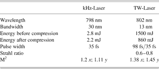
One of the applications of the kHz beam will be the development of non-destructive longitudinal, i.e., temporal, diagnostics of the accelerator electron bunches; the Coulomb field of an electron bunch interacts, through the electro-optic (EO) effect, with a probe laser pulse in an in vacuo non-linear crystal. The temporal evolution of the Coulomb field is encoded as a time-dependent probe polarization (EO-sensing) (Berden et al., Reference Berden, Jamison, MacLeod, Gillespie, Redlich and van der Meer2004; Jamison et al., Reference Jamison, MacLeod, Berden, Jaroszynski and Gillespie2006). The kHz beam also drives a stand-alone THz-source, with the terahertz-radiation produced by either optical rectification or a large area photo-conductive antenna; this THz-source provides radiation pulses that can mimic the relativistic Coulomb field, and hence allow significantly simplified testing of diagnostic concepts (Jamison et al., Reference Jamison, Shen, MacLeod, Gillespie and Jaroszynski2003). The detailed laser parameters—measured during the installation at Daresbury—of the multi-10 TW laser system are given in Table 2.
Table 2. The main laser parameters of the customized (COHERENT) table-top multi-10 TW laser system installed at the Daresbury high field laser laboratory
INVERSE COMPTON SCATTERING
The discovery of the Compton effect (Compton, Reference Compton1923) was a milestone in physics in which the quantum nature of light was finally proved. The Compton effect is the inelastic scattering of X-rays by electrons. The electrons gain energy in the process and the Compton scattered X-rays are observed at lower energy than the incident X-rays. Due to the kinematics of the scattering process, there is a fixed relationship between the energy of the scattered X-rays and the angle through which they are scattered. Soon after the discovery of the Compton effect, it was realized that electron motion during the scattering process Doppler-broadens the emission spectrum—at fixed scattering angle—and it is this Doppler shift that forms the basis of inverse Compton X-ray sources. In inverse Compton scattering, the electrons are highly energetic, and the Doppler shift results in the scattered photons gaining significant energy from the electrons. This process allows low energy photons, e.g., from an intense laser, when scattered by ultra relativistic electrons in an accelerator, to generate hard X-rays. If the energy of the incident photon E ph in the frame of the interaction is much less than m ec 2, the Thomson scattering cross-section (σTh = (8π/3)r e2) can be used to describe the probability of scattering, where m e is the rest mass of the electron, and r e is the classical electron radius. In the laboratory frame, the scattered laser photons are relativistically up-shifted in frequency into the hard X-ray range and are emitted into a narrow cone. Alternatively, the interaction can be viewed in the electron rest mass frame, the incident laser pulse then appears as an electromagnetic undulator of wavelength λu = λph/γ (1−β cos Φ), where γ is the relativistic factor E e/m ec 2, β is the velocity of the electrons relative to the speed of light, and Φ is the angle of incidence between electron and laser beams. The undulating electrons radiate photons, which are frequency up-shifted, in the laboratory frame, by a second factor of 2γ. In the laboratory frame, the X-rays are confined to a narrow cone with opening angle about 1/γ in the electron beam propagation direction. The X-ray energy E γ varies with the observation angle θ in the laboratory frame due to the kinematics of the scattering

where a 0 = eE phλph/2πm ec 2 is the normalized vector potential of the laser field, analogous to the undulator deflection parameter K = eB 0λu/2πm ec 2 of a static field undulator (Brown & Kibble, Reference Brown and Kibble1964; Sarachik & Schappert, Reference Sarachik and Schappert1970; Alferov et al., Reference Alferov, Bashmakov and Bessonov1974; Coisson, Reference Coisson1979; Kim, Reference Kim1989). The peak X-ray energy for transverse interaction (Φ = π/2) is half that for head-on collisions (Φ = π). Due to the similarity between synchrotron radiation and inverse Compton scattering, the radiation is often called laser synchrotron radiation or Compton synchrotron radiation. Using this analogy, and replacing the magnetic undulator with a laser beam of 104 times smaller period, the electrons are deflected many more times and contribute to coherent energy gain with electron beams 100 times less energetic than in a conventional magnetic undulator. When a 0≪1, the interaction is in the linear regime; when a 0 ≥ 1 the interaction is in the nonlinear regime with higher order harmonics, which may be used to extend the tuning range of the X-ray source (Ride et al., Reference Ride, Esarey and Baine1995; Salamin & Faisal, Reference Salamin and Faisal1996). However, ponderomotive scattering (Krafft, Reference Krafft2004, 2005) also starts to occur for a 0 > 1, where the electrons are deflected from the laser focus point before they are able to scatter the photons, therefore decreasing the total flux of emitted X-rays (Chen et al., Reference Chen, Kando, Xu, Li, Koga, Chen, Xu, Yuan, Dong, Sheng, Bulanov, Kato, Zhang and Tajima2008). The precise intensity and spectral profile of the resultant X-ray beam is closely linked to the laser frequency and bandwidth, the electron beam emittance, energy and energy spread, and the interaction and focusing geometries (Hartemann et al., Reference Hartemann, Baldis, Kerman, Le Foll, Luhmann and Rupp2001). A schematic illustration of the inverse Compton scattering geometry is shown in Figure 5.

Fig. 5. (Color online) Interaction geometry of inverse Compton scattering.
From the particles point of view, the photon strikes an electron of energy E e- with an angle of incidence Φ. In the electron's rest frame, the energy of the photon is Doppler up-shifted to E ph′ = γ(1 + cos Φ)E ph and the wavelength of the scattered photon is given by the Compton formula λγ′−λph′ = λc(1−cos θ′), where λc = h/m ec is the Compton wavelength, h is the Planck's constant, and θ′ is the scattering angle in the electron rest frame. The scattered photons gain energy E γ′ = m ec 2 E ph′/[m ec 2 + (1−cos θ′)E ph′], with the contracted angle cosθ′ = (cos θ−β)/(1−β cos θ) satisfying the momentum conservation constraint. Since the relativistic Doppler up-shifted X-ray energy E ph′ is much less that m ec 2 in the electron rest frame, the center of momentum frame is very close to that of the relativistic electron. Using the back transformation in the laboratory frame E γ = γ(1−β cos θ)E γ′ results in the relation between the scattered photon energy E γ and that of the incident laser photon energy E ph and electron energy E e- being given by: E γ = E ph(1−β cos Φ)/[(1−β cos θ) + E ph(1−β cos θ−Φ)/E e-]. The dependence of the energy of the scattered X-ray photons on the collison angle Φ between the laser photons and the electrons, and on the scattering angle θ, is shown in Figure 6, for the laser and electron parameters at Daresbury (Tables 1 and 2).

Fig. 6. (Color online) Dependence of the X-ray energy E γ on the interaction geometry—the collision angle Φ—and the scattering angle θ.
For a fixed laser incidence angle, there is a unique relation between the resultant scattered energy E γ and the photon trajectory of the X-rays. The peak X-ray energy (θ = 0) for head-on collisions (Φ = π) and transverse interaction (Φ = π/2) simplifies—using E ph≪m ec 2, β ≈ 1, and γ = (1–β2)−1/2—to the expected result:

In practice, the X-ray spectrum is broadened by the bandwidth, and the divergence of the laser pulse as well as the energy spread and the divergence of the electron bunch (Table 3). The energy spread depends linearly on the laser energy spread and quadratically on the electron energy spread. The energy spread of the bandwidth-limited laser pulse is given by ΔE ph = chΔλ/λ2, where Δλ is given by the time-bandwidth product τphcΔλ/λ2 = 2 ln 2/π if the pulse shape is Gaussian and where τph is the laser pulse duration. For a 100 fs laser pulse the energy spread is therefore ΔE ph/E ph ≈ 1.2% and ΔE ph/E ph ≈ 3.9% for a 30 fs laser pulse.
Table 3. X-ray energy broadening induced by the wavelength bandwidth and the divergence of the laser pulse as well as the energy spread and the divergence of the electron bunch

Using an off-axis parabolic mirror with a focal length f of 1780 mm for head-on interaction and 1230 mm for transverse interaction, to focus the laser beam gives a focused spot size (2λf/π∅︀) of ≈40 µm with an opening angle of ≈1.4° and a focal length (twice the Raleigh length z R = πw 02/λ) of ≈3 mm for head-on collision, and a spot size of ≈28 µm with an opening angle of ≈2.1° and a focal length of ≈1.5 mm for transverse collision.
The divergence of the electron bunch is ≈0.2° given by the normalized emittance and the spot size σe of the electron focus (Δθe = ɛn/γβσe). In head-on collision, the on-axis X-ray energy spread caused by the divergence of the photons, and of the electrons is negligible (0.004% ∣ 0.0003%) compared with the broadening due to their own energy spread (Fig. 7). For transverse interaction, the divergence of the photons becomes the dominating factor (1.8%) and the energy spread induced by the divergence of the electrons (0.4%) becomes comparable to the energy spread itself.

Fig. 7. (Color online) X-ray energy spread ΔE γ due to the divergence of the laser beam (top) and the detailed distribution of the X-ray energy spread ΔE γ for head-on collision geometry (bottom) as a function of the collision angle Φ and the scattering angle θ.
In the rest frame of the electron bunch, the differential scattering cross-section is described by the Klein–Nishina formula (Klein & Nishina, Reference Klein and Nishina1929):
where ɛ′ is the ratio between the energies of the scattered and incident photon ɛ′ = E γ′/E ph′. If the interaction is such that the recoil momentum of the electron can be disregarded, E ph≪m ec 2 (ɛ′ = 1), then this leads to the well known (cos θ)2 distribution of dipole radiation and the differential Thomson scattering cross-section:
and integrated, gives the Thomson cross-section σTh = (8/3)πr e2 = 0.665 × 10⁻²⁸ m2 for elastic scattering of a photon from an electron. The total cross-section for the non-relativistic ɛ′≫1 and the extremely relativistic ɛ′≪1 cases are given as:

Transforming the differential Klein–Nishina cross-section to the laboratory frame of reference, using the above mentioned boundary condition, as well as its derivative (dθ′sin θ′ = dθ sin θ(1−β2)/(1−β cos θ)2) and the transformations E γ' → E γ and E ph′ → E ph gives the differential Compton cross-section:
![{d\sigma_c \over \sin \theta \, d\theta } = \pi \, r_{\rm e}^2 \, \varepsilon^2 \left[\varepsilon + {1 \over \varepsilon } - 1 + \left({\cos \theta - \beta \over 1 - \beta \cos \theta}\right)^2 \right]\comma \;](https://static.cambridge.org/binary/version/id/urn:cambridge.org:id:binary:20160126040956937-0419:S0263034608000700_eqnU6.gif?pub-status=live)
with ɛ = m ec 2/[m ec 2 + γ (1 + cos Φ)(1−(cos θ−β)/(1−β cos θ))E ph]. The cross-section for scattering a photon into a cone of angle θc in the laboratory frame is then σc(θc) = ∫dθ(dσc/dθ) and the average energy of X-rays backscattered in the laboratory frame is (∫dθ E γ dσc/dθ)/(∫dθ dσc/dθ). Limiting the integration to the opening half-angle of 1/2γ leads to a scattering cross-section independent of the electron energy. Numerically, this is 0.165 barn and of course integrating over the full phase space gives the Thomson cross-section of 0.665 barn.
The total number of X-ray photons N γ scattered per unit time and volume into a cone of angle θc is given by the product of the electron beam four-current j μ(x ν) = ecn e(x ν)u μ/γ and the incident photon four-flux Φμ(x ν) = cn λ(x ν)k μ/ω as d 4N γ(x ν)/d 4x ν =(σc/ec)j μ(x ν)Φμ(x ν) = (cσc/γω)n e(x ν)n ph(x ν)u μk μ (Landau & Lifshitz, Reference Landau and Lifshitz1975). Here, u μ = dx νc dt is the electron four-velocity, k μ = (ω/c, k) is the incident photon four-wave number. The number of X-ray photons per unit time can be expressed in Cartesian coordinates explicitly as d 4N γ(x, y, z, t)/dx dy dz c dt = σcn e(x, y, z, t)n ph(x, y, z, t)(1−β cos Φ) (Hartemann et al., Reference Hartemann, Brown, Gibson, Anderson, Tremaine, Springer, Wootton, Hartouni and Barty2005):
where L = ∫n en ph dx dy dz c dt is the luminosity, which is determined by the collision geometry of the electron bunch and the laser pulses. In order to evaluate the pulse length of the X-rays, we assume that both the electron bunch and the laser pulse satisfy the Gaussian distribution. The density profile of the laser light pulse and the electron bunch are thus given by:
![\eqalign{n_{\rm ph} & = {N_{\rm ph} \over 2\sqrt {2} \, c \pi^{3/2}} {1 \over w_{\rm phx} \lsqb z\rsqb w_{\rm phy} \lsqb z\rsqb \, \tau_{\rm ph}} \cr & \quad \times \exp \left[- {2x^2 \over w_{\rm phx}^2 \lsqb z\rsqb } - {2y^2 \over w_{\rm phy}^2 \lsqb z\rsqb } - {2\lpar t - \lpar z / c\rpar \rpar ^2 \over \tau_{\rm ph}^2} \right]\cr n_{\rm e} & = {N_{\rm e} \over 2\sqrt {2} \, c\pi^{3/2}} {1 \over w_{\rm ex} w_{\rm ey} w_{\rm ez}} \cr & \quad \times \exp \left[- {x^2 \over 2w_{\rm ex}^2} - {y^2 \over 2w_{\rm ey}^2} - {\lpar t + \lpar z / \beta \, c\rpar \rpar ^2 \over 2w_{\rm ez}^2} \right].}](https://static.cambridge.org/binary/version/id/urn:cambridge.org:id:binary:20160126040956937-0419:S0263034608000700_eqnU8.gif?pub-status=live)
w phx,y = (w 02(1 + (z/z r)2))1/2 is the beam waist, N ph is the total number of photons in the laser pulse, τph is the pulse duration—which is related to the bandwidth as Δτph Δω = √2, in the case of a Fourier-transform-limited pulse—w 0 is the 1/e 2 focal radius, N e is the total number of electrons in one bunch, w ex,y are the magnitude of the electron bunch in the x and y directions, and cw ez is the bunch length. For laser pulses crossing the electron bunch at an angle Φ, the luminosity can be described as: L = N eN ph/[2π(w ey2 + w oy2/4)1/2 (cos2 Φ/2 (w ex2 + w ox2/4) + (w ez2 + c 2τph2/4β2) sin2 Φ/2)1/2] (Yang et al., Reference Yang, Washio, Endo and Hori1999). This leads to a luminosity of L = N eN ph/[2π(w ex2 + w ox2/4)1/2(w ey2 + w oy2/4β2)1/2] for head-on collision and, respectively, L = N eN ph/[2π(w ey2 + w oy2/4)1/2(w ez2 + c 2τph2/4β2)1/2] for transverse collision. The total number of scattered X-ray photons N γ per unit time and volume into a cone of angle θc is given as:

where the electron bunch was taken as focused to an spot size of w ex = 35 µm, w ey= 20 µm and w ez = c 350 fs. The laser was assumed to be focused to a spot size of w 0 = 40 µm with a pulse duration of 100 fs.
In head-on geometry, the overlap integral depends only on the spot size; the laser and electron bunch durations play no role. The laser pulse travels through the electron bunch and the X-ray pulse length is determined almost entirely by the length of the electron bunch. In transverse interaction, it is noticeable from the interaction geometry (Fig. 4) that the electron bunch spot size in the x-direction w ex is negligible, but the beam size in the y-direction, w ey must fit with the laser spot size, and the electron bunch length w ez must be comparable to the travel time of the laser pulse through the electron bunch cτph. The length of the X-ray pulse will be dependent on how long the laser and electron beam interact. This indicates that tight electron beam focusing and short laser pulses will result in short X-ray pulses due to a minimized interaction time.The relation between these parameters and the resultant source pulse length τγ is expressed for head-on collision as w ez/cβ and as w ez(w ey2 + c 2τph2/4β2 + w oy2/4)1/2/[c(w ey2 + w ez2 + w oy2 + w oy2/4 + c 2τph2/4β2)1/2], respectively, for transverse collision (Pogorelski et al., Reference Pogorelski, Ben-Zvi, Hirose, Kashiwagi, Yakimenko, Kusche, Siddons, Skaritka, Kumita, Tsunemi, Omori, Urakawa, Washio, Yokoya, Okugi, Liu, He and Cline2000).
The T3 laser beam transport line is shown in Figure 8. The vacuum system pumping, gauges and protection (fast valve) are designed to operate assuming no window between the laser beam transport line and the accelerator vacuum. This is necessary, as a window thick enough to withstand atmospheric let-up would be radiation-damaged by absorption of TW pulses, and a thin window will have the risk of breakage and particle contamination of ALICE in the event of a vacuum failure.

Fig. 8. (Color online) Laser beam transport line through the concrete shielding wall to the interaction region.
The laser beam transport line vacuum design specification is 10−6 mbar and the ALICE machine vacuum is at 10−8 mbar. The ALICE system is protected from a let-up in the laser beamline by a fast-acting valve. The laser beam propagates from the CPA compressor vessel through a concrete shielding wall, passing an optical delay line, periscoped down to the electron beam level, focused via an off-axis parabolic mirror (OAP) and finally turned to the interaction point. For the case of transverse interaction, the periscope is also used to rotate the polarization of the laser. The last vacuum vessel containing the OAP-mirror sits on rails allowing the focal position to be moved through the electron bunch. The focal length of the OAP-mirror is 1780 mm in head-on collision geometry and 1230 mm for transverse geometry. The scattered hard X-rays—in the direction of the electron beam—will be taken through a beryllium window to the diagnostic area (X-ray deep depletion CCD camera, fast X-ray streak camera, fast melting experiment). In head-on geometry, the last turning mirror has a hole of about 5 mm diameter to let the X-rays through and also to take a part of the laser beam out for pump-probe applications. Figure 9 shows the proposed experimental setup for the head-on collision geometry (left) and transverse collision geometry (right).

Fig. 9. (Color online) Experimental setup for head-on collision (left) and transverse collision (right).
In order to model the X-ray output, it was first necessary to simulate the electron bunch spatial, angular and energy distribution at the interaction point. For optimal generation of Compton scattered X-rays, the ALICE lattice is set to focus the electrons to give the highest electron density at the interaction point. This maximizes the X-ray flux and also reduces the size of the X-ray source. The electron trajectories were modeled using the particle tracking code ELEGANT. In excess of 100,000 electrons were tracked through to the focus. The electron distribution produced was then used as input to code written to simulate the inverse Compton scattering. The electron data consisted of the electron position at the focus plane, the electron direction vector (direction cosines), and the electron energy. The laser photons were taken as a Gaussian angular distribution (in the horizontal and the vertical planes) and an energy distribution given by the transform function of the laser Gaussian temporal distribution. For each electron at the focus, a laser photon was generated at random by the code with direction vector and energy randomly chosen according to their assumed distributions. An X-ray was then generated with direction vector selected randomly conforming to the Klein-Nishina cross-section. The calculated X-ray energy as a function of emission angle in head-on scattering geometry is shown in Figure 10 and for transverse scattering geometry in Figure 11, respectively. The spectral brightness of the X-ray source (photons/(mm2 mrad2 s 0.1% bandwidth) is shown in Figure 12. Figure 13 shows the angular distribution of the X-rays for head-on geometry.

Fig. 10. (Color online) X-ray energy E γ versus the emission angle θ in head-on scattering geometry.

Fig. 11. (Color online) X-ray energy E γ as a function of the emission angle θ in side scattering geometry.

Fig. 12. (Color online) Spectral brightness of the X-ray source versus E γ for head-on scattering geometry.

Fig. 13. (Color online) Angular distribution of the X-rays generated in head-on collision geometry; each colour is a 1 keV energy band with 20–21 keV on outside and 30–31 keV at the centre.
CONCLUSION AND OUTLOOK
COBALD is an instrument capable of generating high peak brightness fs X-ray pulses. X-rays generated by the interaction of the table-top multi-10 TW laser with electron bunches have been modeled by Monte Carlo simulation calculating over 105 interactions, and have shown that a brightness in excess of 1021 X-rays/(mm2 mrad2 s 0.1% bandwidth) will be obtained in head-on geometry. It is intended to operate COBALD in both counter-propagating and transverse interaction geometry. The peak X-ray energy is about 30 keV in head-on geometry and approximately 15 keV in transverse scattering geometry. The X-ray pulse length in head-on mode will be about 350 fs and in transverse scattering geometry about 100 fs. The X-ray source size will be of order 20 × 35 µm2. Characterization of the X-ray beam profile and energy spectrum will provide vital information about the spatial and temporal structure of the electron beam in the ALICE. Further studies are planned to examine photon/electron beam synchronization and source stability; this is a necessary requirement prior to any dynamic experiments being performed. The availability of this ultra-short brilliant X-ray source will allow X-ray diffraction and spectroscopy techniques to be extended into the time domain allowing atomic resolution probing of structural dynamics. This type of experiment could be carried out using a pump pulse—for example, by extracting a fraction of the multi-10 TW laser pulse—to start a time-dependent process in a sample. The sample could then be probed, after a short delay, by the X-ray pulse. This would allow, for example, intermediate species to be probed during chemical reactions or strain relaxation following lattice distortion to be measured.
ACKNOWLEDGEMENT
We would like to acknowledge the financial support of STFC and the Northwest Development Agency, through the Northwest Science Fund.


