1. INTRODUCTION
Proton radiography was started at the Los Alamos National Laboratory (LANL) in 1995 (Gavron et al., Reference Gavron, Morris, Ziock and Zumbro1996; Amann et al., Reference Amann, Espinoza, Gomez, Hart, Hogan, Marek, McClelland, Morris, Ziock, Zumbro, Atencio, Hill, Jaramillo, King, Morley, Pazuchanics, Yates, Mottershead, Mueller, Sarracino, Saunders, Hartouni, Prigl, Scaduto and Schwaner1997; Ziock et al., Reference Ziock, Adams, Alrick, Amann, Boissevain, Crow, Cushing, Eddleman, Espinoza, Fife, Gallegos, Gomez, Gorman, Gray, Hogan, Holmes, Jaramillo, King, Knudson, London, Lopez, McClelland, Merrill, Morley, Morris, Mottershead, Mueller, Neri, Numkena, Pazuchanics, Pillai, Prael, Riedel, Sarracino, Stacy, Takala, Thiessen, Tucker, Walstrom, Yates, Zumbro, Ables, Aufderheide, Barnes, Bionta, Fujino, Hartouni, Park, Soltz, Wright, Balzer, Flores, Thompson, Prigl, Scaduto, Schwaner, Saunders and O'Donnell1998). Since then, proton radiography has been an important tool in the weapons program due to its spatial resolution and material identification. Radiographic information is obtained by measuring the intensity of the shadow of an object in a beam of penetrating radiation (Morris et al., Reference Morris, Hopson and Goldstone2006). Proton radiography has many advantages to X-ray radiography in beam spot size, penetration capability, energy spectrum, multiple angles radiography, multiple times radiography, and sensitivity to the density and atomic number of the measured object. With the urgent need of knowing the material information of an object, many institutes or laboratories develop proton radiography technique in the United States (Schwartz et al., Reference Schwartz, Hogan, Kwiatkowski, Rigg, Rightley, Mariam, MarrLyon, Merrill, Morris, Saunders and Tupa2007; Morris et al., Reference Morris, Ables, Alrick, Aufderheide, Barnes, Buescher, Cagliostro, Clark, Clark, Espinoza, Ferm, Gallegos, Gardner, Gomez, Greene, Hanson, Hartouni, Hogan, King, Kwiatkowski, Liljestrand, Mariam, Merrill, Morgan, Morley, Mottershead, Murray, Pazuchanics, Pearson, Sarracino, Saunders, Scaduto, Schach von Wittenau, Soltz, Sterbenz, Thompson, Vixie, Wilke, Wright and Zumbro2011), Russia (Golubev et al., Reference Golubev, Demidov, Demidova, Dudin, Kantsyrev, Kolesnikov, Mintsev, Smirnov, Turtikov, Utkin, Fortov and Sharkov2010; Antipov et al., Reference Antipov, Afonin, Vasilevskii, Gusev, Demyanchuk, Zyat'kov, Ignashin, Karshev, Larionov, Maksimov, Matyushin, Minchenko, Mikheev, Mirgorodskii, Peleshko, Rud'ko, Terekhov, Tyurin, Fedotov, Trutnev, Burtsev, Volkov, Ivanin, Kartanov, Kuropatkin, Mikhailov, Mikhailyukov, Oreshkov, Rudnev, Spirov, Syrunin, Tatsenko, Tkachenko and Khramov2010), Germany (Tahir et al., Reference Tahir, Shutov, Varentsov, Hoffmann, Spiller, Lomonosov, Wieser, Jacoby and Fortov2002; Hoffmann et al., Reference Hoffmann, Blazevic, Ni, Rosmej, Roth, Tahir, Tauschwitz, Udrea, Varentsov, Weyrich and Maron2005), and China (Wei et al., Reference Wei, Yang, He, Long, Zhang, Wang, Yang, Li, Li, Yang, Wang, Shi, Zhang, Deng and Zhang2010; Zhao et al., Reference Zhao, Hu, Cheng, Wang, Peng, Golubev, Zhang, Lu, Zhang, Zhou, Wang, Xu, Ren, Li, Lei, Sun, Zhao, Wang, Wang and Xiao2012). LANL proposed 3 GeV proton radiography which is upgraded from present 800 MeV protons (Garnett et al., Reference Garnett, Merrill, O'Hara, Rees, Rybarcyk, Tajima and Walstrom2012). In addition, new facilities are being constructed in Germany and China. A 4.5 GeV proton radiography beam line with 5.0 × 10¹² particles/pulse will be operated at the Gesellschaft für Schwerionenforschung for FAIR experiments with great discovery potential for plasma physics and high energy density physics research (Merrill et al., Reference Merrill, Golubev, Mariam, Turtikov and Varentsov2009). The Institute of Modern Physics (IMP) of the Chinese Academy of Sciences (CAS) has also proposed two proton radiography setups, one is for 2.6 GeV in Heavy Ion Research Facility (HIRFL) in Lanzhou-Cooler Storage Ring (CSR) and the other is for 9 GeV in the High Intensity heavy-ion Accelerator Facility (HIAF) (Zhao, Reference Zhao2011).
Heavy-ion radiography is very similar to proton radiography with respect to great penetration capability, clear imaging, and less number of ions for detection. A high energy heavy-ion radiography facility is constructed at the IMP-CAS for diagnosing static targets, which is expected to accumulate experiment data for ions interacting with matter, verify the theory and simulation of heavy-ion radiography, and make foundation for proton radiography. This paper presents the heavy-ion radiography setup at IMP, shows the beam optics of the facility and the simulation of radiography as well as the performance test results. Furthermore, two new dedicated radiography beam lines in plan are also designed here.
2. HEAVY-ION RADIOGRAPHY SETUP AT IMP
The heavy-ion radiography setup is based on the HIRFL-CSR, which can provide a variety of ion species from carbon ions to uranium ions and the highest energy is 1 GeV/u for carbon ions. Figure 1 shows the overall layout of the HIRFL-CSR and the heavy-ion radiography beam line.

Fig. 1. Overall layout of the HIRFL-CSR and the heavy-ion radiography beam line.
The heavy-ion radiography beam line with a large field-of-view is modified from an old beam line, and the schematic view is shown in Figure 2. The ions from fast extraction of HIRFL-CSR are first focused onto the object plane with suitable beam parameters by three matching magnets. Then the lens system after the object focuses the transmitting ions onto the image plane to provide image and material identification information. The matching lens system provides the required phase space correlation upstream of the object, and an imaging lens system downstream of the object provides the phase space correlation to maximize the image quality and minimize the chromatic aberration in second order.

Fig. 2. Schematic view of the heavy-ion radiography beam line at IMP.
The Zumbro magnets are usually used for radiography (Mottershead et al., Reference Mottershead and Zumbro1998). They have the useful property of having an “angular focus” at the midpoint, which facilitates to insert a collimator to eliminate the large angle scattered particles to enhance spatial resolution and material identification. But in order to keep the original configuration of the beam line as much as possible, a special radiography beam line is developed at IMP. There is a point-to-point imaging in the first order transfer matrix, so the final position is independent of the initial angle. The second order chromatic correction is achieved by a special position-angle correction at the entrance of the object, that is, x 0′/x 0 = −T 116/T 126 and y 0′/y 0 = −T336/T346 (Merrill et al., Reference Merrill, Campos, Espinoza, Hogan, Hollander, Lopez, Mariam, Morley, Morris, Murray, Saunders, Schwartz and Thompson2011). x 0 and y 0 are initial half beam spot sizes, x 0′ and y 0′ are initial half beam angles; T 116, T 126, T 336, and T 346 are terms of second order transfer matrix in TRANSPORT code. Figure 3 gives the beam optics of the imaging lens system in the first order. Note that, this radiography beam line is not symmetric and doesn't satisfy the Zumbro magnets, but its magnifications are both 1 in the x and y directions.

Fig. 3. Beam optics of the imaging lens system for IMP heavy-ion radiography setup.
In order to achieve clear imaging, the positions and angles of heavy ions at the entrance of the object are measured, and a Lutetium oxyorthosilicate crystal scintillator and a charge-coupled device camera are used for image recording online. Mathematical analysis of the image allows separate determination of the atomic number and thickness for object identification (Ryu et al., Reference Ryu, Song, Lee and Kim2008).
3. SIMULATION AND EXPERIMENTAL RESULTS
The heavy-ion radiography facility at IMP was simulated by Geant4 code with 1 GeV protons. A circular beam spot was first focused onto the object plane by three matching quadrupoles, and then refocused onto the image plane by the imaging lens system. Two detectors were placed at the object plane and the image plane for phase space measurement, respectively. A circular aluminum object (2 cm thickness) with two 10 mm × 3 mm stripes in the center was simulated in the object plane. We use 107 particles to simulate here. Figure 4 shows the beam transmission onto the image plane.

Fig. 4. Beam transmission onto the image plane simulated by Geant4 code; (a) Image in x and y directions; (b) Transmission in the edge of y direction; (c) Gauss fit for derivative of edge transmission.
A performance test has been carried out to characterize the heavy-ion radiography facility at IMP with 600 MeV/u carbon ions and 5.0 × 10⁹ particles/pulse. The pulse length of beam for radiography was 300 ns with a cycle of 20 s. The circular moveable aluminum object mentioned above was placed on the object plane. A high sensitive charge-coupled device camera was placed near the image plane to catch the image in real time. Figure 5 shows the image for the two stripes and its vertical edge transmission in the heavy-ion radiography facility at IMP. The spatial resolution is about 65 µm (1 σ) in vertical direction by fitting Gauss curve.

Fig. 5. (a) Image for the two stripes in x and y directions; (b) Transmission in the edge of y direction; (c) Gauss fit for derivative of edge transmission.
4. PROPOSED DEDICATED PROTON RADIOGRAPHY BEAM LINES
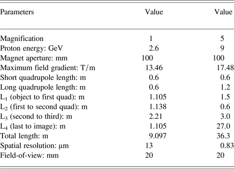
Two new dedicated proton radiography beam lines are proposed at IMP with magnifications of M = 1 and M = 5, and the imaging lens systems inherits the main features of the Zumbro magnet (Yang et al., Reference Yang, Zhang, Wei, He, Long, Shi and Zhang2012). Figure 6 shows the beam optics for the proton radiography with 2.6 GeV at HIRFL-CSR and 9 GeV at HIAF calculated by My-BOC code (Zhang et al., Reference Zhang, Yang and Lv2010). The limiting spatial resolution is proportional to T 126 term of transfer matrix, the angular spread and the momentum spread of the beam, and inversely proportional to the magnification. So the limiting spatial resolutions are expected to be 13 µm for 2.6 GeV proton radiography at HIRFL-CSR and 830 nm for 9 GeV proton radiography at HIAF. The corresponding parameters for the proton radiography beam lines are listed in Table 1.

Fig. 6. Beam optics for proton radiography beam lines with the magnification of M = 1 (a) and M = 5 (b).
Table 1. Parameters of dedicated proton radiography beam lines for M = 1 and M = 5

5. CONCLUSION AND OUTLOOK
The carbon ion radiography experiment for static target has been carried out at IMP-CAS, and the spatial resolution is about 65 µm (1 σ), which agrees with the simulation of 1 GeV proton radiography by Geant4 code. Dedicated proton radiography beam lines with 2.6 GeV at HIRFL-CSR and 9 GeV at HIAF are proposed here, which will enhance the experiment capability in spatial resolution and material identification for thicker object. In addition, sidestep objects will be made radiograph in future. Short-bunch and multi-bunch extraction is improved for HIRFL-CSR at present, and the dynamic experiment can be carried out further.
ACKNOWLEDGEMENTS
This work is supported by the Major State Basic Research Development Program of China (‘973’ Program) No. 2010CB832902 and the National Science Foundation of China under Grant Nos. 11176001 and 10805063. The authors would like to thank the accelerator staffs of IMP and IFP during beam commissioning.


