INTRODUCTION
The upcoming facility for antiproton and ion research (FAIR), currently under construction at the Helmholtz Center for Heavy-Ion Research GSI (Darmstadt, Germany), will offer heavy ion beams at unprecedented intensities (Henning, Reference Henning2004; Spiller & Franchetti, Reference Spiller and Franchetti2006). One of the research pillars within the multi-facetted scientific program at FAIR is the area of dense plasmas (Hoffmann et al., Reference Hoffmann, Blazevic, Ni, Rosmej, Roth, Tahir, Tauschwitz, Udrea, Varentsov, Weyrich and Maron2005). A variety of schemes has been proposed to generate matter at high energy density (HED) conditions using the intense ion pulses delivered by the FAIR accelerator complex (e.g., Tahir et al., Reference Tahir, Deutsch, Fortov, Gryaznov, Hoffmann, Kulish, Lomonosov, Mintsev, Ni, Nikolaev, Piriz, Shilkin, Spiller, Shutov, Temporal, Ternovoi, Udrea and Varentsov2005; Reference Tahir, Stöhlker, Shutov, Lomonosov, Fortov, French, Nettelmann, Redmer, Piriz, Deutsch, Zhao, Zhang, Xu, Xiao and Zhan2010, Tauschwitz et al., Reference Tauschwitz, Novikov, Tauschwitz, Rosmej, Abdallah, Onkels, Jacoby and Maruhn2009). This promises novel and unique approaches for accurate studies of matter under such extreme conditions, complementary to the highly non-equilibrium states produced with X-ray free electron lasers (e.g., Zastrau et al., Reference Zastrau, Burian, Chalupsky, Döppner, Dzelzainis, Fäustlin, Fortmann, Galtier, Glenzer, Gregori, Juha, Lee, Lee, Lewis, Medvedev, Nagler, Nelson, Riley, Rosmej, Toleikis, Tschentscher, Uschmann, Vinko, Wark, Whitcher and Förster2012).
As a feature common to most of these schemes, the targets undergo a large hydrodynamic evolution, mostly in the so-called warm-dense matter (WDM) regime. In this regime, the equation-of-state is rather poorly known and theoretical modelling is a great challenge (e.g., Lomonosov, Reference Lomonosov2007) due to strong-coupling of the ions and partial electron degeneracy (which is, of course, also the main motivation for the experimental study of matter at WDM conditions). For this reason, accurate monitoring of the target density distribution is crucial to verify the target performance. Besides, density is an important plasma parameter, and, for example, conductivity measurements in the WDM regime are meaningless without accurate knowledge of the target density. Finally, in most equation-of-state experiments, the target hydrodynamic evolution, such as shock propagation or isentropic release is the primary observable. Thus, radiography to measure the target density distribution will be an indispensable key enabling diagnostic technique for HED experiments at the future FAIR facility.
In this paper, we explore the potential of radiography based on hard X-rays produced by intense high-energy laser pulses on solid targets for targets proposed for FAIR. This study is in context of current planning for a high-energy high-intensity laser system to be installed near the FAIR plasma physics experimental area, with the purpose to enable advanced diagnostic techniques. Furthermore, we present first test results from recent developments of hard X-ray imaging detectors particularly designed for use in such scenarios.
RADIOGRAPHY OF DENSE PLASMAS USING PW-LASER GENERATED HARD X-RAY BREMSSTRAHLUNG
X-ray radiography using high-energy laser-produced plasmas as sources for intense X-ray radiation of some 10s of keV photon energy (see, e.g., Hicks et al., Reference Hicks, Spears, Braun, Olson, Sorce, Celliers, Collins and Landen2010; Kritcher et al., Reference Kritcher, Döppner, Swift, Hawreliak, Collins, Nilsen, Bachmann, Dewald, Strozzi, Felker, Landen, Jones, Thomas, Hammer, Keane, Lee, Glenzer, Rothman, Chapman, Kraus, Neumayer and Falcone2014) has become a routine tool in many HED laboratories. However, most of the target schemes proposed to produce HED samples at FAIR involve large (mm-size), dense high-Z targets. Photon energies suitable to penetrate such targets are in the 100 keV to MeV spectral range. Intense bursts of such energetic X-rays are emitted from solid targets irradiated by laser-pulses at relativistic intensities (> 1018 W/cm2). In the interaction of laser light at relativistic intensities with overdense plasma, a large fraction of laser energy is converted to hot (supra-thermal) electrons with energies reaching several times the ponderomotive energy E p = 511 keV × (1 + 0.73 × I 18λμ2)1/2, where I 18 × 1018 W/cm2 and λμ × 1 μm are the laser intensity and wavelength, respectively. Traversing the solid density target these fast electrons give rise to intense characteristic line emission (Park et al., Reference Park, Chambers, Chung, Clarke, Eagleton, Giraldez, Goldsack, Heathcote, Izumi, Key, King, Koch, Landen, Nikroo, Patel, Price, Remington, Robey, Snavely, Steinman, Stephens, Stoeckl, Storm, Tabak, Theobald, Town, Wickersham and Zhang2006) and bremsstrahlung. The emission duration is limited by the relaxation of the hot electron population to picoseconds corresponding to the energetic electron stopping range (≈ mm) in matter at solid density (Nilson et al., Reference Nilson, Davies, Theobald, Jaanimagi, Mileham, Jungquist, Stoeckl, Begishev, Solodov, Myatt, Zuegel, Sangster, Betti and Meyerhofer2012). This time is short compared to the hydrodynamic timescale, thus the hard X-ray emission is spatially restricted to the initial target dimensions. Using limited-size targets source sizes down to 10 µm have been demonstrated, allowing simple point-projection radiography schemes (Park et al., Reference Park, Maddox, Giraldez, Hatchett, Hudson, Izumi, Key, Le Pape, MacKinnon, MacPhee, Patel, Phillips, Remington, Seely, Tommasini, Town, Workman and Brambrink2008). Point-projection radiography has the important advantage, that no imaging optics (lens, pinhole) is required, making it applicable up to very high photon energies, where no lenses are available and pinholes become transparent. This radiography scheme has successfully been employed, for example, to image laser-driven shocks (Brambrink et al., Reference Brambrink, Wei, Barbrel, Audebert, Benuzzi-Mounaix, Boehly, Endo, Gregory, Kimura, Kodama, Ozaki, Park and Koenig2009; LePape et al., Reference Le Pape, Neumayer, Fortmann, Döppner, Davis, Kritcher, Landen and Glenzer2010), the isentropic expansion of isochorically heated wire-targets (Hochhaus et al., Reference Hochhaus, Aurand, Basko, Ecker, Kühl, Ma, Rosmej, Zielbauer and Neumayer2013) and direct-drive implosions of CH shells at the Omega Laser facility (Tommasini et al., Reference Tommasini, Hatchett, Hey, Iglesias, Izumi, Koch, Landen, MacKinnon, Sorce, Delettrez, Glebov, Sangster and Stoeckl2011) and is being under consideration for implementation at the National Ignition Facility to measure the areal density of the highly-compressed fuel in inertial confinement fusion experiments. While most experiments were performed on low-Z targets of small areal density, measurements of the hard X-ray yield indicate that a large amount of high-energy (> 100 keV) bremsstrahlung is produced, suitable to penetrate large high-Z targets.
SIMULATIONS OF HARD X-RAY RADIOGRAPHY ON HED TARGETS FOR FAIR
In order to assess the potential of intense-laser driven hard X-ray radiography as diagnostic for HED experiments proposed for FAIR, we have simulated radiographic images. Here we have used results from hydrodynamic calculations presented by Tahir et al. (Reference Tahir, Deutsch, Fortov, Gryaznov, Hoffmann, Kulish, Lomonosov, Mintsev, Ni, Nikolaev, Piriz, Shilkin, Spiller, Shutov, Temporal, Ternovoi, Udrea and Varentsov2005). In the proposed experiments, a solid lead cylinder (diameter 0.6 mm, length 2 mm) is placed in the focus of the heavy ion beam, with the cylinder axis aligned parallel to the ion beam. Transverse homogenous heating is ensured as the ion focal spot diameter is significantly larger than the target. As the stopping range of the relativistic heavy ions largely exceeds the target length energy deposition along the ion beam direction is very homogenous, resulting in a quasi-cylindrical geometry. Using the ion beam parameters anticipated at FAIR Tahir et al. (Reference Tahir, Deutsch, Fortov, Gryaznov, Hoffmann, Kulish, Lomonosov, Mintsev, Ni, Nikolaev, Piriz, Shilkin, Spiller, Shutov, Temporal, Ternovoi, Udrea and Varentsov2005) could show that a wide range of high-entropy states can be accessed, ranging from hot liquid, over the two-phase liquid-gas regime and the region around the critical point, up to the regime of strongly coupled plasmas.
Figure 1a shows a schematic of a possible setup to perform X-ray radiographic measurements on this target. A thin (10 µm) “backlighter” foil is located at a distance of about 1 cm from the target and irradiated by the high-energy high-intensity laser pulse. Employing the radiation emitted from the edge of the foil results in a strongly astigmatic backlighting source. As explained above, emission in the direction normal to the foil is limited to the initial foil thickness (i.e., 10 µm). In the direction along the foil, the emission source size is rather determined by the transverse spreading of the hot electrons, which is typically of the order 100 µm, as inferred from X-ray imaging crystal spectrometers (e.g., Zastrau et al., Reference Zastrau, Audebert, Bernshtam, Brambrink, Kämpfer, Kroupp, Loetzsch, Maron, Ralchenko, Reinholz, Röpke, Sengebusch, Stambulchik, Uschmann, Weingarten and Förster2010) or knife-edge tests (Hochhaus et al., Reference Hochhaus, Aurand, Basko, Ecker, Kühl, Ma, Rosmej, Zielbauer and Neumayer2013). By orienting the backlighter foil edge parallel to the cylinder axis, the transverse and longitudinal source sizes are well-matched to the characteristic length scales of the heavy-ion heated sample. The distance between source (foil edge) and target was chosen to limit the effective increase in transverse source size due to an oblique view onto the backlighter foil toward the edge of the required field-of-view. This distance, at the same time, determines the solid angle subtended by a resolution element in the target, and thereby the number of photons available in the corresponding pixel for image generation. X-ray emission spectra are calculated by a Monte-Carlo electron-photon transport code, using a 1-temperature hot electron spectrum with mean electron energy of 200 keV. In the widely used scaling by Beg et al. (Reference Beg, Bell, Dangor, Danson, Fews, Glinsky, Hammel, Lee, Norreys and Tatarakis1997), this corresponds to a focused intensity of about 1018 W/cm2, and we have found reasonable agreement with our measurements of the hard X-ray yield (Neumayer et al., Reference Neumayer, Aurand, Basko, Ecker, Gibbon, Hochhaus, Karmakar, Kazakov, Kühl, Labaune, Rosmej, Tauschwitz, Zielbauer and Zimmer2010).

Fig. 1. (a) Schematic setup to perform laser-driven X-ray radiography on a heavy-ion heated sample. (b) Calculated bremsstrahlung emission spectrum (blue, solid) generated in the backlighter foil by the laser-generated supra-thermal electrons, and cumulated radiated energy (blue, dot-dashed). For comparison, the transmission through 1 mm of lead is plotted (black).
Figure 1b shows the calculated emission spectrum, the number of photons per keV energy interval and per Joule of energy in the hot electron distribution, emitted into the full solid angle. At about 8 keV the copper K-alpha fluorescence can be observed as the target material used in the simulations was copper. The bremsstrahlung continuum emission initially falls off rather rapidly as dN/dE ∝ e−E/30 keV up to about 100 keV. The slope gradually decreases until for photon energies beyond 1 MeV the slope approaches that of a 1-temperature distribution with a temperature of 200 keV. For comparison, we plot the transmission of a 1 mm thick lead target showing that photon energies well above 100 keV are required to penetrate the target. Also shown is the cumulative energy (normalized to 1) of the X-ray emission. We find close to 30% of the total emitted energy at photon energies between 100 keV and 200 keV, and another 10% into photons at 200–300 keV. The total radiated energy is about 0.8% of the hot electron energy. This value is in good agreement with the conversion efficiencies reported by Tommasini et al. (Reference Tommasini, Hatchett, Hey, Iglesias, Izumi, Koch, Landen, MacKinnon, Sorce, Delettrez, Glebov, Sangster and Stoeckl2011). Here we assume laser pulse energy of 400 J (which is the projected pulse energy for the laser system under consideration for FAIR) and a conversion of 10% of laser energy into the hot electron fraction. This order of magnitude is typically inferred from absolute measurements of the K-alpha emission (e.g., Myatt et al., Reference Myatt, Theobald, Delettrez, Stoeckl, Storm, Sangster, Maximov and Short2007), whereas recent three-dimensional particle-in-cell simulations at intensities beyond 1020 W/cm2 suggest even values exceeding 50% (Kemp et al., Reference Kemp and Divol2012).
To generate synthetic radiographic images, the Abel transform of the radial mass densities reported in Tahir et al. (Reference Tahir, Deutsch, Fortov, Gryaznov, Hoffmann, Kulish, Lomonosov, Mintsev, Ni, Nikolaev, Piriz, Shilkin, Spiller, Shutov, Temporal, Ternovoi, Udrea and Varentsov2005) is performed and tabulated cold opacities are used to calculate the transmission. Use of cold opacities is justified as the attenuation at photon energies beyond the K-edge is dominated by photo-ionization of the K-shell, which is un-affected by the few-eV temperatures reached in the heavy-ion heated target, and Compton scattering, which is only dependent on the total electron density, and thus temperature independent. The simulated images include the shot noise due to the finite number of photons emitted per resolution element, the photon absorption statistics inside the target, and the quantum efficiency of the assumed detector.
Figure 2 shows simulated radiographic images of the lead target at various conditions: (Fig. 2a) heated liquid state, the target has been heated to a temperature of 3400–6000 K, while still at solid density; (Fig. 2b) the target has undergone expansion to conditions around the critical point (T ≈ 4000 K, ρ ≈ 2 g/cm3); and (Fig. 2c) a smaller initial target, and further expansion to the strongly coupled plasma regime (T ≈ 5600 K, ρ ≈ 4 g/cm3). Figure 2d shows the signal behind a calibration wedge, consisting of steps with thicknesses ranging from 20 to 600 µm, manufactured from the same material as the target. This method provides an absolute calibration allowing to directly relate the image exposure to the areal mass density, and has been successfully employed by Brambrink et al. (Reference Brambrink, Wei, Barbrel, Audebert, Benuzzi-Mounaix, Boehly, Endo, Gregory, Kimura, Kodama, Ozaki, Park and Koenig2009). As can be seen, an average transmission of ≈ 50% at the maximum areal density in the target center provides a good image contrast. The signal-to-noise ratio (SNR) of around 60 suggests that absolute measurement of the areal density to a few percent levels can be achieved.
Fig. 2. Attenuation (dot-dashed) and fraction of deposited energy (solid line) within the sensitive layer, for SR-type imaging plate (red) and DRZ screen (blue), determined from Monte-Carlo calculations with GEANT4. Again, the transmission through 1 mm lead is shown (black).
The detector assumed for calculating the radiographs in Figure 2 (top) was an imaging plate detector (SR-type, Fujifilm). Imaging plate detectors are widely used in medical imaging applications involving X- rays with energies up to 100 keV. The imaging plate is doped with a photostimulable phosphor which is excited to a metastable state upon deposition of energy by ionizing radiation, e.g., by X-ray photons. The stored information can later be retrieved by scanning the image plate with a laser scanner which de-excites the phosphor, resulting in emission of fluorescence light.
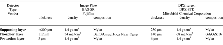
For a realistic assessment of the achievable SNR in the radiographic images, the detector quantum efficiency and response function over the relevant photon energy range (i.e., up to MeV) is needed. A good estimate for the detector quantum efficiency (i.e., the probability of a photon to make an interaction within the sensitive layer) and the detector response (i.e., the average amount of signal generated by a photon) can be obtained from the attenuation and the average amount of deposited energy per photon, respectively. We calculated these by means of Monte-Carlo simulations using the code suite GEANT4 (Agostinelli et al., Reference Agostinelli, Allison, Amako, Apostolakis, Araujo, Arce, Asai, Axen, Banerjee and Barrand2003), detector parameters used in the calculations are listed in Table 1. Figure 3 (red, dot-dashed) shows the results obtained for the image plate. After an initial rise (due to the increasing transmission through the top protective layer) we find the interaction probability close to unity for photon energies up to about 20 keV. For higher photon energies, the interaction probability rapidly drops to about 5% and further to <1% for 100 keV and 200 keV, respectively. The jumps around 13.5 keV and 37 keV are due to the absorption K-edges of Bromine and Barium, respectively.
Table 1. Detector parameters as used in the Monte-Carlo simulations for quantum efficiency and response
Also shown (solid line) is the fraction of energy a photon deposits on average within the sensitive layer. For small energies, this follows closely the interaction probability, as attenuation is dominated by photo-ionization and the resulting photo-electron will be stopped within the sensitive layer. For higher energies (beyond the K-shell absorption edges), the deposited energy fraction falls below the attenuation probability as an increasing amount of the initial photon energy can escape the sensitive layer (e.g., K-shell fluorescence photons, energetic photo electrons). In addition, in Compton scattering only a fraction of the photon energy is transferred in the interaction process.
HARD X-RAY IMAGING DETECTOR DEVELOPMENT AND TESTS
Again, for reference, in Figure 3, the transmission through 1 mm of lead is plotted, showing that photons with energies beyond 100 keV are required for imaging such thick high-Z targets. The significant drop in quantum efficiencies for imaging plate detectors directly translates into to the need for the powerful X- ray source to achieve high-quality images with sufficient SNR. Increasing the detector quantum efficiency in this energy range would improve the photon statistics and thus allow more accurate density measurements, or alternatively, for a less energetic laser system to drive the X-ray source. As the quantum efficiency is directly related to the interaction probability within the sensitive layer, increasing thickness, density, and average atomic number are required.
For comparison, we have calculated response and efficiency of a commercial fluorescent screen (DRZ-Std, Mitsubishi Chemical Corporation, parameters see Table 1) see Figure 3 (blue). We note that in these calculations the X-rays were assumed to enter the screen from the side of the supporting base, to view the fluorescence light through the thin protection layer. This leads to reduced efficiencies below 10 keV, which is uncritical for our envisioned application. In the relevant range above 100 keV, the quantum efficiency is approximately four times larger. Employing this increased efficiency the simulated radiographs (Fig. 2, bottom) show a SNR of approximately 120, an increase of two times as expected.
Fig. 3. Synthetic radiographs from various stages of a heavy-ion heated target (a–c), see text), and calibration wedge (d). The X-ray spectrum from Figure 1b was used with a total energy of 40 J in the hot electron distribution. Upper images assume an image plate detector, lower images using the DRZ-screen show improved signal-to-noise due to the increased quantum efficiency.
Conversion of X-ray radiation to visible light by scintillating screens, in combination with subsequent digitization by charge-coupled device arrays, also addresses another important requirement for detection systems at FAIR. Irradiation of massive targets with intense pulses of relativistic heavy-ions will lead to a considerable amount of activation of the experimental area close to the interaction. Access to the target area will therefore only be granted after a certain decay time (of the order of days) until this activation has “cooled down” to a level that again permits human activities. This is in contrast with the high repetition rates typical for accelerators, and also the laser system planned for FAIR will be designed to deliver several shots per hour. For this reason, detection schemes that require physical removal of the detector to access the experimental data (e.g., film, image plate) are strongly discouraged. Rather, detection systems should allow remote operation and electronic readout.
We have designed a first prototype of such a hard X-ray imaging detector (Fig. 4). Fluorescence from a DRZ screen is imaged by a combination of achromatic lenses (AC508-150-A, AC508-750-A, Thorlabs) onto a 12-bit charge-coupled device camera (Pixelfly QE, PCO). The lens system had been designed to provide a good balance between conflicting requirements for field-of-view, resolution, and light collection efficiency. The DRZ screen, optics, and optical path up to the camera are housed within black-anodized aluminum tubes to suppress background due to visible straylight. X-rays enter the housing through a 5 mm thick aluminum entrance “window,” which acts as a high-pass, suppressing photons < 20 keV. A flat mirror in the optical path brings the charge-coupled device camera out of the direct view to the X-ray source so the sensitive electronics can be heavily shielded.
Fig. 4. Schematic of the prototype hard X-ray imager (details see text).
First tests were performed at the superconducting electron linear accelerator S-DALINAC (University of Darmstadt, Darmstadt, Germany). Electron beams at 6.5 MeV energy produced bremsstrahlung in a thick copper converter target. Using currents of 9.25 µA and an exposure time of 10 s, high quality radiographic images were obtained (Figs. 5a and 5b), even penetrating tantalum blocks up to 24 mm in thickness. At the high-energy laser facility PHELIX (Bagnoud et al., Reference Bagnoud, Aurand, Blazevic, Borneis, Bruske, Ecker, Eisenbarth, Fils, Frank, Gaul, Goette, Haefner, Hahn, Harres, Heuck, Hochhaus, Hoffmann, Javorkova, Kluge, Kuehl, Kunzer, Kreutz, Merz-Mantwill, Neumayer, Onkels, Reemts, Rosmej, Roth, Stoehlker, Tauschwitz, Zielbauer, Zimmer and Witte2010) at GSI, we employed hard X-ray sources as discussed in the previous sections, i.e., driven by the energetic high-intensity laser pulses. Pulses with energies up to 100 J in 500 fs were focused onto thin metal foils and radiographs of various objects (M4 steel nut, Ta-slabs) were obtained.
Fig. 5. Radiographs obtained with the hard X-ray imager at the S-DALINAC electron accelerator (a, b) and at the PHELIX laser facility (c). The test objects are tantalum blocks of various thicknesses (a), M10 steel screw (b), and M4 steel nut and tantalum slabs (c).
In these measurements, the test objects were located close to the scintillator so that the detector's overall spatial resolution could be assessed. We find a resolution of the order of 150 µm. Detailed optical ray-tracing analysis indicates that the resolution is mainly limited by the remaining spherical aberrations. Nevertheless, the useful field-of-view of 40 mm provides more than 250 resolution elements. To achieve a resolution at the target of 10 µm (the lower limit due to the source size) would require a magnification of 15, i.e., a distance of 15 cm between target and detector. The field-of-view of about 2.5 mm matches well the typical target dimensions (cf. Fig. 2).
Our Monte Carlo simulations suggest that lateral spreading of the deposited energy in the scintillating layer is less than or equal to 10 µm, i.e., small compared to the resolution of our optical system. We therefore expect that the overall spatial resolution could be significantly improved by use of coherent fiber bundles with individual fiber diameters of 10 µm to transport the scintillation light from the screen to the charge-coupled device. At the same time, close coupling would increase the light collection efficiency by a factor 100 which would greatly improve the SNR achieved by the charge-coupled device camera.
While the use of a DRZ screen already leads to an increase in quantum efficiency by four times compared to imaging plates, even further increase should be achievable when using thick (≈ 500 µm) heavy scintillation crystals, e.g., BGO (Bismuth germinate) or LYSO (Cerium-doped Lutetium Yttrium Orthosilicate). These options will be explored in future work.
CONCLUSIONS
Through simulated radiographs of heavy-ion heated targets proposed within the FAIR plasma physics experimental program, we have shown that high-energy laser driven hard X-ray sources fulfil the requirements to produce high-quality radiographic images. This provides a strong case for a high-energy high-intensity laser installation at FAIR to enable this indispensable diagnostic capability. At the same time this will allow a variety of other X-ray diagnostic techniques, being routinely employed in state-of-the-art HED facilities, such as X-ray diffraction, absorption spectroscopy or Thomson scattering (Riley et al., Reference Riley, Khattak, Garcia Saiz, Gregori, Bandyopadhyay, Notley, Neely, Chambers, Moore and Comley2007).
Development of high repetition rate X-ray imaging detectors with increased quantum efficiencies at photon energies beyond 100 keV show promising first results. Development towards thicker scintillators and fiber-based image transport is expected to further improve their performance.
ACKNOWLEDGEMENTS
We gratefully acknowledge support by the PHELIX laser and operations team. We thank Prof. Dr. N. Pietralla (TU Darmstadt) for the opportunity to perform tests at the S-DALINAC. This work was supported by the Alliance Program of the Helmholtz Association (HA216/EMMI).