


INTRODUCTION
Members of the genus Dinophysis Ehrenberg have the morphological and ecological characteristics of the dinoflagellates of the order Dinophysales. Dinophysis is the type genus of the order Dinophysales and its species are marine and truly planktonic, armoured and laterally flattened, have a sagittal serrate suture extended throughout the body. The theca is mainly composed of two valves, left and right, a conserved number of plates, usually 18, six epithecal, four cingular, four sulcal, and four hypothecal, and sulcal lists of variable development (Norris & Berner, Reference Norris and Berner1970; Fensome et al., Reference Fensome, Taylor, Norris, Sarjeant, Wharton and Williams1993; Steidinger & Tangen, Reference Steidinger, Tangen and Tomas1997; Gómez, Reference Gómez2005; Hernández-Becerril et al., Reference Hernández-Becerril, Ceballos-Corona, Esqueda-Lara, Tovar-Salazar and León-Álvarez2008).
Species of Dinophysis can be photosynthetic or mixotrophic/heterotrophic (Hallegraeff & Lucas, Reference Hallegraeff and Lucas1988; Schnepf & Elbrächter, Reference Schnepf and Elbrächter1999; Park et al., Reference Park, Kim, Kim, Yi, Kang and Yih2006; Reguera et al., Reference Reguera, González-Gil and Delgado2007), with four distinct types of plastids of origins related to the groups of Cryptophytes (Schnepf & Elbrächter, Reference Schnepf and Elbrächter1988, Reference Schnepf and Elbrächter1999; Qiu et al., Reference Qiu, Huang, Liu and Lin2011), Haptophytes (Hallegraeff & Lucas, Reference Hallegraeff and Lucas1988; Qiu et al., Reference Qiu, Huang, Liu and Lin2011), Prasinophytes (Koike et al., Reference Koike, Sekiguchi, Kobiyama, Takishita, Kawachi, Koike and Ogata2005), or Cyanobacteria (Qiu et al., Reference Qiu, Huang, Liu and Lin2011).
Some Dinophysis species have the ability to produce cysts as part of their life cycle (Mackenzie, Reference MacKenzie1992; Berland et al., Reference Berland, Maestrini and Grzebyk1995; Giacobbe & Gangemi, Reference Giacobbe and Gangemi1997; Reguera & González-Gil, Reference Reguera and González-Gil2001), and mechanisms of sexual reproduction through engulfment of ‘small cells' (Koike et al., Reference Koike, Nishiyama, Saitoh, Imai, Koike, Kobiyama and Ogata2006), which have been suggested as part of complex life cycles of certain species currently recognized (Reguera et al., Reference Reguera, Bravo and Fraga1995; Reguera & González-Gil, Reference Reguera and González-Gil2001). Some of these mechanisms of sexual reproduction (as formation of ‘small cells') (Reguera & González-Gil, Reference Reguera and González-Gil2001), heterotrophic nutrition (that may produce swollen cells) (Hansen, Reference Hansen1991; Park et al., Reference Park, Kim, Kim, Yi, Kang and Yih2006), transitions in the cell cycle, including formation of megacytic and postmitotic cells (Reguera, Reference Reguera2003), and age of the populations, can affect the size and shape of cells in Dinophysis species (Reguera et al., Reference Reguera, González-Gil and Delgado2007), especially the dorso-ventral view, because the general outline of cells in lateral view seems to have slight changes (Park et al., Reference Park, Kim, Kim, Yi, Kang and Yih2006; see Discussion).
Molecular phylogenies of Dinophysis (and its allies), mainly based on rDNA (LSU, SSU, ITS1 and ITS2) (Edvarsen et al., 2003; Papaefthimiou et al., Reference Papaefthimiou, Aligizaki and Nikolaidis2010; Gómez et al., Reference Gómez, López-García and Moreira2011a, Reference Gómez, López-García and Moreirab; Qiu et al., Reference Qiu, Huang, Liu and Lin2011), and more recently based on chloroplasts (Qiu et al., Reference Qiu, Huang, Liu and Lin2011), and mitochondrial cytochrome b (Cob), cytochrome c oxidase (Cox1) (Zhang et al., Reference Zhang, Bhattacharya, Maranda and Lin2008; Waller & Jackson, Reference Waller and Jackson2009; Papaefthimiou et al., Reference Papaefthimiou, Aligizaki and Nikolaidis2010; Qiu et al., Reference Qiu, Huang, Liu and Lin2011), have been produced. However, it has not been possible to establish the most adequate molecular marker (Koumandou et al., Reference Koumandou, Nisbet, Barbrook and Howe2004; Zhang et al., Reference Zhang, Bhattacharya, Maranda and Lin2008; Waller & Jackson, Reference Waller and Jackson2009; Papaefthimiou et al., Reference Papaefthimiou, Aligizaki and Nikolaidis2010; Gómez et al., Reference Gómez, López-García and Moreira2011b), and these phylogenies do not compare or coincide with the taxonomic classification of the species.
The taxonomic classification of Dinophysis proposed by Pavillard (Reference Pavillard1916) consists of five sections: Homunculus, Acuta, Sacculus, Hastata and Sphaerica, whereas Jörgensen (Reference Jörgensen1923) included six sections: Acutae, Ovum, Sphaericae, Homunculus, Hastatae and Scolops, and Kofoid & Skogsberg (Reference Kofoid and Skogsberg1928) proposed only three groups: Acuta, Caudata and Hastata. The section Hastata Pavillard is consistent in all three proposals, and seems to be the most important, in terms of biodiversity and distribution. Members of Hastata are characterized by having the ‘Hypotheca in side view more or less asymmetrically oval or roundish, with one or two terminal spines below, belonging to the right half of the theca. Left longitudinal fin more or less acutely protracted below, and here with more or less powerful and long, often downward curving terminal spine. Structure of more or less small areoles' (Jörgensen, Reference Jörgensen1923).
The section Hastata has been divided into four subsections: Uracanthoides, Phalacromoides, Pusilla and Acutissima (Gómez et al., Reference Gómez, López-García and Moreira2011a). The members of the subsection Uracanthoides are characterized by an ‘elliptical cell body, small epitheca with a funnel-shaped cingular list', and left sulcal list with R3 emerging from the lower half of the hypotheca (Gómez et al., Reference Gómez, López-García and Moreira2011a). The subsection Phalacromoides comprises forms with a ‘flat and wider epitheca', anterior cingular list slightly extended over the epitheca, third rib of the sulcal list emerging from the middle of the ventral side of the hypotheca and not extending beyond the basis of the epitheca, and antapical appendage ventrally deflected (Gómez et al., Reference Gómez, López-García and Moreira2011a). The subsection Pusilla includes the smallest species of the section Hastata, rotund hypotheca, prominent funnel-shaped anterior cingular list, well-developed left sulcal list, and antapical appendage ventrally deflected emerging in ventral posterior position (Gómez et al., Reference Gómez, López-García and Moreira2011a). Finally, the members of the subsection Acutissima are characterized by an elongated hypotheca with a pronounced antapex and a short antapical spine (Gómez et al., Reference Gómez, López-García and Moreira2011a).
Previous investigations dedicated in general to the planktonic dinoflagellates from the Mexican Pacific coasts are relatively abundant (Hernández-Becerril, Reference Hernández-Becerril1988a, Reference Hernández-Becerrilb, Reference Hernández-Becerril1989, Reference Hernández-Becerril1992; Licea et al., Reference Licea, Moreno, Santoyo and Figueroa1995; Okolodkov & Gárate-Lizárraga, Reference Okolodkov and Gárate-Lizárraga2006; Esqueda-Lara & Hernández-Becerril, Reference Esqueda-Lara and Hernández-Becerril2010), but more limited regarding studies on the order Dinophysales, or to the species of the genus Dinophysis (Hernández-Becerril, Reference Hernández-Becerril1992; Hernández-Becerril et al., Reference Hernández-Becerril, Meave del Castillo, Flores-Granados, Barreiro, Meave, Signoret and Figueroa2003, Reference Hernández-Becerril, Ceballos-Corona, Esqueda-Lara, Tovar-Salazar and León-Álvarez2008), and the members of the section Hastata have been studied to lesser detail, consequently few species of the section have been reported. Also in the Gulf of Mexico, studies on the planktonic dinoflagellates have been carried out (Balech, Reference Balech1967a, Reference Balechb; Steidinger et al., Reference Steidinger, Joanne and Williams1967, Reference Steidinger, Faust, Hernández-Becerril, Felder and Camp2009; Hernández-Becerril et al., Reference Hernández-Becerril, Meave del Castillo, Flores-Granados, Barreiro, Meave, Signoret and Figueroa2003; Parra-Toriz, Reference Parra-Toriz2011), and the section Hastata has been subject of detailed studies (Norris & Berner, Reference Norris and Berner1970).
The main goals of this paper are: (i) to characterize the Dinophysis species of the section Hastata from the Mexican Pacific and southern Gulf of Mexico, based on both light and scanning electron microscopy observations; (ii) to annotate those new records; and (iii) to provide a full description of a new species of Dinophysis, Dinophysis conjuncta sp. nov., from both regions (Mexican Pacific and Gulf of Mexico).
MATERIALS AND METHODS
This work is based on the analysis of phytoplankton net (54 µm mesh) samples collected in vertical hauls (up to 100 m) from the Mexican Pacific, including the Gulf of California (sampled once during October 2007, part of the windy season), the Central Mexican Pacific (sampled from various locations during September 2007 and March 2008, rainy and dry seasons, respectively) and the Gulf of Tehuantepec (August 2008, rainy season), and the southern Gulf of Mexico (with samples taken from different oceanographic cruises carried out during 1999–2002, including dry, rainy and windy seasons). All samples were fixed with formaldehyde at final concentration of 4%. Sampling locations are given in Figure 1.

Fig. 1. Map of the sampling locations in the Mexican Pacific Ocean and the southern Gulf of Mexico.
Preliminary observations, species identification, measurements and microphotography were made with a light microscope (Carl Zeiss Axiolab, at 10× and 20×, exceptionally at 40×), using fresh material. Individual cells were isolated (with the aid of a micropipette) and rinsed in several small drops of Mili-Q water. Then they were mounted in aluminium stubs, dried to air and coated with gold for observations in scanning electron microscopy (SEM) (JEOL JMS). Sometimes, a same cell was observed and photographed by light microscopy (LM), and then it was isolated and mounted for SEM observations. The evaluation of the theca ornamentation was made using both SEM observations of current material (at least fifteen ‘mature' specimens from 10 taxa) and previous studies by SEM (Hallegraef & Lucas, 1988; Hernández-Becerril, Reference Hernández-Becerril1992; Parra-Toriz, Reference Parra-Toriz2011); usually some patterns were recognized, with one pore surrounded by a number of poroids (4–7) and these patterns seem to be fairly stable within a species.
All taxa (species and varieties) are described, including dimensions, and local distribution data are given for each taxon. General terminology of dinoflagellates and the order Dinophysales, follows recommendations by Kofoid & Skogsberg (Reference Kofoid and Skogsberg1928), Balech (Reference Balech1980), Fensome et al. (Reference Fensome, Taylor, Norris, Sarjeant, Wharton and Williams1993) and Steidinger & Tangen (Reference Steidinger, Tangen and Tomas1997). General classification adopts proposals by Fensome et al. (Reference Fensome, Taylor, Norris, Sarjeant, Wharton and Williams1993), and specific classification of the genus Dinophysis, section Hastata follows the recent proposals by Gómez et al. (Reference Gómez, López-García and Moreira2011a). Abbreviations for all dimensions are: L, length of cell body (without lists or appendages); E, depth of the epitheca; H, depth of the hypotheca (Balech, Reference Balech, Balech and Ferrando1964, Reference Balech1980, Reference Balech1988)—ribs of the anterior cingular list can be facing only to the epitheca or to the cingulum, or both sides, and this is considered in the descriptions.
RESULTS AND OBSERVATIONS
Eleven taxa (10 species and one variety) of Dinophysis section Hastata were identified and described (especially by SEM observations, in most cases). Additionally, we described a new species, Dinophysis conjuncta sp. nov., and two unidentified species, only represented by one specimen and which might, in the near future, be recognized as a new species.
SYSTEMATICS
Division DINOFLAGELLATA (Bütschli) Fensome et al.
Class DINOPHYCEAE Pascher
Subclass DINOPHYSIPHYCIDAE Möhn ex Fensome et al.
Order DINOPHYSALES Kofoid
Family DINOPHYSACEAE Stein
Genus Dinophysis Ehrenberg
Section Hastata Pavillard
Subsection Uracanthoides Gómez, López-García et Moreira
Dinophysis balechii Norris et Berner
(Figures 2 & Figure 22)
Reference: Norris & Berner, Reference Norris and Berner1970, p. 154, figures 9–22.
A short description based only on LM observations and one single specimen is given. Small-sized species with elliptical body in lateral view (Figure 2) and elliptical shape in dorso-ventral view (Figure 22). Epitheca is low and narrow. Hypotheca has a relatively long dorso-posterior appendage with two lateral spines (Figure 2). The cingulum is convex. The anterior cingular list is wide and has ribs. The left sulcal list is wide, with a straight margin; all ribs (R1–R3) are equidistant, R2 is straight and thin, whereas R3 is thick, straight and clavate. Dimensions: L 40 µm, E 15 µm, H 30 µm, Trd. 10 µm, N = 1.

Figs 2–17. Dinophysis taxa, light microscopy: (Figure 2) Dinophysis balechii in left lateral view, showing the posterior appendage with two lateral spines (arrows); (Figure 3) Dinophysis aff. balechii in right lateral view, with the posterior appendage having two lateral spines (arrows); (Figure 4) a specimen of D. hastata in left lateral view; (Figue 5) another specimen of D. hastata in right lateral view, with a posterior appendage and one central spine (arrow); (Figure 6) D. uracantha in right lateral view, having two lateral spines (arrows) in the posterior appendage; (Figures 7 & 8) two different specimens of D. uracantha var. mediterranea in left and right lateral views, respectively, showing two lateral spines (arrows); (Figure 9) D. uracanthoides in right lateral view, with two lateral spines (arrows) in the posterior appendage; (Figure 10) D. conjuncta sp. nov. in right lateral view; (Figure 11) D. monacantha in right lateral view, with two lateral spines (arrows) in the posterior appendage; (Figure 12) D. phalacromoides in right lateral view, showing one large spine (arrow) in the posterior appendage; (Figures 13 & 14) two different specimens of D. pusilla in left and right lateral views, respectively, with a single spine (arrow) in the posterior appendage; (Figure 15) a specimen of D. schuettii in left lateral view, with two spines (arrows) in the posterior appendage; (Figure 16) another specimen of D. schuettii in right lateral view, with one spine (arrow) in the posterior appendage; (Figure 17) Dinophysis aff. schuettii in right lateral view. All scale bars = 50 µm.
LOCAL DISTRIBUTION
Gulf of California, 28.4°C and salinity 35 (unit of salinity, psu) (Table 1).
Table 1. Distribution of Dinophysis taxa in the study area.

Letters are the stations where taxa were found, as in Figure 1.
REMARKS
Our specimen is similar to those illustrated by Norris & Berner (Reference Norris and Berner1970), but is slightly longer and narrower than in the original description, and additionally it was described as having one central spine.
Dinophysis aff. balechii
(Figures 3, 23, 34, 47 & 62)
This is also a small-sized species with elliptical body in lateral view (Figures 3 & 34) and irregularly elliptical in dorso-ventral view, with one bump in the middle of the cell (Figure 23). The epitheca is low and narrow, whereas the hypotheca shows a long dorsal–posterior appendage with two lateral spines (Figure 3), inclined toward the dorsal portion (Figures 3 & 34). The cingulum is concave and is ornamented with two rows of pores (Figure 47). The anterior cingular list is wide and lacks ribs (Figure 34). The left sulcal list appears wide, also with a straight margin; R2 is closer to R1 than to R3, and is straight and thin, whereas R3 is convex, long and thick. The theca has poroids (depressions) and there is one pore surrounded by four to six poroids (Figure 62). Dimensions: L 35 µm, E 18, H 35 µm, Trd. 9 µm, N= 1.

Figs 18–33. Dinophysis taxa, light microscopy: (Figures 18 & 19) two different specimens of Dinophysis nias in right lateral view, each with two appendages carrying a central spine (arrows); (Figures 20 & 21) two specimens of D. swezyae in left and right lateral views, respectively, showing a central spine (arrow) in the posterior appendage (Figure 21); (Figure 22) D. balechii in ventral view; (Figure 23) Dinophysis aff. balechii in dorsal view; (Figure 24) D. hastata in dorsal view; (Figure 25) D. uracantha in ventral view; (Figure 26) D. uracanthoides in dorsal view; (Figure 27) D. conjucta sp. nov. in dorsal view; (Figure 28) D. monocantha in ventral view; (Figure 29) D. pusilla in ventral view; (Figure 30) D. schuettii in ventral view; (Figure 31) Dinophysis aff. schuettii in ventral view; (Figure 32) D. nias in dorsal view; (Figure 33) D. swezyae in ventral view. All scale bars = 50 µm.
LOCAL DISTRIBUTION
Gulf of Tehuantepec, 23°C and salinity 34.8 (Table 1).
REMARKS
Dinophysis aff. balechii has similar characters to D. balechii (Norris & Berner, Reference Norris and Berner1970), for example, the cell shape in lateral view and the shape of the cingular lists, but the main difference is the shape of the cell in dorso-ventral view, which in D. balechii is oval, and in D. aff. balechii is irregularly oval for it has one bump in the middle of the cell, however additional observations are needed to decide whether that is a constant feature (Table 2). Additionally, the cingulum is definitely concave in D. aff. balechii whereas that in D. balechii is convex, the cingular anterior list has no ribs in contrast with the conspicuous ribs occurring in D. balechii, and the orientation of the appendage, in D. balechii is ventrally orientated whereas that in D. aff. balechii is dorsally orientated. Also, R2 of the left sulcal list is different between the two taxa: the illustrations shown by Norris & Berner (Reference Norris and Berner1970) have R2 in a middle position, between R1 and R3, and it is concave, however D. aff. balechii has R2 closer to R1 that to R3, and it is rather straight. All of these differences in morphology lead us to believe that we deal with a new species, rather than a form within the morphological variation of D. balechii or an overlapping form of other related species; however, as we found only one specimen, we consider it premature to propose a new species as yet.
Table 2. Comparative morphological characters of the Dinophysis taxa studied.

Dinophysis hastata Stein
(Figures 4, 5, 24, 35, 36, 48, 55 & 63)
References: Schiller, Reference Schiller1933, p. 138, figure 131; Böhm, Reference Böhm1936, p. 17, figure 6; Abé, Reference Abé1967, p. 76, figure 25; Norris & Berner, Reference Norris and Berner1970, p. 165, figures 46–59; Dodge, Reference Dodge1985, p. 21; Balech, Reference Balech1988, p. 54, pl. 13, figures 1–3; Esqueda-Lara & Hernández-Becerril, Reference Esqueda-Lara and Hernández-Becerril2010, p. 151, figure 144.
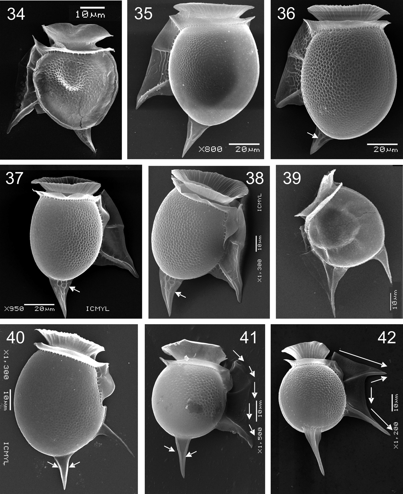
Figs 34–42. Dinophysis taxa, scanning electron microscopy: (Figure 34) Dinophysis aff. balechii in left lateral view; (Figures 35 & 36) two different specimens of D. hastata in left lateral view, arrow in Figure 36 points to the central spine in the posterior appendage; (Figures 37 & 38) same specimen of D. uracantha in right lateral view (two different tilts), arrows point to irregular ornamentation; (Figure 39) D. uracantha var. mediterranea in left lateral view; (Figure 40) D. uracanthoides in right lateral view, with two lateral spines in the posterior appendage (arrows); (Figure 41) D. pusilla in right lateral view; two arrows point to the lateral spines of the posterior appendage, and other arrows in the margin of the left sulcal list show its wavy-shape; (Figure 42) D. schuettii in right lateral view, arrows follow the margin of the sail-shaped left sulcal list (more angled).

Figs 43–56. Dinophysis taxa, scanning electron microscopy: (Figure 43) Dinophysis schuettii, left lateral view; (Figure 44) Dinophysis aff. schuettii in right lateral view, with one spine in the posterior appendage (arrow); (Figure 45) D. nias, left lateral view; the two central spines of the two appendages are arrowed; (Figure 46) D. swezyae in right lateral view, with one spine in the posterior appendage (arrow); (Figure 47) Dinophysis aff. balechii, detail of cingulum; (Figure 48) D. hastata, cingulum and cingular lists; (Figure 49) D. uracantha, cingulum and cingular lists; (Figure 50) D. uracantha var. mediterranea, detail of cingulum and cingular lists; (Figure 51) D. uracanthoides, epitheca and anterior cingular list; (Figure 52) D. pusilla, cingulum and cingular lists; (Figure 53) D. schuettii, cingular lists; (Figure 54) D. nias, detail of cingulum and cingular lists; (Figure 55) Dinophysis hastata, view of the epitheca and anterior cingular list; (Figure 56) D. uracantha, epitheca and anterior cingular list.

Figs 57–71. Dinophysis taxa, scanning electron microscopy: (Figure 57) D. uracanthoides, view of the ephiteca and anterior cingular list; (Figure 58) D. pusilla, epitheca and cingular lists; (Figure 59) D. schuettii, cingular lists; (Figure 60) D. nias, view of the epitheca and anterior cingular list; (Figure 61) D. swezyae, epitheca, cingular lists and part of sulcal lists; (Figures 62–71) various Dinophysis taxa showing theca ornamentation; (Figure 62) Dinophysis aff. balechii; (Figure 63) D. hastata; (Figure 64) D. uracantha; (Figure 65) D. uracantha var. mediterranea; (Figure 66) D. uracanthoides; (Figure 67) D. pusilla; (Figure 68) D. schuettii; (Figure 69) Dinophysis aff. schuettii; (Figure 70) D. nias; (Figure 71) D. swezyae.
A large-sized species with an elliptical body in lateral view (Figures 4, 5, 35 & 36) and in dorso-ventral view (Figure 24). Epitheca low and hypotheca with a rounded or slightly acute margin posterior and a long appendage, which shows variation in its ornamentation: one central spine (Figure 5) or two lateral spines with irregular ornamentation (Figures 4 & 36). Cingulum convex, ornamented with two rows of pores. Cingular lists wide, the anterior cingular list with marked and strong ribs facing to the epitheca and ribs inconspicuous when facing to the cingulum (Figures 48 & 55). The left sulcal list is long, with straight margin, ornamentation variable, and with rations (R1–R3) fairly equidistant; R2 is straight and thick, whereas R3 is thick, straight and clavate. Theca with poroids spread and with one pore surrounded by six to seven poroids (Figure 63). Dimensions: L 65–95 µm, E 36–40 µm, H 55–65 µm, Trd. 30–33 µm, N = 17.
LOCAL DISTRIBUTION
Gulf of California (28–30°C and salinity 34–35), Central Mexican Pacific (29°C and salnity 32) and Gulf of Mexico (25–29°C and salinity 34–36 salinity) (Table 1).
REMARKS
Specimens studied by SEM were similar to the illustration provided by Dodge (Reference Dodge1985).
Dinophysis uracantha Stein
(Figures 6, 37, 38, 49, 56 & 64)
References: Kofoid & Skogsberg, Reference Kofoid and Skogsberg1928, p. 273, figures 35–36; Wood, Reference Wood1954, p. 200, figure 48; Norris & Berner, Reference Norris and Berner1970, p. 185, figures 121–130; Rampi & Bernhard, Reference Rampi and Bernhard1980, p. 83, pl. 38; Steidinger & Tangen, Reference Steidinger, Tangen and Tomas1997, p. 434, pl. 12; Esqueda-Lara & Hernández-Becerril, Reference Esqueda-Lara and Hernández-Becerril2010, p. 155, figure 148.
Medium-sized species with irregularly elliptical body (wider at its equator) in lateral view (Figures 6, 37 & 38) and elliptical in dorso-ventral view. The epitheca appears low and hypotheca has a rounded or slightly acute margin posterior and a long appendage, with two lateral spines (Figure 6) and irregular ornamentation (Figures 37 & 38). The cingulum is convex. Anterior cingular list wide, with ribs facing to the epitheca (Figures 49 & 56). The left sulcal list is long, with a wavy margin, R2 closer to R1 than to R3, and R2 being straight or concave, whereas R3 is convex and thick. Theca shows regular poroids and an apparent pattern of one pore surrounded by seven poroids (Figure 64). Dimensions: L 50–65 µm, E 30 µm, H 50–60 µm, Trd. 23–30 µm, N = 3.
LOCAL DISTRIBUTION
Gulf of California, 28–29°C and salinity 34–35 (Table 1).
REMARKS
We additionally found two specimens slightly smaller (L 40–48 µm, E 24 µm, H 39–42 µm, Trd. 20 µm), with about the same morphology as depicted above, except that the anterior cingular list has no ribs and the left sulcal list shows a wavier margin, and which can also be assigned to Dinophysis uracantha Stein sensu Jörgensen (Figures 6 & 25) (Jörgensen, Reference Jörgensen1923, p. 32, figure 42; Kofoid & Skogsberg, Reference Kofoid and Skogsberg1928, p. 273, figures 35–36; Schiller, Reference Schiller1933, p. 142, figure 134d; Norris & Berner, Reference Norris and Berner1970, p. 185, figures 121–130), thus showing evidence of high morphological variability within the species, or the existence of different morphotypes which can be recognized as different species in the future. This species (both morphotypes) differs from D. hastata mainly for the wavy-shape of the left sulcal list (Table 2).
Dinophysis uracantha var. mediterranea Jörgensen
(Figures 7, 8, 39, 50 & 65)
References: Jörgensen, Reference Jörgensen1923, p. 32, figure 43; Gómez et al., Reference Gómez, López-García and Moreira2011a, p. 397, figure 3al.
Species of medium to large-sized with irregularly elliptical body (wider at the equator level) in lateral view (Figures 7, 8 & 39). The hypotheca has a slightly acute margin posterior and a relatively long dorso-posterior appendage with two lateral spines (Figures 7, 8 & 39). The cingulum is convex and has two longitudinal rows of pores (Figure 50). The cingular lists are wide, the anterior one with few ribs facing to the epitheca. The left sulcal list is wide, long and with a wavy margin; all three ribs (R1–R3) are equidistant, R2 is straight or concave and thin, and R3 is convex and thick. Theca with poroids and one pore surrounded by six to seven poroids (Figure 65). Dimensions: L 45–57 µm, E 24–26 µm, H 38–53 µm, N = 4.
LOCAL DISTRIBUTION
Gulf of California and Central Mexican Pacific, 28°C and salinity 35, and 30.7°C and salinity 32, respectively (Table 1). This is a new record for the Mexican Pacific.
REMARKS
A megacytic specimen (Figure 7) was found, with a similar shape to others that are not megacytic (Figure 8), in lateral view. New observations concerning SEM (Figures 39, 50 & 65) show details of the ribs in the anterior cingular list and the ornamentation of both the cingulum and the theca. This taxon can be basically distinguished from D. hastata and D. uracantha for its acute posterior margin.
Dinophysis uracanthoides (Jörgensen) Gómez, López-García et Moreira
(Figures 9, 26, 40, 51, 57 & 66)
References: Jörgensen, Reference Jörgensen1923, p.32, figure 40; Gómez et al., Reference Gómez, López-García and Moreira2011a, p. 397, figure 3g–i, n.
Large-sized specimens found, with elliptical body in lateral view (Figures 9 & 40) and also in dorso-ventral view (Figure 26). Epitheca is dome-shaped and relatively high (higher than in D. hastata) (Figures 51 & 57). Hypotheca has a posterior margin rounded or slightly acute and a long appendage with two lateral spines (Figures 9 & 40). The cingular lists are wide, with strong ribs facing to the epitheca and less conspicuous when facing to the cingulum (Figures 51 & 57). The left sulcal list is long, with a straight margin, and is ornamented with short ‘ribs-like' running parallel to the ribs; R2 inconspicuous and R3 convex and thick. Poroids regularly spread on the theca, with one pore surrounded by six poroids (Figure 66). Dimensions: L 55–60 µm, E 24–29 µm, H 45–50 µm Trd. 23–27 µm, N = 2.
LOCAL DISTRIBUTION
Gulf of California and Gulf of Mexico, at 28°C and salinity 35, and 29.5°C and salinity 36.5, respectively (Table 1). This is a new record for both the Mexican Pacific and the Gulf of Mexico.
REMARKS
Our two specimens are similar to the specimen illustrated by Jörgensen (Reference Jörgensen1923): R2 was shown closer to R1 than to R3, whereas in our specimens R2 is inconspicuous. Also, specimens shown here have a different cell outline compared to the specimen of Gómez et al. (Reference Gómez, López-García and Moreira2011a), where the epitheca is narrower and the notch below R1 can not be seen, maybe because of the position of the specimen. Dinophysis uracanthoides differs from the previous taxa because it has a radial ornamentation in the left sulcal list and the notch in the dorsal margin, below R1.
Dinophysis conjuncta Parra-Toriz, Esqueda-Lara et Hernández-Becerril sp. nov.
(Figures 10, 27, & 72–83)
Cellula solitaria, crassa. Corpo elliptico, aspectu laterali et elliptico-depresso, aspectu ventrali/dorsali. Epitheca humile, laxa. Hypotheca leviter rotunda et elongata, habens appendicis posteriore, longo et cum spinae fortis. Cingulum latum et convexum. Ala cingulari anteriore prominente, habens multa fortiter costae, ala cingulari posteriore angustiore. Ala sulci sinistra longa, irregularis, leviter convexa, ornata, incontinuus circa R3. R3 conspicuo, forte et longo, disiunctus ex R2. Theca areolata, 1 poro per 4–6 areolae. Cellulae 50–57 µm longae, 45–51 µm latae, appendicis 18–20 µm longo. Chloroplasti carentes. Species marina et planctonica.

Figs 72–83. Dinophysis conjuncta sp. nov., light microscopy (LM) and scanning electron microscopy (SEM): (Figures 72–74) right lateral view of three different specimens (arrowhead in Figure 73 points to the indentation of the left sulcal list), LM; (Figure 75) one cell in left lateral view, LM; (Figure 76) a specimen in dorsal view, LM; (Figures 77 & 78) same cell in right lateral view (in two different tilts), arrowhead in Figure 78 points to the indentation of the left sulcal list, and arrows point to the irregular ornamentation of the posterior appendage, SEM; (Figure 79) detail of a cell in right lateral view, showing the junction of the left sulcal list and the posterior appendage (large arrow), and irregular ornamentation of the posterior appendage (small arrow), SEM; (Figure 80) specimen in dorsal view, showing details of the epitheca and the cingular lists, SEM; (Figures 81 & 82) epithecal view, showing details of the cingular lists (especially the anterior one) and the epithecal pore (arrows), SEM; (Figure 83) details of the theca ornamentation, SEM.
HOLOTYPE
Slide 2014, Phycological Collection (Colección Ficología), Herbario Nacional, Instituto de Biología, Universidad Nacional Autónoma de México (MEXU).
ICONOTYPE
Specimens illustrated in Figures 73 & 77.
TYPE LOCALITY
Oceanic zone of the south-west Gulf of Mexico (21°35′N 92°30′W), from material collected on 8 August 1999.
ETYMOLOGY
The species name refers to the junction between the left sulcal list and the posterior appendage of the hypotheca.
DESCRIPTION
This is a large-sized species with elliptical body in lateral view (Figures 10, 72–75 & 77–79), and dorso-ventral view (Figures 27, 76 & 80). Epitheca is low and wide. Hypotheca has a rounded or slightly acute posterior margin and a long appendage with irregular ornamentation (Figures 10, 77 & 79). Cingulum is convex. Anterior cingular list is wide and with ribs facing to the epitheca (Figures 80–82). The left sulcal list is long and relatively narrow, convex, and with a concavity at the level of the third rib (R3), in addition is ornamented with short and weak ‘ribs-like' running parallel to the rations (Figures 72–75), and is joined to the posterior appendage (Figure 79); R2 is closer to R1 than to R3, and is straight and thin, whereas R3 is convex, thick and slightly longer than the left sulcal list. Theca is covered by regular poroids, having one pore surrounded by four to six poroids (Figure 83). Dimensions: L: 50–57 µm, E 37–43 µm, H 45–51 µm, Trd. 36–39 µm, N = 3.
LOCAL DISTRIBUTION
Gulf of California and Gulf of Mexico, at 30.5°C and salinity 34, and 25°C and salinity 35.2, respectively (Table 1).
REMARKS
The new species proposed here, Dinophysis conjuncta, exhibits a morphological structure relatively similar to others in the subsections Uracanthoides and Phalacromoides, particularly D. hastata and D. monacantha, but diagnostic characteristics include: (1) the shape of cell in lateral and dorso-ventral views; (2) the shape of the epitheca; (3) the shape of the left sulcal list, with a conspicuous indentation close to R3 (Figures 73 & 78) and ornamentation (consisting of other smaller ribs than R1–R3) (Figures 72–75); (4) the shape and orientation of the third rib (R3); and (5) the extension of this list to the posterior appendage (Figure 78) (Table 2). In addition, this species was found in two oceans, the Mexican Pacific and Atlantic (locations in the southern Gulf of Mexico), with the same morphological characters described here, and only slight variations in shape and size.
Subsection Phalacromoides Gómez, López-García et Moreira
Dinophysis monacantha Kofoid et Skogsberg
(Figures 11 & 28)
References: Kofoid & Skogsberg, Reference Kofoid and Skogsberg1928, p. 283, figure 37: 2, 3; Schiller, Reference Schiller1933, p. 144, figure 136; Gómez et al., 2011a, p. 397, figure 3af, ag.
Only observations by LM were made. A large specimen (probably megacytic) was found, with a subtrapezoidal body in lateral view and elliptical in dorso-ventral views (Figures 11 & 28). Epitheca is low and wide. Hypotheca shows an acute margin posterior and a short appendage with two lateral spines (Figure 11). The anterior cingular list is wide and has ribs. The left sulcal list is wide, long, with a straight margin and ornamented with ‘ribs-like' parallel to the main ribs; R2 is closer to R1 than to R3, and is concave and thin, whereas R3 is convex and thick. The left sulcal list is joined to the appendage, character not seen in previous illustrations (Figure 11). The theca is ornamented, although resolution in LM allows no observations of pores and poroids. Dimensions: L 67 µm, E 40 µm, H 53 µm Trd. 33 µm, N = 1.
LOCAL DISTRIBUTION
Gulf of California, 28.4°C and salinity 35 (Table 1). This is a new record for the Mexican Pacific.
REMARKS
The specimen found here is morphologically similar to the illustrations given by Kofoid & Skogsberg (Reference Kofoid and Skogsberg1928), except for the union of the left sulcal list with the posterior appendage, a character not shown previously (Kofoid & Skogsberg, Reference Kofoid and Skogsberg1928). However our specimen is different from that illustrated and sequenced by Gómez et al. (Reference Gómez, López-García and Moreira2011a), which cell shape differs, has no ribs in the anterior cingular list and with the posterior appendage more ornamented than specimens of Kofoid & Skogsberg (Reference Kofoid and Skogsberg1928). The shape of the cell of this species (subtrapezoidal body), together with the radiate ornamentation of the left sulcal list, separate it from the rest of species of the section Hastata.
Dinophysis phalacromoides (Jörgensen) Gómez, López-García et Moreira
(Figure 12)
References: Jörgensen, Reference Jörgensen1923, p. 31, figure 41; Gómez et al., Reference Gómez, López-García and Moreira2011a, p. 397, 404, figure 3z–aa.
Description is based on LM observations. Large-sized species with slightly subtrapezoidal body in lateral view (Figure 12). Epitheca is wide and low. Hypotheca has a posterior margin rounded or slightly acute, with a long and irregularly ornamented appendage (Figure 12). Anterior cingular list is wide and has ribs. The left sulcal list is long and has a straight margin, R2 is closer to R1 than to R3, and is straight and thin, and R3 is convex and thick. Dimensions: L 82 µm, E 47 µm, H 79 µm, N = 1.
LOCAL DISTRIBUTION
Gulf of California, at 30.5°C and salinity 34 (Table 1). This is a new record for the Mexican Pacific.
REMARKS
The specimen detected of D. phalacromoides has a similar morphology than those depicted by Jörgensen (Reference Jörgensen1923) (as D. hastata f. phalacromides) and Gómez et al. (Reference Gómez, López-García and Moreira2011a), thus indicating a little morphological variability. The relatively large size and shape of this species differs remarkably from all other taxa of the section Hastata.
Subsection Pusilla Gómez, López-García et Moreira
Dinophysis pusilla Jörgensen
(Figures 13, 14, 29, 41, 52, 58 & 67)
References: Jörgensen, Reference Jörgensen1923, p. 33, figure 44; Schiller, Reference Schiller1933, p. 137, figure 130b; Norris & Berner, Reference Norris and Berner1970, p. 175, figures 79–91; Balech, Reference Balech1988, p. 53, pl. 13, figure 4; Gómez et al., Reference Gómez, López-García and Moreira2011a, figure aj, ak.
Small-sized species with subrotund to slightly elliptical body in lateral view (Figures 13 & 14) and elliptical in dorso-ventral view (Figure 29). Epitheca is low and narrow. Hypotheca has a posterior margin rounded and a dorso-posterior appendage with one spine (Figures 13, 14 & 41), which is long and slightly inclined toward the ventral portion (Figures 13, 14 & 41). The cingulum is convex and ornamented with two rows of pores (Figure 52). Anterior cingular list is wide, bearing no ribs (Figures 52 & 58). The left sucal list is wide and has a wavy margin (Figure 41); R2 is closer to R1 than to R3, R2 is convex and thin, and R3 is convex and thick. Theca covered with poroids, with one pore surrounded by six to seven poroids (Figure 67). Dimensions: L 30–37 µm, E 14–21 µm, H 29–37 µm, Trd. 10 µm, N = 3.
LOCAL DISTRIBUTION
Gulf of California and Gulf of Mexico, 28.4°C and salinity 35, and 29.4°C and salinity 36.5, respectively (Table 1). This is a new record for the Mexican Pacific.
REMARKS
All specimens we found, in both the Mexican Pacific and the Gulf of Mexico, are similar among them, and also to those illustrated by Jörgensen (Reference Jörgensen1923), Norris & Berner (Reference Norris and Berner1970), Balech (Reference Balech1988) and Gómez et al. (Reference Gómez, López-García and Moreira2011a), with the exception of the spine (our specimens lack this spine) in the appendage shown in Norris & Berner (Reference Norris and Berner1970) and Balech (Reference Balech1988). Dinophysis pusilla is very characteristic and can be separated from all other taxa of the section Hastata because of its small size and lack of ribs in the anterior cingular list.
Dinophysis schuettii Murray et Whitting
(Figures 15, 16, 30, 42, 43, 53 & 68)
References: Jörgensen, Reference Jörgensen1923, p. 34, figure 46; Schiller, Reference Schiller1933, p. 147, figure 140; Taylor, Reference Taylor1976, p. 41, pl. 6, figures 65 & 66; Balech, Reference Balech1988, p. 53, pl. 12, figures 7–9; Hernández-Becerril, Reference Hernández-Becerril1992, p. 107. figures 13–18; Licea et al., Reference Licea, Moreno, Santoyo and Figueroa1995, p. 22, pl. 6, figure 8, pl. 20, figure 16; Esqueda-Lara & Hernández-Becerril, Reference Esqueda-Lara and Hernández-Becerril2010, p. 154, figure 147.
Medium to large-sized species with subrotund to subelliptical body in lateral view (Figures 15, 16, 42 & 43) and elliptical in dorso-ventral view (Figure 30). The epitheca is low and narrow, and the hypotheca has a posterior margin rounded and with a strong dorso-posterior appendage which differs in length and ornamentation (Figures 15, 16, 42 & 43). The cingulum is convex, with two rows of pores. The anterior cingular list has a funnel-shape (Figure 42) and ribs rather facing to the epitheca (Figure 53). The left sucal list is wide with a sail-shaped margin, and all three ribs equidistant; R2 is straight and/or convex and thick, whereas R3 is straight and thick. The theca has poroids, with one pore surrounded by seven poroids (Figure 68). Dimensions: L 40–85 µm, E 8–16 µm, H 37–75 µm, Trd. 12–17 µm, N = 13.
LOCAL DISTRIBUTION
Gulf of California (28.4–30°C and salinity 34–35), Central Mexican Pacific (20.4–29°C and salinity 32–34.5), and Gulf of Mexico (25°C and salinity 35) (Table 1).
REMARKS
Dinophysis schuettii showed an important morphological plasticity in specimens from the Mexican Pacific and the Gulf of Mexico. The specimens from the Mexican Pacific have differences in the left sulcal list, especially the relative length of R2 and R3, which is shorter compared with specimens from the Gulf of Mexico. Besides, Mexican Pacific specimens have the anterior cingular list shorter than specimens of the Gulf of Mexico. In general, the specimens of the Mexican Pacific appear more delicate than those from the Gulf of Mexico and may have the posterior appendage with or without spine. This species has a characteristic sail-shaped margin of the left sulcal list, and R2 and R3 larger than this list, and this character easily separates it from other related species.
Dinophysis aff. schuettii
(Figures 17, 31, 44 & 69)
One specimen of medium-size was found, very similar to the precedent species, having a subelliptical body in lateral view (Figures 17 & 44) and elliptical shape in dorso-ventral view (Figure 31). The epitheca is low and narrow, whereas the hypotheca shows a posterior margin rounded and an appendage with a central spine (Figure 44), similar to the one of D. schuetti (Figures 17 & 44). Cingulum is convex. The anterior cingular list is also funnel-shaped, with ribs facing to the epitheca (Figures 17 & 44). The left sulcal list is wide, long and sail-shaped, with all three ribs equidistant; R2 is convex and thick and R3 is straight and thick. The left sulcal list is united to the posterior appendage (Figures 17 & 44). The theca is covered with poroids and pores, with no discernible pattern (Figure 69). Dimensions: L 45 µm, E 14 µm, H 40 µm, Trd. 13 µm, N = 1.
LOCAL DISTRIBUTION
Gulf of California, at 28.4°C and salinity 35 (Table 1).
REMARKS
This taxon could represent an overlapping form between closely related species, but some specific morphological characters seem to be diagnostic. The cell size and general shape of the left sulcal list and its ratios are characters shared by both Dinophysis schuettii and D. aff. schuettii. However, the epitheca of D. aff. schuettii is more reduced than that of D. schuettii, the left sulcal list in D. aff. schuettii has a conspicuous extension to join to the posterior appendage, and the ornamentation of D. schuettii has smaller depressions (poroids) with pores, while in D. aff. schuettii has larger depressions and lower number of pores. Despite all these differences, we only found one specimen and consider information insufficient to propose a new species. This could be confirmed if more specimens are detected and/or molecular data are obtained in the future.
Dinophysis nias Karsten
(Figures 18, 19, 32, 45, 54 & 70)
References: Schiller, Reference Schiller1933, p. 149, figure 141; Balech, Reference Balech1988, p. 52, pl. 12, figure 4.
This is a large-sized species with irregularly elliptical body in lateral view (Figures 18, 19 & 45) and elliptical in dorso-ventral view (Figure 32). The epitheca is low and narrow and the hypotheca has a posterior margin rounded or slightly acute, with two similar dorso-posterior appendages, each with a central spine (Figures 18, 19 & 45). Cingulum convex, ornamented with two rows of pores. The cingular lists are wide, the anterior one with ribs facing to the epitheca (Figure 54). The left sulcal list is long and sail-shaped, with equidistant ribs (R1–R3); R2 is wavy and thick, R3 is straight or convex and thick. Pattern already described for the theca is also seen in this species: poroids covering it and one pore is surrounded by six to seven poroids (Figure 70). Dimensions: L 52–65 µm, E 24–27 µm, H 48–55 µm, Trd 18 µm, N = 4.
LOCAL DISTRIBUTION
Gulf of California and Gulf of Mexico, 28.4–29°C and salinity 35 salinity, and 25°C and salinity 35, respectively (Table 1). This is a new record for the Mexican Pacific.
REMARKS
Specimens of Dinophysis nias show a low morphological variability, although the posterior appendages have a spine variable in thickness, and additionally the left sulcal list can be slightly ornamented or not. Dinophysis nias differs from the closely related species for the presence of two posterior appendages (Table 2).
Dinophysis swezyae Kofoid et Skogsberg
(Figures 20, 21, 33, 46, 61 & 71)
References: Kofoid & Skogsberg, Reference Kofoid and Skogsberg1928, p. 289, figure 39, pl. 5, figure 9; Norris & Berner, Reference Norris and Berner1970, p. 183, figures 113–120; Taylor, Reference Taylor1976, p. 42, pl. 6, figures 63 & 64; Balech, Reference Balech1988, p.53, pl. 12, figures 5 & 6.
Large-sized species with irregularly elliptical body in lateral view (Figures 20, 21 & 46) and elliptical in dorso-ventral view (Figure 33). Epitheca is low and narrow. Hypotheca has a posterior margin slightly acute and a long appendage with a central spine (Figure 46). Cingulum convex. The cingular lists are narrow and funnel-shaped, the anterior one with ribs which are facing to the epitheca (Figure 61). The left sulcal list is long and sail-shaped and ribs equidistant; there is a conspicuous, lobulate extension of this list (a prolongation just below R3) in this species (Figures 20, 21 & 46). Both R2 and R3 are straight or convex and thick. Theca covered regularly by poroids, with one pore surrounded by six to seven poroids (Figure 71). Dimensions: L 48–55 µm, E 23–24 µm, H 42–45 µm, Trd. 12 µm, N = 4.
LOCAL DISTRIBUTION
Gulf of California and Gulf of Mexico, at 28.4°C and salinity 35, and 25°C and salinity 35, respectively (Table 1). This is a new record for the Mexican Pacific.
REMARKS
The specimens of the species showed a constant shape and a little morphological variation, which was more evident in the posterior appendage, which can vary in size and have or not a spine. The specimens of Dinophysis swezyae isolated for SEM were observed with its lobulated extension of the left sulcal list bent (Figure 46). This species can be distinguished from other related species for the lobulated extension of the left sulcal list.
DISCUSSION
Diversity and morphology
We report the finding of 14 taxa of Dinophysis section Hastata in this paper, with eleven taxa fully identified, one proposed as new species and two taxa remained as unidentified, because only one specimen of each was found and they might well be new species to be confirmed in the near future. From the four subsections in which the section Hastata is presently divided, taxa of three subsections, Uracanthoides, Phalacromoides and Pusilla, were found. The subsection Uracanthoides was best represented with seven taxa: Dinophysis balechii; D. aff. balechii D. hastata, D. uracantha, D. uracantha var. mediterranea, D. uracanthoides and D. conjuncta. Table 2 shows major morphological characters of the Dinophysis taxa studied here.
Dinophysis balechii and D. aff. balechii were species rare with only one specimen detected, whereas specimens of D. hastata and D. uracantha var. mediterranea were more frequent than others of the subsection. All specimens of Dinophysis hastata were similar to those reported in previous works of the Mexican Pacific, using light microscopy (Esqueda-Lara & Hernández-Becerril, Reference Esqueda-Lara and Hernández-Becerril2010), and new observations by SEM of D. hastata and D. uracantha var. mediterranea show details of the ribs in the anterior cingular list and the ornamentation of both the cingulum and the theca.
Dinophysis hastata is the best represented species; despite the recent separation of D. phalacromoides and D. uracanthoides (Gómez et al., Reference Gómez, López-García and Moreira2011a), it keeps its high morphological variability, confirmed here with variable cell shapes and sizes, length and relative complexity of its posterior appendage, and different ornamentation on the theca and sulcal lists. Despite the morphological variability of D. hastata, the new species proposed here, D. conjuncta, exhibits distinctive morphological characteristics, distinguishing it from the former, as mentioned above.
The morphology of Dinophysis conjuncta, relatively similar to D. hastata, suggests its place into the subsection Uracanthoides, especially by the elliptical cell body and left sulcal list with R3 emerging from the lower half of the hypotheca. Additionally, D. conjuncta lacks flat and wide epitheca, as in the subsection Phalacromoides. On the other hand, despite that Dinophysis conjuncta is also morphologically similar to D. monacantha; the latter has a distinctive subtrapezoidal body.
Two species of the subsection Phalacromoides were found in this paper: Dinophysis phalacromoides (type species of the subsection) and D. monacantha, which are also new records for the Mexican Pacific. These species were also rare because only one specimen of each was found. However, our specimens were morphologically very similar to the original illustrations; therefore, the species show little morphological plasticity.
Representatives of the subsection Pusilla are Dinophysis pusilla and D. schuettii (Gómez et al., Reference Gómez, López-García and Moreira2011a), but also the species D. nias and D. swezyae should be considered part of the subsection, due to the close relationship of Dinophysis swezyae with D. schuettii (Taylor, Reference Taylor1976). The specimens belonging to Dinophysis schuettii were more abundant than D. pusilla, and despite the few specimens found of D. pusilla, this species showed a little morphological plasticity.
According to the descriptions and illustrations from other authors (Hallegraef & Lucas, 1988; Hernández-Becerril, Reference Hernández-Becerril1992; Parra-Toriz, Reference Parra-Toriz2011), and our own observations, additional morphological characters are proposed here for the section Hastata. These characters are: (1) there are defined patterns in the theca ornamentation, with a pore surrounded by (four) six to seven poroids; and (2) when ribs occur in the anterior cingular lists, these are facing only to the epitheca. In contrast, size and shape of cell, depth of thecal poroids, ornamentation of the posterior appendages, and ornamentation of the left sulcal list, are characters with high morphological plasticity.
We did not find specimens with poorly-developed cingular lists, as has been reported by other authors for species of Dinophysis, as evidence of sexual dimorphism (Mackenzie, Reference MacKenzie1992; Reguera & González-Gil, Reference Reguera and González-Gil2001; Koike et al., Reference Koike, Sekiguchi, Kobiyama, Takishita, Kawachi, Koike and Ogata2005). Additionally, all specimens observed here, were whole cells, except one specimen of D. hastata, which lacked the anterior portion on the left sulcal list (probably as a consequence of the cell division), however, it was still possible to recognize the species.
Megacytic cells were observed showing basically a similar body outline to the non-megacytic cells, only being wider than the non-megacytic in dorso-ventral view, as previously reported (Jacobson & Andersen, Reference Jacobson and Andersen1994; Reguera, Reference Reguera2003), but keeping their dorsal margin; this in contrast to previous reports in other Dinophysis species not members of the section Hastata, which vary in the dorsal margin during the cell division (Jacobson & Andersen, Reference Jacobson and Andersen1994; Reguera, Reference Reguera2003), or after ingestion of a prey (Park et al., Reference Park, Kim, Kim, Yi, Kang and Yih2006).
We annotate eight new records for Mexican waters: seven in the Mexican Pacific (Dinophysis monacantha, Dinophysis nias, D. phalacromoides, D. pusilla, D. swezyae, D. uracantha var. mediterranea and D. uracanthoides) and one in the Gulf of Mexico (D. uracanthoides) (Table 1). These new records and the two taxa of Dinophysis section Hastata that remain unidentified, reflect the need for further research on this subject.
The low number of specimens for most of the species found and observed in this study are indicative of the low density populations that characterize almost all species of the genera of the order and the genus Dinophysis itself (except for few species, such as Dinophysis caudata, etc.). In the literature, it is not rare to find descriptions of new species of members of the order Dinophysales, based only on observations of one specimen (Kofoid, Reference Kofoid1907; Wood, Reference Wood1954; Taylor, Reference Taylor1976; Hernández-Becerril &Meave del Castillo, Reference Hernández-Becerril and Meave del Castillo1999; Hernández-Becerril et al., Reference Hernández-Becerril, Ceballos-Corona, Esqueda-Lara, Tovar-Salazar and León-Álvarez2008). Hence, data about the natural variability of morphological characters and identification of further ‘new' characters are still needed.
Besides, the studies of molecular phylogeny have not demonstrated to count on adequate molecular makers to discriminate between species of Dinophysis, for example, the rDNA regions might be more appropriate in identifying most species, whereas mtDNA regions are more appropriate in distinguishing D. acuminata from D. ovum (Papaefthimiou et al., Reference Papaefthimiou, Aligizaki and Nikolaidis2010). According to Papaefthimiou et al. (Reference Papaefthimiou, Aligizaki and Nikolaidis2010) ‘this could be the result of the recent divergence of many of the species accompanied by the relative evolutionary rates of individual markers'. Furthermore, different evolutionary histories are suggested by using different molecular markers, revealing a remarkable evolutionary trend toward complexity and innovation dynamics of the dinoflagellate genomes (Koumandou et al., Reference Koumandou, Nisbet, Barbrook and Howe2004; Waller & Jackson, Reference Waller and Jackson2009; Papaefthimiou et al., Reference Papaefthimiou, Aligizaki and Nikolaidis2010). Therefore, a better understanding of the limitations of the currently used molecular markers is also essential.
The challenge for working on dinophysoid dinoflagellates is that these taxa show highest diversity in tropical oceans, where it is usually difficult to obtain more information from the field and asses the morphological variation.
Taxonomy
The present infrageneric classification of the genus Dinophysis into sections and then the section Hastata into subsections has already been described in this paper. Although we do not have yet all biological elements (morphology, ecology, molecular information, life cycles, etc.) to propose a new classification of the genus, we can note that in very diverse genera within the dinoflagellates (e.g. genera Ceratium or Protoperidinium) or in other algal groups such as diatoms (e.g. genera Asteromphalus and Chaetoceros), the use of infrageneric taxa is commonly widespread, but the taxon subgenus is preferably used, then followed by the taxon section.
We suggest that in the future the genus Dinophysis (and eventually another large genus such as Phalacroma, within the Dinophysales) may be divided into subgenera, and then into sections, yielding perhaps a classification following the scheme of subgenus (not section) Hastata, sections (not subsections) Uracanthoides, Phalacromoides, Pusilla and Acutissima.
ACKNOWLEDGEMENTS
We thank Drs Laura Sánchez, Adela Monreal, David Salas-de-León and Maria Luisa Machain for her offer to participate or get samples in/from the oceanographic cruises ‘GOLCA 07', ‘DIPAL II', ‘PROMEBIO' and ‘TEHUA V', respectively. Thanks are also due to Yolanda Hornelas Orozco, for her assistance using the scanning electron microscopy. K.E.-L. and D.P.-T. received fellowships from CONACYT for their doctorate and masters studies, respectively, whereas D.P.-T. received support from the project IN226209-3, financed by PAPIIT (DGAPA, Universidad Nacional Autónoma de México).


