INTRODUCTION
Benthic dinoflagellates have been historically relatively little studied in comparison to their planktonic counterparts. Since the late 1970s, the epiphytic dinoflagellates attached to macroalgae have received more attention because they have been identified as responsible for ciguatera fish poisoning (Yasumoto et al., Reference Yasumoto, Nakajima, Bagnis and Adachi1977). The sand-dwelling dinoflagellates, living in the interstitial waters between the sand grains, have received less attention. The first studies were carried out in the sandy tidal flats of the British Isles (Herdman, Reference Herdman1922, Reference Herdman1924; Saunders & Dodge, Reference Saunders and Dodge1984), France (Balech, Reference Balech1956; Dragesco, Reference Dragesco1965; Dodge & Lewis, Reference Dodge and Lewis1986; Paulmier, Reference Paulmier1992) and Denmark (Larsen, Reference Larsen1985). The studies have largely increased in the last fifteen years with the description of numerous new species from the beaches of the North Sea, Japan, Russian Pacific, Australia, Canadian Pacific and Kuwait (see references in Al-Yamani & Saburova, Reference Al-Yamani and Saburova2010). In recent years, the description of new benthic species has accounted for almost one half of the total new dinoflagellates species (Gómez, Reference Gómez2012). The publications of sand-dwelling dinoflagellates are usually restricted to the description of a single or a few species, and the review of the closer relatives. This implies a disperse literature, with few compilations of the diversity for a given area (Al-Yamani & Saburova, Reference Al-Yamani and Saburova2010).
This study compiles the observations of sand-dwelling dinoflagellates carried out over two years on the shore of Wimereux, north-eastern English Channel, France. This area, located in the southern side of the Straits of Dover (Pas de Calais) is characterized by strong winds and currents (Brylinski et al., Reference Brylinski, Lagadeuc, Gentilhomme, Dupont, Lafite, Dupeuble, Huault, Auger, Puskaric, Wartel and Cabioch1991; Sentchev et al., Reference Sentchev and Yaremchuk2007; Korotenko & Sentchev, Reference Korotenko and Sentchev2011). Planktonic dinoflagellates, usually better adapted to calmer waters (Margalef, Reference Margalef1978), are not favoured in this highly turbulent environment, with few recorded species (Gómez & Souissi, Reference Gómez and Souissi2007, Reference Gómez and Souissi2008; Grattepanche et al., Reference Grattepanche, Vincent, Breton and Christaki2011; Gómez & Artigas, Reference Gómez and Artigas2013). In the north-eastern English Channel, the semidiurnal macrotidal regime (>8 m height) results in important emersion periods affecting wide sandy beaches. During ebb tide, the exposed substratum becomes green or golden brown in colour in irregular areas of the intertidal sand-flats. This is due to a superficial accumulation of enormous numbers of protists. These organisms dwell in the sediments during tidal inundation and move up onto the surface sands during tidal exposure. These vertical-migration rhythms have been described worldwide for sand-dwelling protists such as dinoflagellates (Herdman, Reference Herdman1924; Horiguchi & Pienaar, Reference Horiguchi and Pienaar1988), euglenoids (Palmer & Round, Reference Palmer and Round1965; Kingston & Gough, Reference Kingston and Gough2009) and diatoms (Janssen et al., Reference Janssen, Hust, Rhiel and Krumbein1999; Consalvey et al., Reference Consalvey, Paterson and Underwood2004; Mitbavkar & Anil, Reference Mitbavkar and Anil2004). These discolorations reach high primary production and respiration rates on the beach of Wimereux (Spilmont et al., Reference Spilmont, Migné, Lefebvre, Artigas, Rauch and Davoult2005; Hubas et al., Reference Hubas, Lamy, Artigas and Davoult2007). Two previous studies provided the first molecular data of the dinoflagellate genera Amphidiniopsis and Sinophysis (Gómez et al., Reference Gómez, López-García and Moreira2011, Reference Gómez, Moreira and López-García2012). However, the composition of the species has not yet been examined.
This study presents a first inventory of the benthic dinoflagellates in the region, with micrographs of several species that have not yet been described. Several species are illustrated for the first time in the area, since their original descriptions in the Pacific Ocean. This constitutes the first observations of several species in the Atlantic Ocean and European seas.
MATERIALS AND METHODS
This study was undertaken in the soft sandy sediments of the shore of Wimereux, France (50°46′12″N 1°36′42″E) from March to October in 2010 and 2011. There were two sampling sites, the border of a large pool (~50 m diameter, ~1 m depth), and several smaller pools and moist sands with a faint brownish discoloration, in front of the LOG laboratory (MREN ULCO and Marine Station of Wimereux UL1). The upper centimetre of sand was collected with a spoon and deposited into a bottle. There, the sand was rinsed with seawater and stirred vigorously, and the suspension settled in a composite settling chamber. The settled material was examined with a Nikon inverted microscope (Nikon Eclipse TE2000-S) and photographed with a Nikon Digital Sight DS-2M camera.
For scanning electron microscopy, aliquots of the agitated sand samples were fixed with glutaraldehyde (5%) and filtered onto a 0.8 µm pore size Nucleopore membrane filter, washed with distilled water, fixed with osmium, dehydrated with a graded series of ethanol and critical-point-dried with CO2. Filters were mounted on stubs, sputter-coated with gold and viewed using a Hitachi S4800 scanning electron microscope (SEM). Images were presented on a black background using Adobe Photoshop CS3.
RESULTS
The sand discolorations on the beach of Wimereux were more conspicuous during sunny periods. There were two main types: green patches tended to occur in the upper limit of the eulittoral zone associated with monospecific proliferations of Euglena sp., and greenish-brown patches were observed in moist sands in the middle of the eulittoral zone and on the bottom of tidal pools. These patches were due to diatoms and photosynthetic dinoflagellates, mainly proliferations of Togula britannica, T. cf. compacta, Polykrikos lebouriae, Amphidinium herdmanii, or more sporadically Spiniferodinium. A total of 70 species were found (Table 1). See video at: http://www.youtube.com/watch?v=BEjD-1wvBTs. A brief description of each species is provided below.
Table 1. List of sand-dwelling dinoflagellate species from the shore of Wimereux, France.

Amphidinium sensu lato
Conspicuous patches in the sandy sediments of Wimereux were due to photosynthetic species that were formerly described under the genus Amphidinium. This unarmoured genus of dorso-ventrally flattened species was traditionally defined by its small episome size, not exceeding one third of the total cell length. Currently Amphidinium sensu stricto is valid for species with minute irregular triangular or crescent-shaped episome, deflected to the left and overlaid in the anterior ventral part of the hyposome (as defined by Flø Jørgensen et al., Reference Flø Jørgensen, Murray and Daugbjerg2004). Other Amphidinium species have been transferred into the genus Gymnodinium (i.e. G. venator), new genera (Ankistrodinium, Apicoporus, Bispinodinium, Testudodinium, Togula) and other species also need to be placed into a separate genus (i.e. Amphidinium scissum).
Amphidinium sensu stricto
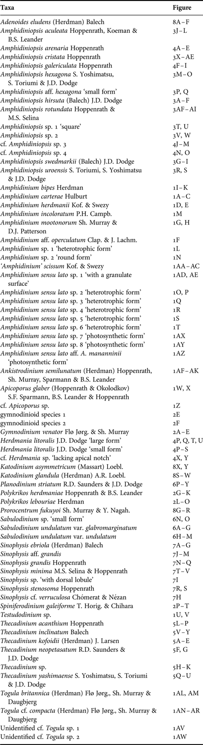
The photosynthetic species Amphidinium carterae, A. operculatum and A. herdmanii are characterized by a small episome, crescent-shaped and deflected towards the left. The cells of Amphidinium carterae are oval, more or less elongated, 13–15 µm long and 9–13 µm wide (Figure 1A–C). When compared to A. operculatum and A. herdmanii, the shape of the hyposome of A. carterae is more asymmetrical, with the episome closer to the right side of the cell (Figure 1A–C). The species A. carterae, A. massartii and A. thermaeum are highly morphologically similar, overlapping completely in size range and in shape. According to Murray et al. (Reference Murray, Garby, Hoppenrath and Neilan2012) these three species can be distinguished on the basis of the shape of the plastid—which is reticulate and distributed throughout the whole cell area in A. carterae, but generally more sparse, with several lobes, in A. massartii and A. thermaeum—and the slightly lower position of flagellar insertion in A. massartii and A. thermaeum compared to A. carterae.
The cells of A. operculatum and A. herdmanii are oval, oblong to egg-shaped from the ventral side, length about 25 µm and width about 20 µm. The species A. herdmanii has been illustrated in the literature with different morphologies (Schiller, Reference Schiller1933, figure 238a–f). We consider that A. herdmanii corresponds to the first illustration by Herdman (Reference Herdman1922, figure 8a). The episome of A. herdmanii is symmetrical and protrudes over the hyposome. The hyposome is slightly asymmetrical, with the left side longer than the right one, appearing indented at the antapex by the sulcus (Figure 1D, E). Consequently, A. herdmanii shows a slightly bi-lobulated antapex in comparison to A. operculatum. The specimens of Figure 1F show the cell shape of A. operculatum. However, they are named Amphidinium aff. operculatum because the episome is wider than in the original description of A. operculatum (Figure 1F). The cells of A. mootonorum differ from other species of Amphidinium sensu stricto in the episome, which does not project beyond the outline of the cell. The cingulum is V-shaped and encircles the episome (Figure 1G, H). The nucleus is usually posteriorly located in the species of Amphidinium sensu stricto. However, the nucleus of A. mootonorum is located in the middle of the cell (Figure 1G, H). This species can be easily confused with Testudodinium testudo.
Fig. 1. Micrographs of unarmoured sand-dwelling dinoflagellates from the shore of Wimereux: (A–C) Amphidinium carterae; (D, E) Amphidinium herdmanii; (F) Amphidinium aff. operculatum – the arrow points to an orange spot; (G, H) Amphidinium mootonorum; (I–K) Amphidinium bipes; (L) unidentified heterotrophic Amphidinium; (M) Amphidinium incoloratum; (N) unidentified round Amphidinium; (O–T) several unidentified heterotrophic species of Amphidinium sensu lato (see Table 1); (U, V) Testudodinium sp.; (W, X) Apicoporus glaber; (Z) cf. Apicoporus sp.; (AA–AC) ‘Amphidinium’ scissum; (AD, AE) unidentified species with granulate surface; (AF–AK) Ankistrodinium semilunatum; (AL, AM) Togula britannica; (AO–AR) Togula cf. compacta; (AS) Togula inside a hyaline capsule; (AT, AU) ameboid cells of Togula; (AV–AY) unidentified photosynthetic species of Amphidinium sensu lato (see Table 1); (AZ) photosynthetic species close to Amphidinium mananninii. Scale bar: 10 µm.
There is less information on the heterotrophic species of Amphidinium when compared to the photosynthetic ones, because they do not form large proliferations and because of the difficulties of establishing cultures. Amphidinium bipes is one of the most distinctive heterotrophic species of Amphidinium because the hyposome is divided into two rounded antapical lobes (Figure 1I–K). The differentiation among other heterotrophic species of Amphidinium is more difficult. Figure 1L illustrates a specimen of similar size to A. bipes, with a similar episome that protrudes over the hyposome. The main difference is the oblong rectangular shape of the hyposome (Figure 1L). We are unable to assign this specimen to any known species.
Amphidinium incoloratum shows an oval hyposome, asymmetrical, relatively straight, with the right side convex (Figure 1M). The small episome slightly protrudes over the hyposome. The cell is about 32 µm long and 20 µm wide (Figure 1M). Figure 1N illustrates a specimen examined by scanning electron microscopy. It was not observed by light microscopy, and we were unable to determinate the presence of plastids. The episome is flat, 6 µm wide, and it protrudes over the hyposome, as typical in Amphidinium sensu stricto (Figure 1N). The most distinctive character is a round cell body (diameter of 20 µm). We are unable to assign this specimen to any known species.
HETEROTROPHIC SPECIES OF AMPHIDINIUM SENSU LATO
Several heterotrophic species show a flat episome that slightly protrudes over the hyposome. The shape of the hyposome varies from ovate (Figure 1O, P), or rounder with the antapical margin slightly divided into two rounded lobes (Figure 1Q). Other heterotrophic specimens show an episome almost embedded in the hyposome (Figure 1R–T). The shape of the hyposome ranges from oval (Figure 1R, S) to elongate with a slightly pointed antapex (Figure 1T). We are unable to assign these specimens to known species.
GENUS TESTUDODINIUM
The genus Testudodinium has been recently proposed for a species with a small episome that does not project beyond the outline of the cell. The cingulum is V-shaped and encircles the episome. The sulcus is about one-third of the total cell length and is located at the centre of the hyposome. This morphology is close to the species Amphidinium mootonorum which has the nucleus located in the middle of the cell (Figure 1G, H). We found a species that fits the characteristics of the genus (Figure 1U, V). The small episome is embedded in the hyposome, protruding from the hyposome on the ventral side. The hyposome is elongated with a slightly pointed margin. The specimen shows a brownish pigmentation (Figure 1U, V).
GENUS APICOPORUS
The heterotrophic species Apicoporus glaber (=Amphidinium gabrum) is characterized by a small, low, wide, beak-shaped, asymmetrical episome. The cells shows an oblong shape (Figure 1W, X). The specimen in Figure 1Z shows a low episome and a smooth cell surface, as in Apicoporus. The cell shows a pointed antapical margin, as in Amphidinium scissum (Figure 1Z).
AMPHIDINIUM SENSU LATO: GENUS OF ‘AMPHIDINIUM’ SCISSUM
The species of Amphidinium sensu stricto have a smooth cell surface. The shape of the episome of A. scissum does not match with the recent definition of Amphidinium sensu stricto, and it also differs in the surface striation, with fine longitudinal striations that resemble the genus Gyrodinium (Figure 1AA–AC).
AMPHIDINIUM SENSU LATO: UNIDENTIFIED GENUS WITH GRANULATE SURFACE
Figure 1AD illustrates a specimen found in a sample examined by scanning electron microscopy. The hyposome is slightly bifurcated. The most characteristic feature is a cell surface covered with homogeneous rounded granules (Figure 1AE). To the best of our knowledge, this type of surface ornamentation is unknown for species of Amphidinium sensu lato.
GENUS ANKISTRODINIUM
The cells of Ankistrodinium semilunatum are oval to oblong-shaped in lateral view, strongly laterally compressed. The cingulum rises from its origin towards the dorsal side of the cell, then tilts downwards slightly. The cell size varies from 30 to 50 µm long and from 20 to 30 µm wide. This heterotrophic species usually shows food particles (Figure 1AF–AK).
GENUS TOGULA
The most conspicuous discolorations in the moist sands of Wimereux are due to species of the genus Togula. The cells are highly pigmented with irregular greenish-yellow chloroplast lobes radiating from the centre towards the periphery. The episome and hyposome are asymmetrical. The cingulum originates posteriorly to centrally, taking an anterior straight course, turning left from the apex and descending in a sigmoid-straight fashion dorsally, descending in a straight course upon reaching the ventral side.
The genus currently accounts for three species: Togula britannica, T. compacta and T. jolla. Togula britannica (Figure 1AL, AM) can be identified by its larger size, and by its slimmer appearance when compared to the smaller species T. cf. compacta (Figure 1AN–AR). The distinction between T. compacta, T. jolla and Amphidinium ovum is difficult. The motile cells of Togula cf. compacta are broadly ellipsoidal in the dorso-ventral view, with the width of the episome a little less than the width of the hyposome, and golden-brown to dark-brown in colour. The apex and antapex are broadly rounded, but the antapex can appear slightly pointed in the dorsal view (Figure 1AN–AR). During cell division in the genus Togula the two daughter cells initially separate at the antapex, and the epicones are the last parts to split (Figure 1AL, AP). It was common to observe immotile cells surrounded by a hyaline sheath. These immotile cells tended to be broader than the motile cells (Figure 1AS). A short period of metabolic movement occurred during the change from immotile to motile cell (Figure 1AT, AU).
UNIDENTIFIED PHOTOSYNTHETIC SPECIES OF AMPHIDINIUM SENSU LATO
Figure 1AV–AY represents several unidentified species that superficially resemble Togula. Much research is needed to facilitate the distinction between these species under routine microscopical analysis. Figure 1AZ illustrates a small cell (20 µm long) with yellowish pigmentation resembling the heterotrophic species Amphidinium mananninii. The cell body is broadly ellipsoidal, the episome angular and the hyposome cordate with a rounded point at the antapex. The morphology of the episome does not match with the current definition of Amphidinium sensu stricto. The species A. mananninii is described as colourless, while this specimen shows yellowish-brown pigmentation that suggests the occurrence of plastids (Figure 1AZ).
Family Gymnodiniaceae
The cells of the heterotrophic species Gymnodinium venator (=Amphidinium pellucidum) are oval to oblong from the ventral side. Coloured food particles and colourless lipid globules were often present. The length is variable from 30 to 35 µm and the width from 15 to 25 µm (Figure 2A–E). A closely related species, Amphidinium flexum, is described by Herdman as rectangular in shape. The species Amphidinium latum shows a cone-shaped episome with a pointed apex. The shorter and flattened episome is the main characteristic of G. venator when compared to Amphidinium latum. Figure 2F shows two specimens of different sizes which correspond to an unidentified species.
Fig. 2. Micrographs of unarmoured sand-dwelling dinoflagellates from the shore of Wimereux: (A–D) Gymnodinium venator; (E) Gymndoinium venator (small) and an unidentified gymnodinioid species; (F) unidentified gymnodinioid species; (G–K) Polykrikos herdmaniae; (L–O) Polykrikos lebouriae; (P–T) Spiniferodinium galeiforme. Scale bar: 10 µm.
GENUS POLYKRIKOS
A photosynthetic species of Polykrikos was also responsible for discolorations in the moist sands of the beach at Wimereux, and was more usually found in the bottom of tidal pools during low water. Polykrikos is a distinctive genus of pseudo-colonies consisting of eight zooids, sometimes four zooids. Currently, there are two benthic species within this genus: Polykrikos lebouriae and Polykrikos herdmaniae. The morphological distinction is the presence of plastids in P. lebouriae. Both species contained red coloured food bodies of variable number and size that were positioned in different areas of the pseudo-colony. The transparency of the heterotrophic P. herdmaniae (Figure 2G–K) facilitated observation of the taeniocyst–nematocyst complexes that were less prominent in the photosynthetic P. lebouriae (Figure 2L–O).
The photosynthetic P. lebouriae shows a high variability in cell size, ranging from 40 to 90 µm long and from 20 to 50 µm wide (Figure 2L–O). The pseudo-colonies were ovate in shape and obliquely flattened, with terminal zooids about half as wide as central zooids. The heterotrophic P. herdmaniae (Figure 2G–K) did not form proliferations, and mainly appeared in the proliferations of Togula and Polykrikos lebouriae.
GENUS SPINIFERODINIUM
This genus was highly distinctive in its immotile stage because it formed a spine shell and was strongly attached to the substrate (Figure 2Q–T). In contrast, the motile form can be easily confused with typical photosynthetic Gymnodinium-like cells (Figure 2P). Spiniferodinium appeared in sands permanently covered by water during low tide, such as the bottom of tidal pools. The presence of Spiniferodinium was sporadic. However, its presence was always associated with large proliferations, where Spiniferodinium was the main photosynthetic species. The observations of live cells under the microscope revealed that the switch from the swimming form to the immotile cells is very fast. In the settling chamber, the motile cells stopped swimming, settled on the bottom, and in less than 2 s the cell acquired a flattened round shape covered by a transparent, spiny helmet shell. The cell strongly attached itself to the surface of substratum. Without doubt, this is a mechanism to avoid re-suspension in the water column.
It was quite easy to identify the genus Spiniferodinium based on the observations of the immotile spiny stages. However, it was difficult to differentiate between the two species described to date (the type Spiniferodinium galeiforme and S. palauense). According to the original description, S. palauense shows a thinner cell body, the cingulum is slightly displaced and the episome is smaller than the hyposome. The cingulum of S. galeiforme is not displaced and is located almost in the middle of the cell (as reported by Horiguchi et al., Reference Horiguchi, Hayashi, Kudo and Hara2011) (Figure 2R–T). The specimens observed on the beach of Wimereux have been assigned to S. galeiforme (Figure 2P–T).
Family Amphidiniopsidaceae
GENUS AMPHIDINIOPSIS
Amphidiniopsis is the most speciose genus of armoured sand-dwelling dinoflagellates, with 15 marine species, and all known species are devoid of chloroplasts. The general appearance is the typical Amphidinium-like shape with a relatively small epitheca and a larger hypotheca. The cingulum is nearly horizontal, with the left ventral part running posteriorly into the sulcus (=slightly ascending cingulum). The sulcus shows a characteristic curved left side and reaches the antapex.
The genus Amphidiniopsis is separated into two groups: dorso-ventrally (Figure 3) and laterally (Figure 4) compressed species, the latter containing the type species. The dorso-ventrally compressed species were more common in the samples of Wimereux, and more easily recognized as members of Amphidiniopsis. The laterally compressed species were less abundant, the morphology of the sulcus was less evident, and the identification as members of the genus Amphidiniopsis was more difficult. Some laterally flattened cells can be confused with heterotrophic species of Thecadinium (Figure 5).
Fig. 3. Micrographs of dorso-ventrally flattened species of Amphidiniopsis from the shore of Wimereux: (A–F) Amphidiniopsis hirsuta; (G–I) A. swedmarkii; (J–L) A. aculeata; (M–O) A. hexagona; (P, Q) Amphidiniopsis aff. hexagona ‘small form’; (R, S) Amphidiniopsis uroensis; (T–W) unidentified species of Amphidiniopsis (see Table 1); (X–AE) Amphidiniopsis cristata; (AF–AI) Amphidiniopsis rotundata. Scale bar: 10 µm.
Fig. 4. Micrographs of Herdmania, laterally flattened species of Amphidiniopsis and unidentified species from the shore of Wimereux. The arrows point the ventral or antapical spine: (A–E) Amphidiniopsis arenaria; (F–I) Amphidiniopsis galericulata; (J–O) several unidentified species close to Amphidiniopsis (see Table 1); (P–W) Herdmania litoralis; (X, Y) cf. Herdmania sp. ‘lacking the apical notch’. Scale bar: 10 µm.
Fig. 5. Micrographs of Thecadinium from the shore of Wimereux: (A–E) Thecadinium kofoidii; (F, G) Thecadinium neopetasatum; (I–K) Thecadinium sp.; (L–P) Thecadinium acanthium; (Q–U) Thecadinium yashimaense; (V–Y) Thecadinium inclinatum. Scale bar: 10 µm.
Dorso-ventral flattened species of Amphidiniopsis
The genus Amphidiniopsis on the shore of Wimereux was largely dominated by three species: Amphidiniopsis hirsuta, followed by A. aculeata and A. swedmarkii. These species are rounded in the posterior margin, the epitheca, slightly narrower than the hypotheca, is cap-like and pointed at the front part. An apical hook or crest-like process is absent in these three species (Figure 3A–L). The most common species, Amphidiniopsis hirsuta, shows a row of irregular spines at its antapical cell margin (Figure 3A–F). Due to the difficulties in observing the spines, some specimens lacking the spines and having a more rounded hyposome may correspond to Amphidiniopsis swedmarkii sensu Yoshimatsu et al. (Reference Yoshimatsu, Toriumi and Dodge2000) or the recently described species A. konovalovae. Two prominent antapical spines are the distinctive characteristic of Amphidiniopsis swedmarkii (Figure 3G–I). Amphidiniopsis aculeata is characterized by prominent spines that are especially visible at the lateral contour of the cell at mid focus (Figure 3J–L).
The species Amphidiniopsis hexagona is characterized by its hexagonal shape and the absence of spines. We found specimens of two different sizes, both with a hexagonal shape. The theca was covered with small spines. The larger specimen was 35 µm length (Figure 3M–O), while the smaller specimen (20–µm in length) showed a more marked hexagonal shape (Figure 3P, Q). The latter is named Amphidiniopsis aff. hexagona ‘small form’.
Another group of dorsal-ventrally flattened species was characterized by a prominent apical hook or crest-like process (Figure 3R–W). We found specimens with this characteristic of different sizes and shapes that ranged from oval (Figure 3R, S) to square (Figure 3T, U), and specimens with an intermediate shape and more irregular contour (Figure 3V, W). The specimens with an oval hypotheca are here assigned to Amphidiniopsis uroensis (Figure 3R, S). The accurate identification of other specimens remains uncertain (Figure 3T–W).
Amphidiniopsis cristata was one of the most distinctive taxa among the dorso-ventrally flattened species with an apical hook. The cells are roughly square in shape and smaller than the other dorso-ventrally compressed species of Amphidiniopsis. The epitheca is slightly narrower than the hypotheca and shows a conspicuous apical crest-like process. There are several conspicuous but irregular antapical spines, of which the two most lateral are the largest (Figure 3X–AE).
Amphidiniopsis rotundata was other dorso-ventrally flattened species. It is difficult to recognize it as a member of Amphidiniopsis due to the rounded shape and the lack of other distinctive characteristics, lack of the apical hook and no antapical spines of any kind. Under routine light microscopy observations of live specimens, the sulcus curving to the left side and reaching the antapex was the main character that related this species to the genus Amphidiniopsis (Figure 3AF–AI).
Laterally flattened species of Amphidiniopsis
A common characteristic for the laterally compressed Amphidiniopsis species is a list-like flange that runs down into the sulcus. This group of species includes the type of Amphidiniopsis, A. kofoidii, and one of the problems in identification is the lack of reliable records for the type species. Some records in the literature of A. kofoidii are currently considered to be A. arenaria. Some lateral flattened species of Amphidiniopsis may be also misidentified with heterotrophic species of Thecadinium (i.e. Thecadinium acanthium and Amphidiniopsis dentata).
Amphidiniopsis arenaria is nearly rectangular (Figure 4A–E), with a characteristic list-like flange that runs down into the sulcus (Figure 4C), and a conspicuous ventral spine (Figure 4E). Amphidiniopsis galericulata is smaller than other laterally flattened species of Amphidiniopsis (about 25 µm in length). The epitheca is slightly narrower than the hypotheca and shows a helmet-like appearance, with a conspicuous apical, hook-like spine pointing towards the dorsal side. There is a ventral antapical spine and a row of short irregular spine-like projections (Figure 4F–I). We were unable to identify several specimens that show a Phalacroma-like shape (Figure 4J–O). It is uncertain whether they belong to the genus Amphidiniopsis.
GENUS HERDMANIA
The cells of the monotypic heterotrophic genus Herdmania are flattened dorso-ventrally and rounded in ventral view, with a distinct small, hook-like apical notch pointing to the left lateral side of the cell. The incomplete cingulum starts at the left ventral side, runs around the dorsal side just above the celĺs middle and ends at the right ventral side (Figure 4P–W). The specimens of Herdmania showed different sizes and were grouped into two populations that often coexisted (Figure 4P–U). These two groups of specimens were of about 25 µm (Figure 4R, S) and about 35 µm in diameter (Figure 4T, U), respectively. This suggests the occurrence of, at least, two separate species. We include here an unidentified species that resembles Herdmania, but lacks the distinctive apical notch (Figure 4X, Y).
Family Thecadiniacae: Thecadinium
The cells of Thecadinium are flattened laterally with an oval shape, and the typical Amphidinium-like shape, a relatively small epitheca and a larger hypotheca. Most of the species are heterotrophic, with the exception of a few photosynthetic species: Thecadinium yashimaense, T. arenarium and the type T. kofoidii.
The cells of Thecadinium kofoidii are strongly laterally flattened, roughly elliptical in side view and slightly pointed in the antapical margin, and show a reduced epitheca. The cell contains a number of yellowish chloroplasts that radiate from a central pyrenoid (Figure 5A–E).
The cells of Thecadinium neopetasatum are rather similar in shape to T. kofoidii. Thecadinium neopetasatum is heterotrophic and slightly larger (30 µm length) and rounder than T. kofoidii (Figure 5F, G). The specimen of Figure 5H–K is a heterotrophic species of large size (45 µm), with a large episome and an asymmetrical hypotheca. In some way, the shape resembles the photosynthetic species Pseudothecadinium campbellii. We are unable to provide an accurate identification of this species (Figure 5H–K).
The cells of Thecadinium acanthium are oval to almost rectangular (55 µm long and 35 µm wide). The very small epitheca is cap-like, and there is a ventral antapical spine and a row of regular tooth-like projections (Figure 5L–P). It could be confused with Amphidiniopsis dentata.
The cells of Thecadinidum yashimaensis are pigmented golden-brown. The shape is broadly ovoid and slightly laterally compressed (Figure 5Q–U). In lateral view, the cells range from ovoid to squarish-ovoid. The cingulum is deeply excavated, descending and displaced approximately half of the cell length deep (Figure 5Q–U).
The cells of Thecadinium inclinatum are heterotrophic, frequently with large orange-red food bodies. The cell shape is oval to slightly rectangular, with a laterally asymmetrical epitheca smaller than the hypotheca, about one fourth to one-third of the cell length. The cells are larger than other species of Thecadinium, ranging from 55 to 80 µm long and 40 to 60 µm deep (Figure 5V–Y).
Family incertae: Genus Sabulodinium
The cells of Sabulodinium undulatum are laterally flattened, more or less oval with a truncated apex in lateral view, and elongated–elliptical in ventral view (Figure 6A–O). The lower dorsal margin of the cell is either slightly irregular (undulating) with rounded edges, or has more pronounced undulations containing one spiny dorsal edge or two spiny edges – one dorsal and one posterior. The cingulum is deep and not displaced, and the sulcus is positioned on the right lateral side of the cells. This is a monotypic genus, although the morphological differences between the specimens suggests the occurrence of undescribed species. There are two main morphologies in the large specimens: Sabulodinium undulatum var. glabromarginatum for more round specimens with smooth/straight dorsal hypotheca border (Figure 6A–G) and Sabulodinium undulatum var. undulatum, where the antapical contour could form one spiny dorsal edge or two spiny edges—one dorsal and one posterior (Figure 6H–M). A third morphology was found—Sabulodinium sp. was a smaller species with three spiny edges (Figure 6N, O).
Fig. 6. Micrographs of Sabulodinium and Planodinium from the shore of Wimereux: (A–G) Sabulodinium undulatum var. glabromarginatum; (H–M) Sabulodinium undulatum var. undulatum; (N, O) Sabulodinium sp. ‘small form’ – the arrows point to the spiny edges; (P–Y) Planodinium striatum. Scale bar: 10 µm.
Family incertae: Planodinium
The cells of this heterotrophic genus are laterally compressed with an oval shape. The cingulum is horizontal. The epitheca is much reduced and narrower than the hypotheca. The surface of the hypotheca is ridged (Figure 6P–Y).
Family of Sinophysis: Genus Sinophysis
These species show dinophysoid morphology, laterally flattened, with a minute crown-like epitheca that resembles the planktonic genus Dinophysis. The genus Sinophysis contains seven species; five species have been described from temperate waters, and two species from tropical coasts. The species of temperate waters show a smooth thecal surface, with the exception of S. verruculosa. All the species described from temperate waters were found in our sampling area (Figure 7). The cells of Sinophysis ebriola (about 35–45 µm) are roughly oval. The small episome is about 9–12 µm wide (dorso-ventrally), surrounded by a wide collar and slightly tilted back to the dorsal side (Figure 7A–G). The species S. verruculosa is distinguished from S. ebriola by the verrucose ornamentation of the theca. This diagnostic characteristic is not easy to observe under light microscopy. We glimpsed some kind of thecal ornamentation in the specimen of Figure 7H. This specimen, with a more circular cell shape and wider episome than S. ebriola, we identified as S. cf. verruculosa (Figure 7H). Sinophysis sp. corresponded to a specimen with an antapical lobule in the dorsal side (Figure 7I).
Fig. 7. Micrographs of Sinophysis from the shore of Wimereux: (A–G) Sinophysis ebriola; (H) Sinophysis cf. verruculosa; (I) Sinophysis sp. ‘with dorsal lobule’; (J–M) Sinophysis aff. grandis; (N–Q) Sinophysis grandis; (R, S) Sinophysis stenosoma; (T–V) Sinophysis minima. Scale bar: 10 µm.
The cells of Sinophysis grandis are large (about 50–60 µm), roughly rectangular, with roundish edges. The episome is the largest found in Sinophysis, surrounded by a weakly developed smooth girdle list, and less tilted to the dorsal side (Figure 7J–Q). The specimens can be divided into two groups according to their size. The larger specimens (55–60 µm in length) are closer to the original description of S. grandis, with more quadrangular cell shape and wider episome (Figure 7N–Q). Other specimens identified as Sinophysis aff. grandis are smaller (50–55 µm in length) with a more oval cell shape and narrower episome (Figure 7J–M).
The cells of Sinophysis stenosoma are oblong ellipsoidal, epitheca cylindrical, crown-like, and 5–8 µm wide. The small cylindrical, crown-like episome is notably small compared with the other Sinophysis species. It is surrounded by a well-developed smooth girdle list of the hypotheca and slightly tilted back to the dorsal side (Figure 7R, S). The cells Sinophysis minima are very small (20 µm long), roughly rectangular to almost square, with more or less round edges. The cylindrical epitheca is small, 5–7 µm deep, constituting about one-third of the hypotheca depth, and is notably asymmetric (Figure 7T–V). This species can go easily unnoticed due to its small size and transparency.
Family incertae: Genus Adenoides
The cells of Adenoides eludens are asymmetrical, oval to round, slightly flattened laterally (30–35 µm). The minute epitheca is cup-shaped, depressed and scarcely visible. The cingulum is almost at the anterior end of the cell, completely encircling the epitheca and meeting without displacement. The sulcal area is on the anterior third of the cell, neither extending onto the epitheca nor reaching the antapex, and is slightly depressed (Figure 8A–F). The cytoplasm contains brown chloroplasts (Figure 8A–D).
Prorocentraceae sensu stricto: Genus Prorocentrum
The genus Prorocentrum contains numerous benthic species, mainly epiphytes in macroalgae. However, the diversity in the sands of Wimereux was low, restricted to a common species, Prorocentrum fukuyoi. The cells are oval to oval–elongate (40 µm long and 35 µm wide). The left valve shows a broad noticeable depression in the anterior region. The right valve is asymmetrical; its right upper corner is obliquely truncate and has a long wedge-shaped indentation oblique to the right, with a small flange on the right side (Figure 8G–R). This flange usually appears under light microscope as a spine 3–6 µm long (Figure 8R). The valve surface is smooth. Two pore sizes are present, one approximately of 0.3 µm, and a second approximately of 0.1 µm. The smaller-sized pores are present around the periphery of the valve and scattered in the centre. The larger pores are scattered over the valve, sometimes in short (of three pores) radial rows toward the centre (Figure 8N–Q). It was common to find specimens with deformations in the shape of the valve (Figure 8J–M), and specimens under division inside a hyaline capsule (Figure 8I). This species is closely related to Prorocentrum emarginatum, which is rounder, with a wider apical region and with a left valve less deeply indented than P. fukuyoi. Both species show a smooth valve surface with small and large pores but the pattern generally radiates more clearly from the centre in P. emarginatum than in P. fukuyoi (as explained in Murray et al., Reference Murray, Nagahama and Fukuyo2007).
Fig. 8. Micrographs of Adenoides, Prorocentrum and Katodinium from the shore of Wimereux: (A–F) Adenoides eludens; (G–R) Prorocentrum fukuyoi; (S–W) Katodinium glandula; (X, Y) Katodinium asymmetricum. Scale bar: 10 µm.
Family incertae: Genus Katodinium
The genus Katodinium contains small cells which characteristically have a much longer and wider episome than hyposome. They were first classified as unarmoured dinoflagellates. However, the cell is covered by a delicate theca which contains thin plates. In the sands, they appear sporadically and in huge numbers. The identification of these small and fast-moving cells from live material is difficult.
The cells of Katodinium glandula are ovoid and flattened, somewhat asymmetrical. The episome is helmet-shaped with the apex produced into a sharp point, which is bent backwards so as to lie closely along the surface. The cingulum is post-median, deeply impressed; its ends meeting without displacement. The hyposome is only half the height of the episome. The cells are colourless with some refractive granules or red bodies (Figure 8S–W). The cells of Katodinium asymmetricum are smaller and with an irregular contour of the hyposome (Figure 8X, Y).
DISCUSSION
The thecate sand-dwelling dinoflagellates tend to show flattened morphologies, usually rounded or oval shapes and lack long spines or protuberances. In other groups such as euglenoids, the metaboly movement favours migration between the sand grains (Palmer & Round, Reference Palmer and Round1965). Another important aspect is the adhesion to the substrate in order to avoid re-suspension in the water column. This phenomenon is evident for Spiniferodinium, which is able to change its morphology in a few seconds and to adhere strongly to the substrate. When compared to observations of planktonic species, sand-dwelling dinoflagellates such as Sinophysis or Sabulodinium are able to strongly attach to the bottom of the settling chamber. In sand-dwelling diatoms the production of extracellular polymeric substances has been described, which favour adhesion to the sand grains, and that also plays an important role in the cohesion of the sediments on sandy beaches (Underwood et al., Reference Underwood, Boulcott, Raines and Waldron2004). The mechanism of adhesion to the sand particles remains under-investigated for most of the sand-dwelling dinoflagellates.
When compared with the planktonic dinoflagellates, the primary production of the sand-dwelling photosynthetic dinoflagellates is restricted to shorter periods (during the low tide in daylight). The photosynthetic species (Togula brittanica, T. cf. compacta, Amphidinium herdmanii) form dense proliferations concentrated in the upper few millimetres of the moist sands (see video at: http://www.youtube.com/watch?v=BEjD-1wvBTs). High primary production rates can be attributed to them, considering the high rates already recorded in the sands of Wimereux (Spilmont et al., Reference Spilmont, Migné, Lefebvre, Artigas, Rauch and Davoult2005; Hubas et al., Reference Hubas, Lamy, Artigas and Davoult2007). The sandy shore of Wimereux seems to be a high favourable environment for microphytobenthic primary production, and adaptation to the photosynthetic metabolism. For example, all the planktonic species of Polykrikos are well adapted to heterotrophy, with the development of specialized ejective organelles for prey capture. The only known species with plastids, Polykrkios lebouriae, is found in the sands (Figure 2L–O).
This study reports numerous species that have never been reported in the area. The first finding of a species is often confused with the arrival of an invasive species. In the last decades, marine invasive species have received increasing attention. In a recent review, Dewarumez et al. (Reference Dewarumez, Gevaert, Massé, Foveau and Grulois2011) identified 40 non-indigenous marine species in the eastern English Channel and the southern North Sea, among which 30 were from Asia. They are mainly macroscopic species, and the few examples of invasive protists are doubtful (Gómez, Reference Gómez2008; Gómez & Souissi, Reference Gómez and Souissi2010). While it is easier to track the occurrence of newcomers amongst the macroscopic species, it is more difficult to do so for protist species, due to the lack of previous inventory studies. It is even more difficult to evaluate the arrival of newcomers in the case of the sand-dwelling dinoflagellates. While marine biology studies on the coast of Wimereux began in 1874, until now, the sand-dwelling dinoflagellates had not been examined. In this study the species Amphidiniopsis hexagona, A. rotundata, A. uroensis and Sinophysis minima are reported for the first time in the Atlantic Ocean and the European seas. To date these species were only known from the Pacific Ocean. However, in no way should these species be considered exotic; they might just have been unnoticed until now due to the scarce coverage of studies in the microbenthos. For example, Amphidiniopsis rotundata is difficult to recognize as a member of Amphidiniopsis, and Sinophysis minima is very small and transparent and easily goes unnoticed for non-trained observers. Amphidiniopsis hexagona or A. uroensis can be confused with more common Amphidiniopsis species such as A. hirsuta. Most of the literature on sand-dwelling dinoflagellates is restricted to the original descriptions, with detailed illustrations of the species by scanning electron microscopy. The literature is dispersed, with few light microscopy pictures and often from fixed material.
Other species were also first described in the Pacific Ocean, although recently reported in the Atlantic Ocean. They are candidates to be included in the list of non-indigenous species in Europe based only on the exotic etymology of the species epithets. For example, Prorocentrum fukuyoi was described in 2007 from Japanese and Australian coastal waters (Murray et al., Reference Murray, Nagahama and Fukuyo2007), but it is also a common species on the European Atlantic coasts (Laza-Martínez et al., Reference Laza-Martínez, Orive and Irati2011). In the pioneer studies on sand dwelling dinoflagellates, Herdman (Reference Herdman1924) reported this species as Exuviaella marina, and it was further illustrated by Dragesco (Reference Dragesco1965) and Paulmier (Reference Paulmier1992) from French coasts. Other species first described from the Pacific Ocean, such as Spiniferodinium galeiforme and Thecadinium yashimaense, are responsible for important proliferations. However, these proliferations are sporadic and, consequently, they might easily have been by the few previous studies in European coastal waters. Amphidinium mootonorum was described from the South Pacific Ocean (Murray & Patterson, Reference Murray and Patterson2002); on European coasts this species can be easily confused with Testudodinium testudo, which was described from the British Isles by Herdman (Reference Herdman1924).
Although the number of species of sand-dwelling dinoflagellates has largely increased in the last 15 years, this seems to be only the tip of the iceberg of the still unknown diversity. For example, the genus Amphidiniopsis accounted for four marine species in 1999, while there are currently more than 16 described species (Gómez, Reference Gómez2012). The usual morphology of the sand-dwelling dinoflagellates results in a high percentage of monotypic genera, restricted to a single species, such as Sabulodinium or Herdmania. However, the evidences reveal that these genera are not monotypic (Figures 4PW, 6N, O). The records of Sinophysis grandis may pool two different species (Figure 7J–Q). The taxonomy of the unarmoured dinoflagellates of Amphidinium sensu lato has been revised in recent years, with the erection of several new genera. This split is still incomplete (i.e. Amphidinium scissum), and numerous heterotrophic species still need to be described (Figure 1). In addition, it is necessary to apply a standardized methodology for the ecological studies. The present study constitutes a first step in investigating the diversity, ecological role and adaption mechanisms of the sand-dwelling dinoflagellates.
ACKNOWLEDGEMENTS
We appreciate the improvements of an anonymous referee.
FINANCIAL SUPPORT
F.G. was supported by a UL1 post-doctoral grant and a CNRS convention of research. F.G. is currently supported by the Brazilian Conselho Nacional de Desenvolvimento Científico e Tecnológico (grant number BJT 370646/2013-14).