


Introduction
The squat lobsters belonging to the superfamilies Chirostyloidea Ortmann, 1892 and Galatheoidea Samouelle, 1819 are represented in Indian waters by six families, 15 genera and 69 species (Baba et al., Reference Baba, Macpherson, Poore, Ahyong, Bermudez, Cabezas, Lin, Nizinski, Rodrigues and Schnabel2008; Komai et al., Reference Komai, Chakraborty, Paramasivam and Gidda2019; Macpherson et al., Reference Macpherson, Chan, Kumar and Rodríguez-Flores2020; Padate et al., Reference Padate, Tiwari, Cubelio and Takeda2020). The R.I.M.S. ‘Investigator’ expeditions in the mid-to-late 1800s resulted in the discovery of 37 new species, including seven chirostyloids and 30 galatheoids (Wood-Mason & Alcock, Reference Wood-Mason and Alcock1891; Alcock, Reference Alcock1894, Reference Alcock1901; Alcock & Anderson, Reference Alcock and Anderson1894, Reference Alcock and Anderson1899; Anderson, Reference Anderson1896; McArdle, Reference McArdle1901; MacGilchrist, Reference MacGilchrist1905). Balss (Reference Balss1913) described four new species from the waters off the Andaman and Nicobar archipelagos collected during the Valdivia expedition in 1898–1899. Tirmizi & Javaid (Reference Tirmizi and Javaid1993) described two new, reef-associated Galathea species collected in the Bay of Bengal during the International Indian Ocean Expedition (1962–1965). Baba (Reference Baba2005) described one new species of Uroptychus Henderson, 1888 collected during the Galathea Expedition (1950–1952). Ahyong (Reference Ahyong2014) described one new species of deep-sea squat lobster belonging to the Munidopsis serricornis species-complex based on specimens collected during the R.I.M.S. ‘Investigator’ expedition. Recent studies based on material collected from commercial trawlers and dedicated cruises on-board the Fishery Oceanographic Research Vessel Sagar Sampada (FORVSS) resulted in the discovery of two chirostyloids and one galatheoid new to science (Komai et al., Reference Komai, Chakraborty, Paramasivam and Gidda2019; Macpherson et al., Reference Macpherson, Chan, Kumar and Rodríguez-Flores2020; Padate et al., Reference Padate, Tiwari, Cubelio and Takeda2020).
The Centre for Marine Living Resources and Ecology, Kochi, India, has conducted extensive faunistic surveys in deeper waters of the Indian Exclusive Economic Zone on-board FORVSS under the aegis of the Ministry of Earth Sciences, Government of India since the 10th plan period commencing with 2002. The surveys were funded by ‘Monitoring and Modelling of Marine Ecosystems’ and ‘Resource Exploration and Inventorization System’ research projects in recent years and resulted in small but important collections of decapod crustaceans.
The present study provides systematic accounts of seven species of squat lobsters, including three new species, namely Trapezionida samudrika sp. nov., Paramunida travancorica sp. nov. (Munididae) and Munidopsis bhavasagara sp. nov. (Munidopsidae).
Materials and methods
The samples examined for this study were collected by FORVSS during cruises 236 (Bay of Bengal, July 2005), 279 (Bay of Bengal, August–September 2010), 281 (Arabian Sea, October 2010), 291 (Bay of Bengal, November 2011), 318 (Arabian Sea, August 2013), 321 (Arabian Sea, December 2013), 353 (Bay of Bengal, November 2016), 366 (Arabian Sea, October 2017), 392 (Bay of Bengal, November 2019), 396 (Arabian Sea and Gulf of Mannar, February 2020), 398 (Arabian Sea and Bay of Bengal, February–March 2020) and 399 (Arabian Sea, September 2020) using High Speed demersal Trawl II net – Crustacean Version and Naturalists dredge (Figure 1). The specimens were cleaned on board, photographed and examined using a Nikon D9 camera. Microscopic morphological characters were photographed with a Leica M80 stereo-zoom microscope equipped with Leica MC170 HD microscope camera and Leica Application Suite imaging software. Measurements of the carapace and the appendages were made with a Vernier calliper and the scaling tool provided in the Leica Application Suite imaging software. Maps used in the study were plotted using ODV mapping software (Schlitzer, Reference Schlitzer2021).

Fig. 1. Map indicating collection sites of present specimens (black triangular dots). (1) Eumunida multispina Komai et al., Reference Komai, Chakraborty, Paramasivam and Gidda2019; (2) Trapezionida samudrika sp. nov.; (3) Paramunida travancorica sp. nov.; (4) Munidopsis bhavasagara sp. nov.; (5) Munidopsis scobina Alcock, Reference Alcock1894; (6) Munidopsis stylirostris Wood-Mason & Alcock, Reference Wood-Mason and Alcock1891; (7) Munidopsis unguifera Alcock & Anderson, Reference Alcock and Anderson1894.
Terminology used in the description mainly follows that of Baba et al. (Reference Baba, Macpherson, Lin and Chan2009, Reference Baba, Ahyong, Macpherson, Poore, Ahyong and Taylor2011), except for the use of ‘dorsal’ and ‘ventral’ for ‘extensor’ and ‘flexor’ margins, respectively, of the third maxilliped and ambulatory legs. Specimen size is indicated by postorbital carapace length measured on the midline from the rostral base to posterodorsal margin of the carapace and maximum width of the carapace. Material examined during this study is deposited in the reference voucher collection in the Referral Centre of the Centre for Marine Living Resources and Ecology, Kochi, India (CMLRE), which is the regional node of Ocean Biodiversity Information System (OBIS) for the Indian Ocean. The occurrence data associated with these specimens will be available at the OBIS portal (https://obis.org/). Abbreviations used are: CW, carapace width; FORVSS, Fishery Oceanographic Research Vessel Sagar Sampada; G1 and G2, first and second gonopods; HSDT II (CV), High Speed demersal Trawl II net (Crustacean Version); IO/SS/ANO/, Indian Ocean/Sagar Sampada/Anomura; Mxp3, third maxilliped; P1, P2, P3, P4 and P5, first to fifth pereopods, respectively; and PCL, postorbital carapace length.
For comparative purposes in the description of Munidopsis bhavasagara sp. nov., the following specimens deposited in the National Museum of the Philippines, Manila (NMCR) and Taiwan Ocean University, Keelung (NTOU) were examined.
Munidopsis dentifalx Osawa et al., Reference Osawa, Lin and Chan2007. Holotype: NMCR, ovigerous female (24.9 mm PCL), Bohol Sea off Baligasag, Philippines, Panglao 2005 station CP2386 (8°49′N 123°02′E), 2149–2217 m depth, 29 May 2005. Paratype: NTOU A00834, 1 female (14.9 mm PCL), Bohol Sea off Baligasag, Philippines, Panglao 2005, station CP2387 (8°51′N 122°59′E), 2150–2307 m depth, 29 May 2005.
Results
Systematics
Order DECAPODA Latreille, 1802
Infraorder ANOMURA MacLeay, 1838
Superfamily CHIROSTYLOIDEA Ortmann, Reference Ortmann1892
Family EUMUNIDIDAE A. Milne-Edwards & Bouvier, Reference Milne-Edwards, Bouvier and Milne Edwards1900
Genus Eumunida Smith, 1883
Eumunida multispina Komai et al., Reference Komai, Chakraborty, Paramasivam and Gidda2019
(Figures 2A, 3A, 4 & 5)
Eumunida funambulus: Pillai et al., Reference Pillai, Kunhikoya and Koya2014: 116, figure 1. ( = not Eumunida funambulus Gordon, Reference Gordon1930)
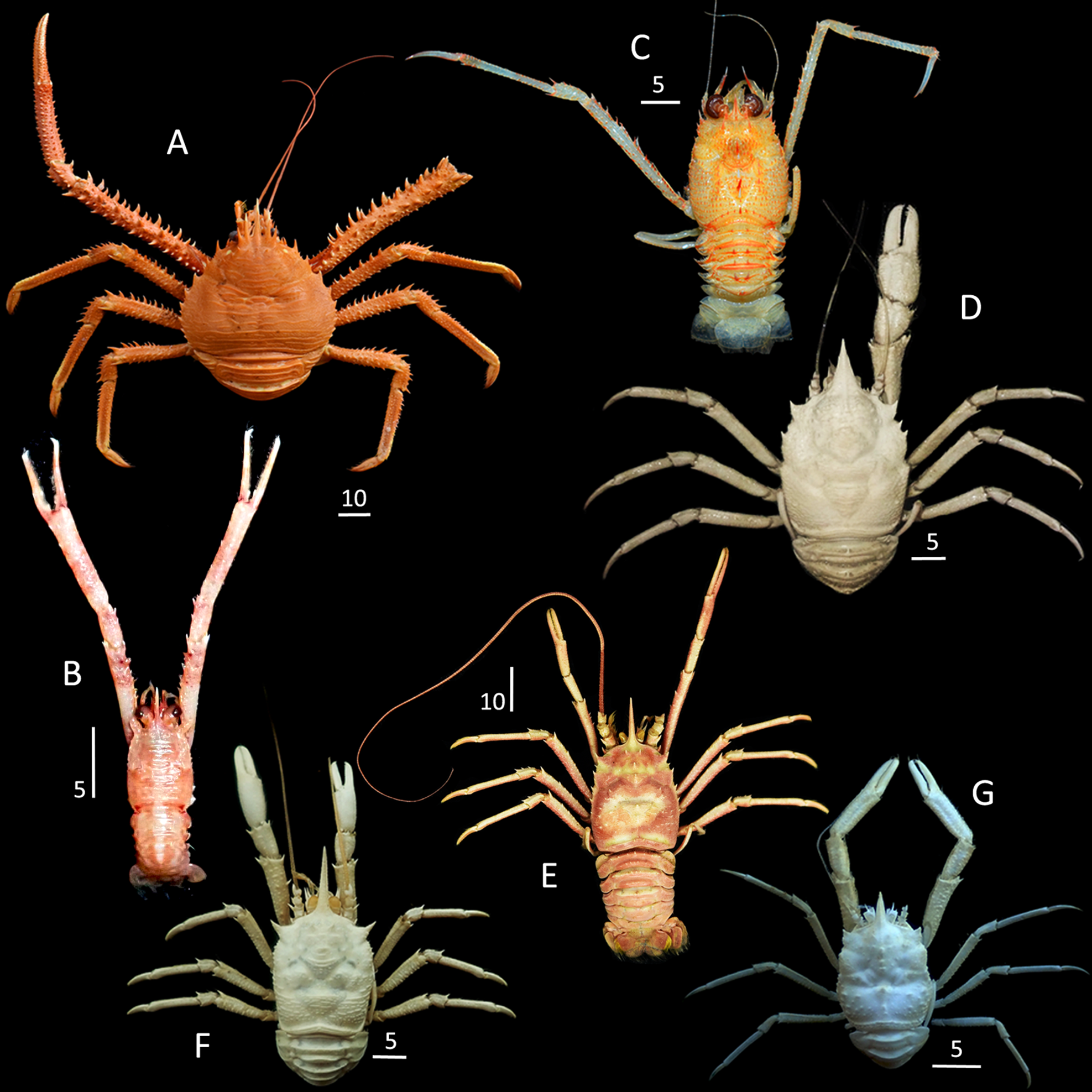
Fig. 2. Habitus, dorsal view. (A) Eumunida multispina Komai et al., Reference Komai, Chakraborty, Paramasivam and Gidda2019 (IO/SS/ANO/00098, male, 46.5 mm PCL); (B) Trapezionida samudrika sp. nov. (IO/SS/ANO/00040, holotype, male, 5.2 mm PCL); (C) Paramunida travancorica sp. nov. (IO/SS/ANO/00061, holotype, male, 10.9 mm PCL); (D) Munidopsis bhavasagara sp. nov. (IO/SS/ANO/00124, holotype, female, 19.5 mm PCL); (E) Munidopsis scobina Alcock, Reference Alcock1894 (IO/SS/ANO/00066, female, 20.9 mm PCL); (F) Munidopsis stylirostris Wood-Mason & Alcock, Reference Wood-Mason and Alcock1891 (IO/SS/ANO/00118, male, 12.9 mm PCL); (G) Munidopsis unguifera Alcock & Anderson, Reference Alcock and Anderson1894 ((IO/SS/ANO/00102, male, 11.0 mm PCL). Scale bars in millimetres.

Fig. 3. Carapace, dorsal view. (A) Eumunida multispina Komai et al., Reference Komai, Chakraborty, Paramasivam and Gidda2019 (IO/SS/ANO/00098, male, 46.5 mm PCL); (B) Trapezionida samudrika sp. nov. ((IO/SS/ANO/00040, holotype male, 5.2 mm PCL); (C) Paramunida travancorica sp. nov. (IO/SS/ANO/00061, holotype male, 10.9 mm PCL); (D) Munidopsis bhavasagara sp. nov. (IO/SS/ANO/00124, holotype female, 19.5 mm PCL); (E) Munidopsis scobina Alcock, Reference Alcock1894 (IO/SS/ANO/00066, female, 20.9 mm PCL); (F) Munidopsis stylirostris Wood-Mason & Alcock, Reference Wood-Mason and Alcock1891 (IO/SS/ANO/00118, male, 12.9 mm PCL); (G) Munidopsis unguifera Alcock & Anderson, Reference Alcock and Anderson1894 (IO/SS/ANO/00102, male, 11.0 mm PCL). Scale bars in millimetres.

Fig. 4. Eumunida multispina Komai et al., Reference Komai, Chakraborty, Paramasivam and Gidda2019 (IO/SS/ANO/00098, male, 46.5 mm PCL). (A) carapace, dorsal view; (B) carapace and pterygostomian flap, left lateral view; (C) thoracic sternum, ventral view; (D) right antennal peduncle, dorsal view; (E) right antennal peduncle, ventral view; (F) pleon, dorsal view; (G) telson, external view; (H) right Mxp3, lateral view; (I–K) right P2–4, lateral view; (L–N) right P2–4 dactyli, lateral view. Scale bars in millimetres.

Fig. 5. Eumunida multispina Komai et al., Reference Komai, Chakraborty, Paramasivam and Gidda2019 (IO/SS/ANO/00098, male, 46.5 mm PCL). Left P1. (A) dorsal view; (B) ventral view; (C) mesial view; (D) lateral view. Scale bars in millimetres.
Eumunida multispina Komai et al., Reference Komai, Chakraborty, Paramasivam and Gidda2019: 442, figures 1–8 (type locality: south-eastern Arabian Sea, off Sakthikulangara Fishing Port, Kollam, Kerala State, India, 08°56′60″N 76°32′34″E, 250–400 m).
Material examined
CMLRE IO/SS/ANO/00098, 1 male (46.5 mm PCL, 54.9 mm CW), south-eastern Arabian Sea, off Kerala, FORVSS station 39903 (8.36°N 76.30°E), HSDT II (CV), 265 m depth, coll. Aneesh Kumar K. V, 25 September 2020.
Taxonomic remarks
The present specimen agrees with the original description of Eumunida multispina by Komai et al. (Reference Komai, Chakraborty, Paramasivam and Gidda2019) particularly in the carapace armature as follows: two pairs of submedian epigastric spines, each pair is situated mesially to the anterior-most hepatic spine adjacent to the base of lateral supra-ocular spine; four lateral marginal spines anterior to the posterior cervical groove; one small extra spine slightly inferior to the lateral margin, located in the notch formed by the anterior cervical groove; and eight spines on the branchial margin posterior to the posterior cervical groove, as well as the morphology of the pleon and telson (Figures 3A, 4A, B, F, G). However, it differs from the type material (cf. Komai et al., Reference Komai, Chakraborty, Paramasivam and Gidda2019: figures 1–3, 5–7) in the following details.
(1) Rostrum is 0.4 times PCL (the rostrum is abnormally short in the holotype, and 0.3 times PCL in the paratype); the posterior median epigastric carapace spine is not developed, whereas it is well developed in the holotype and the paratype.
(2) Antennal article 3 has a long distomesial spine, overreaching the distal margin of the article 4, as compared with short distomesial spine, not reaching the distal margin of the article 4 in the holotype.
(3) Antennal article 4 lacks a small spine near the base of the ventrodistal spine and a minute subdistal spine on the mesial surface; these spines are present in the holotype.
(4) P1 (chelipeds, right chela and carpus are missing in the present specimen) are proportionally shorter and more slender, and have less developed ornamentation than those of the holotype and a paratype: the merus is 1.3 (left) and 1.4 (right) times PCL (1.6 times PCL in the holotype); the length of palm is 5.6 times width, the surfaces are moderately setose (in the holotype, the palm length is 3.0 times width, the surfaces are covered with dense setae); the fingers are not gaping proximally, the occlusal margins are each with a row of closely spaced, small subacute denticles along the entire length (in the holotype, the dactylus has somewhat differentiated proximal cluster of five denticles and corresponding faint concavity on the fixed finger).
(5) P2 merus is 1.3 times the propodus length (in the holotype, 1.1 times).
(6) P4 merus is 0.7 times longer than P2 merus (in the holotype, about 0.8 times); the carpus has a row of seven spines on the dorsal margin, as compared with six dorsal spines in the holotype; the propodus is armed with 16 spiniform setae on the ventral margin, as compared with 17 spiniform setae in the holotype; the dactylus has 10 spiniform setae, as compared with nine spiniform setae in the holotype.
Owing to the availability of only one specimen, we provisionally regard these differences as rather minor and to represent intraspecific variation of E. multispina. Further examination of additional material in future, supplemented with molecular analysis, could reveal its taxonomic status more satisfactorily.
Distribution
South-eastern Arabian Sea, off Kerala (depths of 250–400 m) and Kavaratti Island, Lakshadweep (300 m) (Komai et al., Reference Komai, Chakraborty, Paramasivam and Gidda2019; present study, Figure 1).
Superfamily GALATHEOIDEA Samouelle, Reference Samouelle1819
Family MUNIDIDAE Ahyong et al., Reference Ahyong, Baba, Macpherson and Poore2010
Genus Trapezionida Macpherson & Baba in Machordom et al., Reference Machordom, Ahyong, Andreakis, Baba, Buckley, García-Jiménez, McCallum, Rodríguez-Flores and Macpherson2022
Trapezionida samudrika sp. nov.
(Figures 2B, 3B, 6 & 7)
Type material
Holotype: CMLRE IO/SS/ANO/00040, male (5.2 mm PCL, 4.6 mm CW), south-eastern Arabian Sea, off Kerala, FORVSS station 39622 (8.56°N 78.33°E); Naturalists dredge, 32 m depth, coll. Usha V. Parameswaran, 12 February 2020.

Fig. 6. Trapezionida samudrika sp. nov. (IO/SS/ANO/00040, holotype, male, 5.19 mm PCL). (A) carapace, dorsal view; (B) carapace and pterygostomian flap, left lateral view; (C) thoracic sternum, ventral view; (D) right antennular article 1 and antennal peduncle, ventral view; (E) left antennal peduncle, ventral view; (F) pleonal somite 6, telson and uropods, external view; (G) right Mxp3, lateral view; (H) right G1, dorsal view; (I) right G2, dorsal view; (J) right P1 carpus, ventral view; (K) left P1 chela, dorsal view; (L) left P1 carpus and merus, dorsal view; (M) right P1, dorsal view; (N) right P1 carpus and merus, dorsal view. Scale bars in millimetres.

Fig. 7. Trapezionida samudrika sp. nov. (IO/SS/ANO/00153, paratype, male, 4.3 mm PCL). (A) P3 merus, lateral view; (B) P3 carpus to dactylus, lateral view; (C) P4 merus and carpus, lateral view; (D) P4 propodus and dactylus, lateral view; (E) P4 dactylus, distal portion, lateral view.
Paratypes: CMLRE IO/SS/ANO/00153, 3 males (3.9–4.8 mm PCL, 3.6–4.8 mm CW), Bay of Bengal, off Puducherry, FORVSS station. 23630 (11.99°N 80.08°E); Smith McIntyre grab, 52 m depth, coll. C. K. Smitha, 16 July 2005.
Etymology
The species is named after the ancient Indian science of physiognomy or observation of facial characters.
Diagnosis
Carapace with 5 pairs of epigastric spines and 1 median epigastric spine followed by small scale; hepatic region with 1 or 2 spines; parahepatic, anterobranchial and postcervical regions each with 1 spine; frontal margin strongly oblique; anterolateral spine short; branchial margin with 5 spines. Rostrum long, half of PCL; supraocular spines short and slender, one-fourth length of rostrum, parallel in dorsal view, not exceeding eyes. Anterior margin of thoracic sternite 4 as wide as and subparallel to sternite 3. Pleonal tergites unarmed; tergites 2–4 each with deep median transverse groove, region anterior and posterior to median groove each with transverse ridge (uninterrupted on tergites 2 and 3, interrupted on tergite 4). Antennular article 1 not reaching distal corneal margin, with distomesial and distolateral spines subequal in length. Antennal peduncle not reaching distal margin of cornea; article 1 with distomesial spine just reaching distal margin of article 2; article 2 with distomesial spine overreaching distal margin of article 3, but not reaching distal margin of article 4, distolateral spine nearly reaching distal margin of article 3; article 3 with small distolateral spine; article 4 unarmed. Mxp3 merus with 4 spines on ventral margin and dorsodistal spine. P1 4.7 times PCL; merus 6.2 times length of width measured on distal margin (exclusive of spines); palm 4.2 times longer than wide; fingers 0.7–0.8 length of palm, fixed finger with 0–1 proximal spine and 2 subdistal spines on lateral margin, dactylus with 1 proximal and 1 distal spine on mesial margin and 3 spines proximally on dorsal surface. P3 and P4 meri each with only distal spine on dorsal margin; carpi each with prominent distal spine followed by small spines on dorsal margin; dactyli long, 0.8–0.9 lengths of propodi, unarmed on distal two-fifths of ventral margin.
Description of holotype
Carapace (PCL) 1.1 times CW; dorsal surface slightly convex transversely; main transverse ridges mostly entire; ridges and striae with dense short setae and few longer setae. Gastric region with 5 pairs of epigastric spines and median epigastric spine followed by small transverse scale. Cervical grooves distinct. Hepatic region with 1 or 2 spines; parahepatic, anterobranchial and postcervical regions each with small spine; lateral part of posterior branchial region with 8 transverse ridges including posterior submarginal ridges. Intestinal region without distinct transverse ridge. Frontal margins strongly oblique. Lateral margins slightly convex in dorsal view. Anterolateral spine short, falling far short of sinus between rostrum and supraocular spines, followed by 1 or 2 distinctly shorter spines on anterolateral margin. Branchial margins with 5 spines, posteriormost spine smallest (Figures 2B, 3B & 6A, B). Rostrum spiniform, half of PCL, directed slightly upwards in lateral view; lateral margins slightly crenulated; supraocular spines short and slender, parallel in dorsal view, one-fourth length of rostrum (Figures 2B, 3B & 6A, B).
Pterygostomian flap with long and short transverse ridges (Figure 6B).
Thoracic sternum slightly longer than wide. Sternite 3 about 3.6 times as wide as long, 0.4 width of sternite 4, with few short striae on surface; anterior margin undulate and granulate, with deep V-shaped median notch. Sternite 4 with some short transverse striae, anterior margin as wide as and contiguous to sternite 3. Sternites 5 and 6 smooth, each with row of short setae on anterior ridge and very short striae near lateral margin. Sternites 7 and 8 smooth on lateral parts (Figure 6C).
Pleon smooth in general. Tergites 2–4 each with deep median transverse groove and 1 transverse ridge anterior and posterior to median groove, transverse ridges uninterrupted on tergite 2–3 and interrupted on tergite 4. Somite 6 with 2 large, squamiform ridges (Figure 6A). Telson distinctly wider than long, with short squamiform ridges (Figure 6F).
Eye moderately large. Cornea dilated; maximum diameter 2.8 times distance between rostrum and supraocular spine, about 0.4 times distance between anterolateral spines, 0.2 times PCL. Ocular peduncle with 2 striae on dorsal surface; eyelash long (Figure 6A, D).
Antennular article 1 about 1.5 times longer than wide, not reaching distal corneal margin; distomesial and distolateral spines subequal in length; lateral margin with 2 spines, distal spine distinctly overreaching distal spines, proximal spine very short, located anterior to midlength of article; ventral surface with scattered, short squamiform ridges (Figure 6D).
Antennal peduncle not reaching distal margin of cornea. Article 1 with distomesial spine just reaching distal margin of article 2; distolateral angle granulate. Article 2 with distomesial spine distinctly overreaching distal margin of article 3, but not reaching distal margin of article 4; distolateral spine nearly reaching distal margin of article 3. Article 3 with small distolateral spine. Article 4 short, unarmed (Figure 6 D, E).
Mxp3 ischium about 1.7 times merus length measured along dorsal margin, ventrodistally terminating in spine. Merus with 4 spines on ventral margin and dorsodistal spine. Carpus to dactylus unarmed on margins.
P1 4.7 times PCL, surfaces covered with numerous squamiform ridges of various size, those bearing short plumose setae. Ischium unarmed. Merus 6.2 times longer than width measured on distal margin (exclusive of spines), with 11–15 spines arranged in 2 irregular rows on dorsolateral surface, spines increasing in size distally; distal margin with 4 spines (dorsomesial, dorsolateral, ventrolateral, ventromesial; dorsomesial spine strongest, ventromesial spine smallest). Carpus 2.1 times length of width measured on distal margin (exclusive of spines), 0.4 palm length; dorsolateral margin with 3 or 4 spines on right P1 and 5 spines on left P1; dorsal surface with 2 or 3 spinules; ventrolateral distal angle produced to rounded lobe, unarmed (Figure 6J). Palm slightly widened distally, length 4.2 times width measured at base of fingers; dorsal surface with median row of 5 or 6 small spines and 1 spine at dactylar articulation; dorsomesial margin with 6 or 7 small spines; dorsolateral margin with 1 or 2 spines. Fingers 0.7–0.8 times palm length, terminating in sharp claws crossing distally; dorsal surfaces with scattered setae; mesial and lateral margins with stiff setae; occlusal margins denticulate, with 1 or 2 rounded teeth on proximal one-third. Fixed finger with no (left) or 1 (right) proximal spine and 2 subdistal spines (1 spine damaged) on lateral margin. Dactylus with 3 small spines proximally on dorsal surface, dorsomesial margin with 1 small spine each on proximal and distal parts (Figure 6K–N).
Epipods absent from P1–4. P2–5 missing.
Variations in paratypes
All paratypes lacking P1and P2, but other morphological characters agreeing well with those of holotype in general. P3 and P4 2.0–2.3 and 1.7–2.0 times PCL, respectively; margins of merus to propodus bearing iridescent setae. Meri 0.6–0.8 (P3) and 0.5–0.6 (P4) PCL, 4.0–6.2 (P3) and 4.5–4.7 (P4) times longer than high; lateral surface with several squamiform ridges; dorsal margins each with distal spine only; ventral margins each armed with 1 strong distal spine followed by 4 or 5 spinules and short transverse ridges (Figure 7A, C). Carpi each with strong distal spine followed by 4 (P3) and 2 (P4) small spines on dorsal margin, distal spine strongest; ventral margin with 1 distal spine (Figure 7B, C). Propodi 5.7–6.5 (P3) and 4.9–5.2 (P4) times longer than high; dorsal margins unarmed; ventral margins each with row of 12 movable spines (including 2 spines on distoventral margin) (Figure 7B, D). Dactyli 0.8–0.9 (P3) and 0.9 (P4) lengths of propodi; ventral margins with 6 or 7 movable spines on proximal 0.6, distal 0.4 length smooth (Figure 7B, D); unguis corneous, with appressed seta-like spine at base (Figure 7E).
Taxonomic remarks
The holotype from the south-eastern Arabian Sea lacks all ambulatory legs (P2–4), whereas all three specimens examined from the Bay of Bengal lack chelipeds and first ambulatory legs (P1 and P2). The specimens from the Bay of Bengal agree well with the holotype in the morphological characters of the body and anterior appendages, and thus we treat these specimens to be conspecific and the specimens from the Bay of Bengal as paratypes of the new species.
Trapezionida samudrika sp. nov. is morphologically close to T . clinata (Macpherson, Reference Macpherson and Crosnier1994) known from a wide area ranging from Indonesia to New Caledonia in the south-western Pacific and T. munin (Komai, Reference Komai2011) known only from Japan. Shared characters are: carapace with strongly oblique frontal margin and five spines on the branchial margin posterior to anterior cervical groove; smooth thoracic sternum; unarmed pleonal tergites; antennular article 1 with distal spines subequal in length; Mxp3 merus with dorsodistal spine, and P1 dactylus bearing a row of spines on the dorsomesial margin. The new species appears more similar to T. munin than T. clinata in the less elongate supraocular spines of the carapace, only reaching the level of the midlength of the eyes, rather than reaching the distal corneal margins; the distomesial spine of the antennal article 2 at most reaching, instead of clearly overreaching, the distal margin of the article 4; the presence of a small distolateral spine on the antennal article 3; and the P1 dactylus is shorter, rather equal to, the palm. Nevertheless, T. samudrika sp. nov. is distinguishable from T. munin by the following details (for the latter species, cf. Komai, Reference Komai2011: figures 8A–B, F, 9C, D).
(1) Carapace is shorter (1.1 times longer than wide) with more convex lateral margins, while it is 1.2 times longer than wide in T. munin.
(2) Median epigastric spine of carapace is followed by a small transverse scale, which is absent in T. munin.
(3) Hepatic region of the carapace has one or two small spines, which are absent in T. munin.
(4) Anterior margin of thoracic sternite 4 is equal in width to sternite 3, whereas it is wider than sternite 3 in T. munin.
(5) P1 is comparatively longer (4.7 times PCL) with more elongated palm (length is 4.2 times width), while it is at most 3.4 times PCL and the palm is 2.8 times longer than wide in T. munin. P1 fixed finger has only 0–1 proximal spine and two subdistal spines on the lateral margin, whereas the margin is armed with 4–5 spines including one proximal and 1–2 subdistal spines in T. munin.
(6) P3 merus possesses only 1 distal spine on dorsal margin, whereas it is armed with 6–8 dorsal spines in T. munin.
(7) P3 carpus has dorsal marginal spines much smaller than in T. munin.
(8) P3 and P4 dactyli are proportionally longer (0.8–0.9 lengths of propodi) and unarmed on distal 0.4 of ventral margin, whereas they are 0.6 propodal lengths and unarmed on distal 0.25 of ventral margin in T. munin.
Trapezionida foresti (Macpherson and de Saint Laurent, Reference Macpherson and de Saint Laurent2002) from Reunion Island (cf. Macpherson and de Saint Laurent, Reference Macpherson and de Saint Laurent2002: figure 1) and T. llenasi (Macpherson, Reference Macpherson, Richer de Forges and Justine2006) from the Austral Archipelago (cf. Macpherson, Reference Macpherson, Richer de Forges and Justine2006: figure 14) are also similar to T. samudrika sp. nov. in the general morphology of the carapace, thoracic sternum and pleon, but differ in the antennular article 1 with the distomesial spine distinctly longer than, rather subequal in length to, the distomesial spine; the Mxp3 merus with two or three, instead of four ventral marginal spines, and the less elongate P1 palm (the length is 1.6, instead of 4.2, times the width).
Distribution
Known from the south-eastern Arabian Sea and the Bay of Bengal, 32–52 m depth (Figure 1).
Genus Paramunida Baba, Reference Baba1988
Paramunida travancorica sp. nov.
(Figures 2C, 3C & 8)
Type material
Holotype: CMLRE IO/SS/ANO/00061, male (10.9 mm PCL, 11.4 mm CW), south-eastern Arabian Sea, off Tamil Nadu, FORVSS station 39632 (7.01°N 77.32°E), Naturalists dredge, 275 m depth, coll. Usha V. Parameswaran, 13 February 2020.

Fig. 8. Paramunida travancorica sp. nov. (IO/SS/ANO/00061, holotype, male, 12.0 mm PCL). (A) carapace, dorsal view; (B) carapace dorsal margin, right lateral view; (C) left pterygostomian flap, lateral view; (D) left eye, dorsal view; (E) thoracic sternum, ventral view; (F) right antennular article 1 and antennal peduncle, ventral view; (G) right Mxp3, lateral view; (H) right G2, dorsal view; (I) pleon, dorsal view; (J) telson, external view; (K) right P2, lateral view; (L) left P4, lateral view; (M) right P2 dactylus, lateral view; (N) left P4 dactylus, lateral view; (O) right P5 dactylus, lateral view. Scale bars in millimetres.
Paratype: CMLRE IO/SS/ANO/00062, 1 female (6.8 mm PCL, 6.9 mm CW), location and collection details same as in holotype.
Etymology
The species is named after the erstwhile princely state of Travancore (located in southern Kerala), with a history of patronage for natural history studies.
Diagnosis
Carapace with median row of short spines on epigastric region, row of 3 distinct median spines each on mesogastric and cardiac regions; posterior branchial region distinctly spinose; posterior transverse ridge with prominent median spine; anterolateral spine exceeding sinus between rostral and supraocular spines; margin between rostral and supraocular spines distinctly concave. Rostrum spiniform, larger than supraocular spines, with thin dorsal carina. Thoracic sternites 5–7 with few striae on lateral sides. Pleonal tergites 2 and 3 each with 4 distinct spines on anterior ridge, 2 submedian spines on posterior ridge; tergite 4 with 4 large and 2 small spines on anterior ridge, single median spine on posterior ridge. Antennular article 1 lateral margin with row of spinules distally on inflated proximal portion, distal portion 0.4 length of proximal portion. Anterior prolongation of antennal article 1 spiniform, overreaching antennular article 1 by two-fifths length; dorsal margin without row of spines; article 2 about twice as long as wide, distomesial spine slightly mucronated, reaching distal end of antennal peduncle, shorter than rest of article 2, distolateral spine not reaching distal margin of article 3; article 3 1.5 times longer than wide. Mxp3 ischium 1.4 times length of merus, with long distoventral spine; merus with well-developed median spine on ventral margin, dorsal margin unarmed. P2 3.4 times PCL; P4 merocarpal articulation much overreaching tip of anterior prolongation of antennal article 1; P2 and P4 propodi 11–14 times as long as high, 1.7–1.8 times longer than dactyli.
Description of holotype
Carapace (PCL) 0.9 times CW. Dorsal surface covered with numerous spines and spinules, each arising from very short arcuate striae and bearing short uniramous setae. Epigastric region with median row of short spines behind rostral spine, prominent epigastric spine posterior to supraocular spine. Mesogastric region with median row of 3 prominent spines, progressively decreasing in size posteriorly. Cervical groove distinct. Cardiac region with median row of 3 prominent spines, progressively decreasing in size posteriorly. Posterior branchial region with longitudinal row of moderately large spines adjacent to cardiac region. Posterior transverse ridge with row of small spines and median prominent spine. Frontal margin gently concave. Lateral margins gently convex. Anterolateral spine clearly exceeding sinus between rostral and supraocular spines, reaching slightly short of distal end of supraocular spine. Rostrum spiniform, larger than supraocular spines, with thin dorsal longitudinal carina; margin between rostral and supraocular spines distinctly concave (Figures 2C, 3C & 8A, B).
Pterygostomian flap with oblique striae, anterior margin subacute (Figure 8C).
Thoracic sternite 3 bilobed (anterior margin of each lobe sinuous), 4.1 times as wide as long, 0.4 times sternite 4 width, with 2 pairs of short striae. Sternite 4 with few arcuate striae. Sternites 5–6 with few striae on each lateral side. Sternite 7 with 2 short striae on each lateral side (Figure 8E).
Pleonal tergites 2 and 3 each with 4 distinct spines and some much smaller spines on anterior ridge, 2 distinct submedian spines on posterior ridge. Tergite 4 with 4 large and 2 small spines on anterior ridge; single median spine on posterior ridge (Figures 2C & 8I).
Maximum corneal diameter 0.4 times distance between bases of anterolateral spines (Figures 2C & 8A).
Antennular article 1 about 2.0 times longer than wide, overreaching anterior margin of cornea; distomesial spine much smaller than distolateral spine; mesial margin with fringe of setae; lateral margin with row of spinules distally on inflated proximal portion; distal slender portion about 0.4 times as long as proximal portion (Figure 8F).
Antennal peduncle slightly overreaching distal margin of cornea. Article 1 mesially with anterior prolongation spiniform, overreaching antennular article 1 by two-fifths length. Article 2 1.8 times length of article 3, about 2.0 times (excluding spines) as long as wide; ventral surface with few scales laterally; distomesial spine, slightly mucronated, shorter than rest of article 2, stronger than distolateral spine, reaching distal margin of antennal peduncle, reaching midlength of anterior prolongation of article 1; distolateral spine not reaching distal margin of article 3. Article 3 1.5 times longer than wide, unarmed. Article 4 short, unarmed (Figure 8F).
Mxp3 ischium about 1.4 times merus length measured along dorsal margin, distoventrally bearing long spine. Merus with well-developed median spine on ventral margin, dorsal margin unarmed (Figure 8G).
P1 and P3 missing; only right P2 and left P4 present. P2 and P4 long, slender; lateral surfaces of meri, carpi and propodi covered with numerous squamiform striae bearing short setae (Figure 8K, L). P2 3.4 times PCL; merus 1.3 times PCL, 10.8 times longer than high, 3.6 times carpus length, 1.5 times propodus length; propodus 11.4 times longer than high, 1.7 times dactylus length. P4 slightly longer than P2; merus 1.4 times PCL, 9.9 times longer than high, 3.5 times carpus length, 1.4 times propodus length; merocarpal articulation much overreaching tip of anterior prolongation of antennal article 1; propodus 13.7 times longer than high, 1.8 times dactyl length (Figure 2C).
Variations in paratype
Female paratype lacking P1–4, but agreeing with holotype, except lack of lateral spinules on anterior ridges of pleonal tergites 2–3.
Taxonomic remarks
Paramunida travancorica sp. nov. appears close to P. bineeshi Macpherson et al., Reference Macpherson, Chan, Kumar and Rodríguez-Flores2020 from the Andaman Sea, P. mozambica Cabezas et al., Reference Cabezas, Macpherson and Machordom2010 from off Mozambique, and P. stichas Macpherson, Reference Macpherson and Crosnier1993 from the wide area of the western Pacific and south-western Australia. Shared characters include: rostrum larger than supraocular spines; mesogastric carapace region with a row of three prominent spines; lateral margin of antennular article 1 with distal slender portion less than half as long as proximal inflated portion; anterior prolongation of antennal article 1 spiniform, without row of spines on dorsal margin; distomesial spine of antennal article 2 at least reaching end of antennal peduncle, shorter than rest of article 2, and mucronated distally (not straight); distolateral spine of antennal article 2 not reaching end of antennal article 3; antennal article 3 about 1.5 times longer than wide; and P2–P4 propodi 7–15 times as long as high (the holotype of the present new species lacks P3). Further, the new species may be more allied to P. mozambica and P. stichas in the rostrum being slender and spiniform, rather than triangular in P. bineeshi.
Nevertheless, P. travancorica sp. nov. is distinguished from P. mozambica by the following particulars (cf. Cabezas et al., Reference Cabezas, Macpherson and Machordom2010: figure 7A, D–E).
(1) The cardiac spines on the carapace are much stronger in P. travancorica sp. nov. than in P. mozambica.
(2) The distolateral spine of the antennular article 1 is much larger than, instead of subequal to, the distomesial spine.
(3) The distomesial spine of the antennal article 2 is only slightly mucronated in P. travancorica sp. nov., instead of clearly mucronated in P. mozambica.
(4) Mxp3 merus is unarmed on the dorsal margin in the new species, whereas it has a distinct dorsodistal spine in P. mozambica.
From P. stichas, the new species differs in the following characters (cf. Macpherson, Reference Macpherson and Crosnier1993: figure 9a, c–d, g; Cabezas et al., Reference Cabezas, Macpherson and Machordom2010: figures 15E, 18G).
(1) Rostrum dorsal carina is sharp, rather blunt in P. stichas.
(2) Thoracic sternites 5–6 have some moderately long striae on the lateral parts, instead of nearly smooth in P. stichas.
(3) Distolateral spine of antennular article 1 is stout, rather long and slender in P. stichas.
(4) Distomesial spine of antennal article 2 is only slightly mucronated, rather than clearly mucronated in P. stichas.
(5) P2 propodus may be proportionally longer, about 11.0 rather than 9.0 times longer than in the type material of P. stichas; although Cabezas et al. (Reference Cabezas, Macpherson and Machordom2010) noted the rate as 15.0 times in the diagnosis of the species.
From P. bineeshi, the present new species is differentiated by the following (cf. Macpherson et al., Reference Macpherson, Chan, Kumar and Rodríguez-Flores2020: figure 4A, C–E).
(1) Anterolateral carapace spine clearly overreaches the sinus between the rostral and supraocular spines, compared with slightly overreaching the sinus in P. bineeshi.
(2) Thoracic sternites 5 and 6 have a few striae on the lateral parts, compared with nearly smooth in P. bineeshi.
(3) Distal slender portion of the antennular article 1 is 0.4 times length of the proximal inflated portion, compared with 0.5 times length of the inflated portion in P. bineeshi.
(4) Antennal article 2 (excluding distal spines) is 2.0 times as long as wide, compared with 1.5 times as long as wide in P. bineeshi.
(5) Distomesial spine of antennal article 2 reaches the distal end of the antennal peduncle, compared with overreaching the distal margin of the antennal peduncle in P. bineeshi.
(6) Mxp3 ischium is 1.4 times the merus length, compared with about 2.0 times in P. bineeshi.
Distribution
So far only from the type locality, south-eastern Arabian Sea, 275 m depth (Figure 1).
Family MUNIDOPSIDAE Ortmann, Reference Ortmann, Gerstäcker and Ortmann1898
Genus Munidopsis Whiteaves, Reference Whiteaves1874
Munidopsis bhavasagara sp. nov.
(Figures 2D, 3D & 9)
Type material
Holotype: CMLRE IO/SS/ANO/00124, female (19.5 mm PCL, 15.7 mm CW), south-eastern Arabian Sea, off Kerala, FORVSS station 32118 (8.42°N 75.92°E), HSDT II (CV); 1221 m depth, coll. Vinu Jacob, 11 December 2013.

Fig. 9. Munidopsis bhavasagara sp. nov. (IO/SS/ANO/000124, holotype, female, 19.5 mm PCL). (A) carapace, dorsal view; (B) carapace and pterygostomian flap, left lateral view; (C) rostrum and right antennal peduncle, dorsal view; (D) left antennular article 1 and antennal peduncle, ventral view; (E) thoracic sternum, ventral view; (F) left Mxp3, lateral view; (G) telson, external view; (H) right P1 merus and carpus, dorsal view; (I) right P1 chela, dorsal view; (J–L) right P2–P4, lateral view; (M) left P2 propodus, distal margin, ventral view; (N–P) right P2–P4 dactyli, lateral view; (Q) right P5 dactylus, lateral view. Scale bars in millimetres.
Etymology
This species is named after ‘Bhavasagara’, the recently established Referral Centre at the Centre for Marine Living Resources and Ecology, Kochi. ‘Bhavaságara’ in Sanskrit means ‘the ocean of worldly life’. Used as a noun in apposition.
Diagnosis
Carapace covered with setiferous, interrupted or scale-like ridges; epigastric lobes distinctly convex, without spines or elevated, anteriorly directed ridges; anterolateral spine strong, directed anteriorly, followed by 1 or 2 moderately large spines on anterior branchial margin. Rostrum without distinct spines on lateral margins. Thoracic sternite 4 not contiguous to posterior margin of sternite 3, greatest width about 3.0 times that of sternite 3. Pleonal tergites 2–4 each with median spines on anterior transverse ridge. Ocular peduncles movable, with subglobular corneas, no eye-spines. Antennal peduncle overreaching distal margin of cornea by half-length of article 4; article 1 with distomesial process slightly overreaching half-length of article 2. P1 moderately long and stout, 1.7 times PCL; fixed finger without denticulate carina on distolateral surface. P2 not exceeding tip of P1. P2–4 moderately slender, unarmed on dorsal margins of meri and propodi; meri not distinctly crested on dorsal margins; propodi uniform in height proximal to distal, 4.5–5.5 times as long as high; dactyli equal in length to or slightly shorter than propodi, ventral margins each with 9–11 subtriangular teeth on proximal half and another subdistal tooth distant from proximal teeth. Epipods present on P1–3.
Description
Carapace (PCL) 1.2 times CW; dorsal surface transversely convex, covered with numerous, short interrupted or scale-like tuberculate ridges and small tubercles bearing short plumose setae; regions well-defined by grooves. Epigastric lobes rounded, distinctly convex, with short squamiform ridges. Posterior cardiac region subtriangular, elevated, preceded by deep transverse depression. Posterior margin preceded by smooth elevated ridge. Lateral margins subparallel; anterolateral spine strong, directed anteriorly, followed by 1 (anterior spine obsolete on left side) or 2 moderately large spines on anterior branchial margin, blunt projection on posterior branchial margin; lateral ends of anterior and posterior cervical grooves each with small notch. Frontal margin weakly concave behind ocular peduncle, leading to low blunt lobe, then concave toward anterolateral spine; antennal spine absent (Figures 2D, 3D & 9A–B). Rostrum subtriangular, moderately narrow, 0.3 times width between anterolateral spines of carapace (measured between lateral bases of ocular peduncles), 0.4 times PCL, gently depressed, slightly upturned distally in lateral view, terminating in acute tip; lateral margins nearly straight, slightly serrated; dorsal surface transversely convex, with blunt median longitudinal carina extending onto epigastric lobes (Figures 2D, 3D & 9A–D); ventral surface with rounded, longitudinal ridge in midline.
Pterygostomian flap with numerous short ridges of small tubercles; anterior margin bluntly angular (Figure 9B).
Thoracic sternum (measured in midline) as long as wide, maximum width at sternite 7. Sternite 3 3.1 times width of length; anterior margin divided by median notch into 2 roundly rectangular lobes. Sternite 4 not contiguous to posterior margin of sternite 3; greatest width 3.2 times that of sternite 3; anterolateral margins oblique, each with row of small tubercles and setae; surface depressed in midline, with scattered, short setae. Sternites 5–7 each with row of setae on anterior tuberculate ridge and scattered short setae on surface (Figure 9E).
Pleon with scattered, short plumose setae. Tergites 2–4 each with anterior transverse ridge sharply elevated and bearing median spine; tergites 2 and 3 each with uninterrupted posterior ridge, tergite 4 lacking posterior ridge (Figure 9A). Tergite 5 without distinct transverse ridges, slightly convex on median surface. Tergite 6 flattish, posteromedian margin gently convex and slightly exceeding posterolateral lobes. Telson composed of 12 calcified plates, each posterior plate slightly wider than long (Figure 9G).
Ocular peduncle comparatively small, reaching proximal 0.3 length of rostrum, movable, unarmed; cornea subglobular, subequal in length with remaining peduncle; distinct spine ventral to frontal carapace margin between ocular and antennal peduncles (Figure 9A–C).
Antennular article 1 with dorsolateral spine subequal in length to ventrolateral spine; lateral margin unarmed, convex; distomesial margin with row of small tubercles, unarmed; ventral surface bearing short striae (Figure 9D).
Antennal peduncle overreaching distal margin of cornea by only half-length of article 4. Article 1 with strong distomesial process not reaching distal margin of article 2; distolateral angle produced, with subacute tubercle. Article 2 with distolateral spine reaching proximal one-third length of article 3, distomesial margin unarmed. Articles 3 and 4 unarmed (Figure 9C–D).
Mxp3 ischium 0.7 times length of merus measured along dorsal margin; dorsal margin terminating in blunt process; ventral margin sharply ridged, terminating in blunt process; crista dentata composed of row of 20 or 21 small corneous teeth. Merus with scattered, short tuberculate ridges on lateral surface; ventral margin with 2 spines; dorsal margin protuberant, with small distal spine. Carpus protuberant on dorsal surface. Propodus and dactylus smooth (Figure 9F). Exopod distinctly overreaching distal margin of merus. Epipod elongated.
P1 moderately long and stout, 1.7 times PCL, 1.3 times carapace length including rostrum; surfaces covered with numerous, short tuberculate ridges bearing short plumose setae; marginal setae longer. Ischium with small spine on ventromesial distal margin. Merus 2.4 times length of width measured at distal end (exclusive of spines), with 4 strong distal spines (dorsomesial, dorsolateral, ventrolateral, ventromesial; dorsomesial spine smallest) (Figure 9H). Carpus 1.3 times length of width measured at distal end (exclusive of spines), with 3 small distal spines (dorsal, dorsolateral, dorsomesial); dorsal surface shallowly concave in midline; ventrolateral distal margin unarmed (Figure 9H). Palm somewhat compressed dorsoventrally, unarmed; length 1.2 times width measured at base of fingers, subequal to carpus length; dorsal surface convex medially, with broad concavity along mesial margin; lateral margin weakly concave at base of fixed finger (Figure 9I). Fingers 1.3 times palm length; occlusal margins nearly straight, narrowly gaping, distally spooned; prehensile edges each with row of low teeth; distal margins each with row of small subacute teeth; fixed finger without denticulate carina on distolateral surface (Figure 9I).
P2–4 moderately slender, subcylindrical, somewhat compressed laterally; surfaces of ischium to propodus with numerous, short tuberculate ridges bearing short plumose setae (Figure 9J–L); P2 longest (distal tip of dactylus broken), overreaching base but not clearly reaching tip of P1 fingers. Meri slender, subrectangular in lateral view; dorsal surface rounded, unarmed; ventrolateral margin with strong distal spine (spine on P4 smallest); ventromesial margin unarmed. Carpi with elevated lateral ridge subparallel to dorsal ridge; dorsal ridge with strong distal spine on P2 and P3, unarmed on P4. Propodi uniform in height from proximal to distal, length (exclusive of distal rounded projection) 4.7–5.8 times height; dorsal and ventral surfaces weakly convex; distoventral margin with 2 lateral and 1 mesial, short corneous spines (Figure 9M). Dactyli 0.9–1.0 times length of propodi, slender, curved, each terminating in long, corneous claw (distally broken in P2); dorsal surface nearly smooth; ventral margin with 9–11 subtriangular teeth decreasing in size proximally on proximal half, each tooth accompanied by slender, small corneous spine, and with another low subdistal tooth distant from proximal teeth (Figure 9N–P).
Epipods present on P1–3.
Taxonomic remarks
Munidopsis bhavasagara sp. nov. resembles M. dentifalx Osawa et al., Reference Osawa, Lin and Chan2007 known from the Philippines and Papua New Guinea (Osawa et al., Reference Osawa, Lin and Chan2007; Macpherson et al., Reference Macpherson, Chan, Kumar and Rodríguez-Flores2020). Shared characters include: carapace covered with setiferous (short plumose setae) interrupted ridges, but without distinct epigastric spines; pleonal tergites 2–4 each with a median spine on anterior transverse ridge; ocular peduncles with subglobular corneas, lacking eye-spines; antennal article 2 with only distolateral spine, article 3 unarmed; P2 not reaching tip of P1; P2–4 relatively slender, P2–4 meri not distinctly crested on dorsal margin; P2–4 propodi uniform in height from proximal to distal; P2–4 dactyli each ventrally with a low subdistal tooth being far distant from row of teeth on proximal half, and epipods present on P1–3. Among these characters, the arrangement of the ventral teeth on the P2–4 dactyli may be unusual in the genus Munidopsis and known only for these two species in the published accounts of congeneric species. However, the new species differs from M. dentifalx in many characters as shown below (cf. Osawa et al., Reference Osawa, Lin and Chan2007: figures 1A, C, F, 2A, D–G).
(1) Epigastric lobes of the carapace lack a pair of elevated, anteriorly directed, short ridges, which are present in M. dentifalx.
(2) Thoracic sternite 4 is comparatively wider; the greatest width is 3.2 times that of the sternite 3, whereas it is 2.8–2.9 times in M. dentifalx.
(3) Antennal peduncle overreaches the distal margin of the cornea by only half-length of the article 4, compared with overreaching the corneal margin by full length of article 4 in M. dentifalx.
(4) Antennal article 1 has a distomesial process which does not reach the distal margin of article 2, compared with reaching it in M. dentifalx.
(5) P1 is proportionally shorter. P1 length is 1.7 times PCL and 1.3 times CL (including rostrum) (1.9–2.1 times PCL and 1.4–1.6 times CL in M. dentifalx); the merus length is 2.4 times width (2.6–3.2 times in M. dentifalx); the carpus length is 1.3 times width (1.5–1.7 times in M. dentifalx); and the palm length is 1.2 times width (1.5–1.7 times in M. dentifalx) and 1.0 times carpus length (1.2–1.4 times in M. dentifalx). The fingers are comparatively longer, 1.3 times palm length (equal in length to the palm in M. dentifalx). The P1 fixed finger lacks a short denticulate carina on the distolateral surface, whereas such carina is present in M. dentifalx.
(6) P2–4 are also proportionally shorter. Particularly, the propodi (exclusive of distal rounded projection) are 4.5–5.5 times longer than high (6.2–8.1 times in M. dentifalx).
(7) P2–4 propodi each have two lateral and one mesial, short corneous spines on the distoventral margin, whereas in M. dentifalx, the distoventral margin possesses only 1 corneous spine mesially.
(8) P2–4 dactyli each has 9–11 subtriangular teeth on the ventral margin, exclusive of a subdistal spine near the base of the terminal claw, instead of 13–15 teeth in M. dentifalx.
Distribution
Known only from the type locality, south-eastern Arabian Sea, off Kerala, India, 1241 m depth (Figure 1).
Munidopsis scobina Alcock, Reference Alcock1894
(Figures 2E, 3E & 10)
Munidopsis scobina Alcock, Reference Alcock1894: 330 [type locality: Bay of Bengal, ‘Investigator’ Station 120, 15°56′5″N 81°30′30″E, 240 fathoms ( = 439 m)]; Alcock and Anderson, Reference Alcock and Anderson1894: 167; Alcock and Anderson, Reference Alcock and Anderson1895: plate 13, figure 1; Alcock, Reference Alcock1901: 254; Baba, Reference Baba1988: 162, figure 64; Baba, Reference Baba2005: 295 (synonymies); Macpherson, Reference Macpherson2007: 97; Baba et al., Reference Baba, Macpherson, Poore, Ahyong, Bermudez, Cabezas, Lin, Nizinski, Rodrigues and Schnabel2008: 158 (compilation); Thirumilu, Reference Thirumilu2011: 1, figures 1 & 2; Vaitheeswaran, Reference Vaitheeswaran2017: 407, figures 2–6; Widyastuti and Lin, Reference Widyastuti and Lin2021: 114, figure 2G.

Fig. 10. Munidopsis scobina Alcock, Reference Alcock1894. (A–H, K–T) IO/SS/ANO/00066, female, 20.9 mm PCL; (I, J) IO/SS/ANO/00065, male, 14.7 mm PCL. (A) carapace, dorsal view; (B) carapace, right lateral view; (C) right pterygostomian flap, lateral view; (D) thoracic sternum, ventral view; (E) pleon, dorsal view (F) telson, external view; (G) right antennular and antennal peduncles, ventral view; (H) right Mxp3, lateral view; (I) right G1, dorsal view; (J) right G2, dorsal view; (K–L) left and right P1, dorsal view; (M) left P1 finger, dorsal view; (N–P) right P2–P4, lateral view; (Q–S) right P2–P4 dactyli, lateral view; (T) right P5 dactylus, lateral view. Scale bars in millimetres.
Munidopsis scobrina [sic]: Lloyd, Reference Lloyd1907: 2 (list).
Munidopsis (Munidopsis) scobina: Tirmizi, Reference Tirmizi1966: 222, figure 35.
Munidopsis (Munidopsis) scobina indica Rao, Reference Rao1974: 304, figure 1d–i, plate I.
Material examined
CMLRE IO/SS/ANO/00065–66, 37 males (14.7–17.2 mm PCL, 11.8–14.9 mm CW), 26 females (13.8–20.9 mm PCL, 11.9–17.5 mm CW), south-eastern Arabian Sea, off Tamil Nadu, FORVSS station 39801 (8.24°N 76.49°E), Expo model trawl, 610 m depth, coll. Vishnu K.V., 29 February 2020. CMLRE IO/SS/ANO/00068–69, 2 males (11.5–12.3 mm PCL, 9.4–10.3 mm CW) 9 females (12.4–13.6 mm PCL, 11.2–12.4 mm CW), north-western Bay of Bengal, off Andhra Pradesh, FORVSS station 35327 (16.04°N 81.68°E), Naturalists dredge, 507 m depth, coll. S.S. Cubelio, 19 November 2016. CMLRE IO/SS/ANO/00070–71, 5 males (12.3–15.4 mm PCL, 9.9–12.5 mm CW), 13 females (3.4–15.7 mm PCL, 11.5–13.6 mm CW), south-eastern Arabian Sea, off Kerala, FORVSS station 31801 (12.47°N 74.15°E), Expo model trawl; 449 m depth, coll. Rajool Shanis C.P., 24 August 2013. CMLRE IO/SS/ANO/00073, 1 female (16.0 mm PCL, 13.8 mm CW), south-western Bay of Bengal, off Pondicherry, FORVSS station 39803 (10.85°N 80.36°E), HSDT II (CV), 518 m depth, coll. Vishnu K.V., 6 March 2020. CMLRE IO/SS/ANO/00076–77, 3 males (11.4–12.0 mm PCL, 9.7 mm CW), 7 females (11.8–15.7 mm PCL, 10.2–13.6 mm CW), south-western Bay of Bengal, off Pondicherry, FORVSS station 39210 (11.95°N 80.15°E), Naturalists dredge, 439 m depth, coll. V. P. Padate, 29 November 2019. CMLRE IO/SS/ANO/00084, 13 females (6.3–17.8 mm PCL, 14.2–14.9 mm CW), north-western Bay of Bengal, off Andhra Pradesh, FORVSS station 27924 (17.05°N 83.3°E), Expo model trawl, 550 m depth, coll. R. Raghu Prakash, 2 September 2010. CMLRE IO/SS/ANO/00085, 1 female (17.4 mm PCL, 15.1 mm CW), south-western Bay of Bengal, off Pondicherry, FORVSS station 39805 (10.99°N 80.43°E), HSDT II (CV), 944 m depth, coll. Vishnu K.V., 7 March 2020. CMLRE IO/SS/ANO/00086, 1 male (17.7 mm PCL, 14.9 CW), south-western Bay of Bengal, off Pondicherry, FORVSS station 27905 (11.13°N 80.2°E), HSDT II (CV), 540 m depth, coll. R. Raghu Prakash, 27 August 2010. CMLRE IO/SS/ANO/00104–105, 23 males (11.8–14.4 mm PCL, 9.5–12.2 mm CW), 19 females (11.0–16.3 mm PCL, 9.0–13.1 mm CW), south-eastern Arabian Sea, off Kerala, FORVSS station 28102 (10.07°N 75.62°E), HSDT II (CV), 400 m depth, coll. Vinu Jacob, 14 October 2010. CMLRE IO/SS/ANO/00109–110, 3 males (10.4–19.1 mm PCL, 9.0–16.9 mm CW), 7 females (14.0–19.8 mm PCL, 12.4–16.6 mm CW), south-western Bay of Bengal, off Pondicherry, FORVSS station 29111 (12.12°N 80.33°E), HSDT II (CV), 785 m depth, coll. Rajeesh Kumar M.P., 5 November 2011.
Taxonomic remarks
The type material of M. scobina deposited in the Zoological Survey of India, Kolkata (registration numbers 6902/9 and 6903/9) was unavailable for examination, however, the present specimens agree with the published descriptions (Alcock, Reference Alcock1894, Reference Alcock1901) and an illustration (Alcock & Anderson, Reference Alcock and Anderson1895: plate 13, figure 1) of the type material, with the exception of lesser number of lateral marginal and posterior marginal spines of the carapace.
The armature of the carapace and thoracic appendages varies in specimens from India and other localities. The lateral carapace margin has 4–8 spines (including the anterolateral spine) in the present Indian material, whereas only 4–5 spines are present in Indonesian specimens reported by Baba (Reference Baba1988). The branchial region has some small but distinct spines dorsally in the present specimens, whereas these spines are absent in the Indonesian specimens reported by Baba (Reference Baba1988). The present Indian specimens also possess higher numbers of spines on the anterior transverse ridges of the pleonal tergites 2–4 (5–13, 6–10 and 1–9 spines, respectively), compared with the South Arabian specimens (4–8, 4–8 and 1–4 spines) reported by Tirmizi (Reference Tirmizi1966) and Indonesian specimens (1–3, 1–3 and 0–2 spines) reported by Baba (Reference Baba1988) and Widyastuti and Lin (Reference Widyastuti and Lin2021). The Mxp3 merus of our Indian specimens has two subequal spines on the ventral margin and a blunt dorsodistal spine, whereas in the Indonesian specimens reported by Baba (Reference Baba1988), the proximal spine on the ventral margin is much larger than the distal spine, and the dorsodistal angle lacks the spine. Moreover, P1 chela is armed with one or two spines on the dorsomesial margin in the present Indian specimens, whereas it is unarmed in the Indonesian specimens (Widyastuti & Lin, Reference Widyastuti and Lin2021: figure 2G). The above morphological differences suggest the existence of more than one species across the known distributional range of M. scobina. However, we consider these differences as intraspecific variations at present until the DNA sequence information is available to confirm if these specimens represent a single or separate species.
Distribution
Bay of Bengal, 200–944 m (Alcock, Reference Alcock1894; Alcock & Anderson, Reference Alcock and Anderson1894; Alcock, Reference Alcock1901; Thirumilu, Reference Thirumilu2011; Vaitheeswaran, Reference Vaitheeswaran2017; present study); south-eastern Arabian Sea, 275–610 m (present study); South Arabian coast, 1046 m (Tirmizi, Reference Tirmizi1966); Molucca Sea, Makassar Strait, Banda Sea, Kei Islands, south-western Java, Indonesia, 183–525 m (Baba, Reference Baba1988; Macpherson, Reference Macpherson2007; Widyastuti & Lin, Reference Widyastuti and Lin2021). This is the first record of the species from the eastern Arabian Sea.
Munidopsis stylirostris Wood-Mason & Alcock, Reference Wood-Mason and Alcock1891
(Figures 2F, 3F & 11)
Munidopsis stylirostris Wood-Mason in Wood-Mason & Alcock, Reference Wood-Mason and Alcock1891: 201 [type locality: Laccadive Sea, ‘Investigator’ station 105, 15°2′N 72°34′E, 740 fathoms ( = 1354 m)]; Alcock, Reference Alcock1894: 328; Alcock & Anderson, Reference Alcock and Anderson1894: 166; Alcock & Anderson, Reference Alcock and Anderson1895: plate 13, figure 6, 6a; Anderson, Reference Anderson1896: 99; Alcock, Reference Alcock1901: 256; Baba, Reference Baba2005: 296 (synonymies); Macpherson, Reference Macpherson2007: 108, figure 50; Baba et al., Reference Baba, Macpherson, Poore, Ahyong, Bermudez, Cabezas, Lin, Nizinski, Rodrigues and Schnabel2008: 163 (compilation); Macpherson et al., Reference Macpherson, Rodríguez-Flores and Machordom2017: 44 (table 2), 56 (table 3).

Fig. 11. Munidopsis stylirostris Wood-Mason, 1891 (IO/SS/ANO/00118, male, 12.86 mm PCL). (A) carapace, dorsal view; (B) carapace and pterygostomian flap, lateral view; (C) thoracic sternum, ventral view; (D) left antennular article 1 and antennal peduncle, ventral view; (E) right Mxp3, lateral view; (F) right G1, dorsal view; (G) right G2, dorsal view; (H) telson, external view; (I) right P1 merus and carpus, dorsal view; (J) right P1 chela, dorsal view; (K) right P1 merus, ventral view; (L–N) right P2–P4, lateral view; (O–Q) right P2–P4 dactyli, lateral view; (R) right P5 dactylus, lateral view. Scale bars in millimetres.
Munidopsis (Munidopsis) stylirostris: Tirmizi, Reference Tirmizi1966: 224, figure 36.
Munidopsis (Munidopsis) stylirostris var. africana Doflein and Balss, Reference Doflein, Balss and Chun1913: 154, figures 19, 20 (type locality: Gulf of Aden, 1840 m).
Material examined
CMLRE IO/SS/ANO/00093, 1 male (15.6 mm PCL, 12.7 mm CW), south-eastern Arabian Sea, off Kerala, FORVSS station 36602 (8.08°N 76.12°E); HSDT II (CV), 1420 m depth, coll. Usha V. Parameswaran, 24 October 2017. CMLRE IO/SS/ANO/00118–119, 1 male (12.9 mm PCL, 15.9 mm CW), 6 females (13.2–19.0 mm PCL, 12.2–16.4 mm CW), south-eastern Arabian Sea, off Tamil Nadu, FORVSS station 32116 (08.01°N, 76.43°E), 1154 m depth, coll. Vinu Jacob, 10 December 2013.
Taxonomic remarks
This species was originally described on the basis of two females collected from the Arabian Sea off Goa, India, and subsequently also reported from the eastern Arabian Sea (Alcock & Anderson, Reference Alcock and Anderson1894; Anderson, Reference Anderson1896; Alcock, Reference Alcock1901). Doflein & Balss (Reference Doflein, Balss and Chun1913) described a new variety M. stylirostris var. africana from the Gulf of Aden due to the absence of setae on the carapace, the higher number of spines on the gastric and branchial carapace regions, and the presence of one or two spines on the mesial margin of P1 merus (instead of a row of four spines in the original description). However, Macpherson (Reference Macpherson2007) concluded that M. stylirostris var. africana was synonymized with M. stylirostris because of the variability in the number and shape of spines on the gastric region and anterior branchial margin in his material examined from the Gulf of Aden.
The type material of M. stylirostris deposited in the Zoological Survey of India, Kolkata (registration number 543/7) was unavailable for examination, however, the present specimens agree with the published descriptions (Wood-Mason & Alcock, Reference Wood-Mason and Alcock1891; Alcock, Reference Alcock1894, Reference Alcock1901) and an illustration (Alcock & Anderson, Reference Alcock and Anderson1895: plate 13, figure 6) of the type material, with the exception of the presence of spinules on the lateral carapace margin behind the strong anterolateral spine (compared with it citing ‘slightly rugose anteriorly’ in the type material). The present specimens also agree with the material from the Gulf of Aden in having one or two ventromesial spines at the midlength of P1 merus (Figure 11K).
Distribution
Arabian Sea, south-eastern Indian Ocean and the Gulf of Aden, 1035–2282 m (Wood-Mason & Alcock, Reference Wood-Mason and Alcock1891; Doflein & Balss, Reference Doflein, Balss and Chun1913; Macpherson, Reference Macpherson2007; Macpherson et al., Reference Macpherson, Rodríguez-Flores and Machordom2017; present study: Figure 1).
Munidopsis unguifera Alcock & Anderson, Reference Alcock and Anderson1894
(Figures 2G, 3G & 12)
Munidopsis unguifera Alcock & Anderson, Reference Alcock and Anderson1894: 172 [type locality: Bay of Bengal, ‘Investigator’ station 162, 13°51′12″N 80°28′12″E, 145–250 fathoms (265–458 m)]; Alcock & Anderson, Reference Alcock and Anderson1895: plate 11, figure 4; Alcock, Reference Alcock1901: 253; Baba, Reference Baba2005: 298 (synonymies); Baba et al., Reference Baba, Macpherson, Poore, Ahyong, Bermudez, Cabezas, Lin, Nizinski, Rodrigues and Schnabel2008: 168 (compilation).

Fig. 12. Munidopsis unguifera Alcock & Anderson, Reference Alcock and Anderson1894 (IO/SS/ANO/00102, male, 10.9 mm PCL). (A) carapace, dorsal view; (B) carapace and pterygostomian flap, right lateral view; (C) thoracic sternum, ventral view; (D) right antennular article 1, dorsal view; (E) left antennular article 1 and antennal peduncle, ventral view; (F) pleon and telson, external view; (G) right Mxp3, lateral view; (H) right G1, dorsal view; (I) right P1 merus and carpus, dorsal view; (J) right P1 chela, dorsal view; (K–M) right P2–P4, lateral view; (N–P) right P2–P4 dactyli, lateral view; (Q) right P5 dactylus, lateral view. Scale bars in millimetres.
Material examined
CMLRE IO/SS/ANO/00102–103, 1 male infected with rhizocephalan (11.0 mm PCL, 10.1 mm CW), 1 female (10.1 mm PCL, 8.8 mm CW), south-eastern Arabian Sea, off Kerala, FORVSS station 28102 (10.07°N 75.62°E), HSDT II (CV), 400 m depth, coll. Vinu Jacob, 14 October 2010. CMLRE IO/SS/ANO/00125, 1 female (11.3 mm PCL, 8.4 mm CW). South-western Bay of Bengal, off Pondicherry, FORVSS station 39210 (11.95°N 80.15°E), Naturalists Dredge, 439 m depth, coll. Vinay Padate, 29 November 2019.
Diagnosis
Carapace with pair of small submedian epigastric spines; frontal margins smoothly sloping, without any lobe or spine behind antennal peduncle; lateral margin with strong anterolateral spine and with few small, posterior branchial spines. Rostrum narrow triangular, without lateral spines, width 0.3 distance between anterolateral spines. Pleonal tergites all unarmed. Cornea well exposed, visible in dorsal view; single eye-spine papilla-like, dorsomesial in position. P1 about 2 times PCL, slender; fixed finger without distolateral denticulate carina. P2–4 slender; propodi uniform in height from proximal to distal; dactyli each ending in talon-like, long claw, unarmed on ventral margin; P2 not reaching tip of P1. Epipods absent from P1–4.
Description of present material
Carapace (PCL) slightly longer than wide; dorsal surface moderately convex transversely, with short striae more numerous on gastric and branchial regions than on cardiac regions, striae on lateral branchial regions somewhat risen; cervical groove distinct. Gastric region elevated, with coarse, small spinulose tubercles and spines, including 1 pair of small submedian epigastric spines and 1 median spine each on protogastric and mesogastric regions, metagastric region with elevated median striae; cervical groove distinct. Cardiac region subtriangular, armed with distinct median spine. Intestinal region armed with 1 spinule and short elevated stria medially. Rostrum narrow, width 0.3 distance between anterolateral spines, 0.3 times PCL, horizontal, carinated dorsally; lateral margins smooth, unarmed. Frontal margin weakly concave behind ocular peduncle, sloping, without antennal spine or distinct lobes, then slightly concave toward anterolateral spine. Anterolateral spine strong, directed anteriorly; lateral margin somewhat constricted at ends of anterior and posterior cervical grooves; anterior branchial margin moderately convex, without distinct spines; posterior branchial margin with 1 or 2 small but distinct spines and few, much smaller spines. Posterior submarginal ridge composed of row of spinulose tubercles (Figures 2G, 3G & 12A, B).
Pterygostomian flap with numerous short ridges; surface with scattered short ridges and median depression; anterior margin bluntly angular (Figure 12B).
Thoracic sternum longer than wide, maximum width at sternite 7. Sternite 3 transversely rectangular in general outline; slightly bilobed on anterior margin by shallow median notch, 3.5 times as wide as long, 0.5 times width of sternite 4. Sternite 4 not contiguous to posterior margin of sternite 3; subtriangular, anteriorly narrow, laterally with setose, oblique ridge on each anterior side. Sternites 5–7 each with pair of setose ridges anteriorly (Figure 12C).
Pleon smooth, tergites unarmed dorsally. Tergites 2–4 each with 2 elevated transverse ridges. Tergite 5 with anterior bulge. Tergite 6 smooth, with posteromedian margin rounded, slightly exceeding weakly produced posterolateral lobes. Telson composed of 8 calcified plates, each posterior plate wider than long (Figure 12F).
Ocular peduncle movable; eye-spine papilla-like, dorsomesial in position, directed anterolaterally; cornea subglobular, slightly longer than remaining peduncle; no spine ventral to frontal carapace margin between ocular and antennal peduncles (Figure 12A).
Antennular article 1 with large distodorsal spine; lateral margin with 3 small but distinct spines distally; distomesial ventral margin dentate; ventral surface with few small tubercles distolaterally (Figure 12D).
Antennal peduncle reaching distal margin of cornea by distal margin of article 2. Article 1 with distinct distomesial spine, distolateral angle denticulate. Article 2 with short, spinulose distomesial projection, distolateral spine obliquely directed, not reaching half-length of article 3. Articles 3–4 unarmed (Figure 12E).
Mxp3 ischium as long as merus measured along dorsal margin, distoventrally bearing acute spine, dorsal margin with distal spinule. Merus with 4 spines progressively smaller distally on ventral margin; dorsal margin crenulate, bearing small distal spine. Carpus slightly undulate on dorsal surface. Propodus and dactylus nearly smooth on surfaces (Figure 12G). Exopod distinctly overreaching distal margin of merus. Epipod short, filamentous.
P1 2.3 times PCL, covered with numerous, somewhat elevated, short squamiform striae bearing short plumose setae. Coxa with 2 ventral spines. Ischium with 1 lateral spine distally. Merus distinctly overreaching tip of rostrum, with irregular row of coarse, spinulose striae each on dorsolateral and dorsomesial margins; distal margin with 3 distinct spines (dorsomesial, ventrolateral and ventromesial; ventrolateral spine strongest) and coarse spinules. Carpus moderately long, half of merus length; mesial surface with subdistal spine dorsally and randomly scattered spinules; distal margin with 2 spines (dorsolateral and ventrolateral) (Figure 12I). Chela slender, 3.9 times as long as broad; fingers 0.9 times palm length, occlusal margins dentate along entire length, distal tips spooned; fixed finger without distolateral denticulate carina (Figure 12I).
P2–4 slender, decreasing in length posteriorly, with scattered short striae more numerous on lateral surfaces; dorsal and ventral margins with sparse, short plumose setae. P2 only overreaching distal margin of P1 carpus; merus 6.2 times longer than high, 2.4 and 1.2 times longer than carpus and propodus, respectively; propodus 7.4 times longer than high; dactylus 0.7 times propodus length. P3 merus 5.8 times longer than high, 2.3 times and 1.1 times longer than carpus and propodus, respectively; propodus 8.4 times longer than high; dactylus 0.6 times propodus length. P4 merus 5.9 times longer than high, 3.2 and 1.0 times longer than carpus and propodus, respectively; propodus 10.5 times longer than high; dactylus 0.6 times propodus length. Coxae with few coarse small tubercles. Meri blunt and unarmed on each dorsal margin; ventrolateral margin with irregular row of small tubercles and spines, those stronger on P3 and P4 than on P2; lateral surface with coarse, small tubercles adjacent to ventrolateral margin. Carpi each with small distal spine on dorsal margin, lateral surface with low granulated crest dorsally along midline. Propodi unarmed, nearly smooth. Dactyli gently narrowed and strongly curved distally, each ending in talon-like, long claw; ventral margin sharp, unarmed (Figure 12K–P).
Epipods absent from P1–4.
Taxonomic remarks
The type material of Munidopsis unguifera deposited in the Zoological Survey of India, Kolkata (registration number 4226/7 to 4231/7) was unavailable for examination, however, the present specimens generally agree with the published descriptions (Alcock & Anderson, Reference Alcock and Anderson1894; Alcock, Reference Alcock1901) and an illustration (Alcock & Anderson, Reference Alcock and Anderson1895, plate 11, figure 4) of the species. The present material slightly differs from the published descriptions in having the Mxp3 merus with four instead of three ventral spines, and slightly longer P1, 2.3 times PCL (compared with ~1.9 times PCL judging the illustration of a female specimen by Alcock & Anderson, Reference Alcock and Anderson1895). The material has enabled us to provide the detailed morphological characters of this poorly known species, including those of the thoracic sternum, antennule and antenna, which were not included in the accounts of Alcock & Anderson (Reference Alcock and Anderson1894) and Alcock (Reference Alcock1901) and supplement these reports.
Munidopsis unguifera resembles M. hemingi Alcock & Anderson, Reference Alcock and Anderson1899 from the Arabian Sea, M. leptotes Macpherson, Reference Macpherson2007 from the Gulf of Aden, M. ornata Faxon, Reference Faxon1893 from the Galapagos Islands, M. scabra Faxon, Reference Faxon1893 from the eastern Pacific, and M. tanneri Faxon, Reference Faxon1893 from the Gulf of Panama, in having the narrow rostrum (the width is less than 0.3 times the anterior CW), the unarmed pleonal tergites, a small dorsomesial eyespine, and the P2 not reaching the tip of P1. Among these species, M. unguifera may be closest to M. hemingi in the absence of an antennal spine on each frontal margin of the carapace, the smooth lateral margins of the rostrum, and no distinct spines on the dorsal margins of P2–4 meri. Nevertheless, M. hemingi and M. unguifera are distinguished by the following characters (Alcock & Anderson, Reference Alcock and Anderson1895, plate 11, figure 4; Alcock, Reference Alcock1901; Alcock & McArdle, Reference Alcock and McArdle1901, plate 55, figure 4; present study).
(1) Frontal margins of the carapace have a blunt tooth on each side in M. hemingi, whereas such tooth is absent in M. unguifera.
(2) Anterior branchial margin of the carapace is armed with two spines in M. hemingi, instead of no distinct spines in M. unguifera.
(3) Mxp3 merus has two spines on the ventral margin in M. hemingi, instead of three or four spines in M. unguifera.
(4) P2–4 of M. unguifera are much more slender than those of M. hemingi.
(5) Epipods are present on P1–4 in M. hemingi, whereas they are all absent in M. unguifera.
The very broad thoracic sternite 3 (about half as wide as the sternite 4), absence of spines on the pleonal tergites 2–4, the slender, arcuate and ventrally unarmed P2–4 dactyli, and the absence of epipods from P1–4 also link M. unguifera to M. babai Osawa et al., Reference Osawa, Lin, Chan, Ahyong, Chan, Corbari and Ng2013 from the Philippines, M. bruta Macpherson, Reference Macpherson2007 from the western Pacific, M. granulata Miyake & Baba, Reference Miyake and Baba1967 from Japan, M. palmatus Khodkina, Reference Khodkina1973 from the eastern Pacific, M. polita (Smith, Reference Smith1883) from the western Atlantic, M. shulerae Vázquez-Baderi et al., Reference Vázquez-Baderi, Garcia and Lemaitre2014 from the western Atlantic, M. truculenta Macpherson & Segonzac, Reference Macpherson and Segonzac2005 from Congo, and M. vesper Taylor et al., Reference Taylor, Ahyong and Andreakis2010 from north-western Australia. However, M. unguifera is readily distinguished from M. babai by the narrower rostrum (the width is 0.3 instead of 0.4 times distance between the anterolateral spines) and the lack of a distinct spine on the anterior branchial carapace margin, and from all of M. bruta, M. granulata, M. polita, M. shulerae, M. truculenta and M. vesper by the small papilla-like dorsomesial eye-spine which is absent in the latter six species. Further, the absence of spines or tuberculate processes on the dorsal margins of P2–4 meri separate M. unguifera from M. granulata, M. palmatus and M. vesper.
The sole male specimen examined (IO/SS/ANO/00102) is parasitized by rhizocephalan on the inner surface of the pleon. It has an abnormally shaped G1 (Figure 12H), compared with other species of genus Munidopsis, and lacks G2. Comparison with intact males would be helpful in confirming if these characters are normal.
A specimen recorded from the Gulf of Mannar and referred to M. ceres Macpherson, Reference Macpherson2007 by Vaitheeswaran (Reference Vaitheeswaran2020: figures 1 and 2) may be morphologically close to M. unguifera instead in having a narrow rostrum and relatively slender P2–4, judging from the photo images of the specimen. Nevertheless, it differs from the present material of the latter species in the possession of a small but distinct spine on the anterior branchial carapace margin. The true identity of Vaitheeswaran's (Reference Vaitheeswaran2020) specimen needs verification.
Distribution
Bay of Bengal and Andaman Sea, 265–897 m (Alcock, Reference Alcock1901); south-eastern Arabian Sea, 400 m (present study: Figure 1). The present material represents the third occurrence record of the species and the first record from the Arabian Sea, showing the distributional range to westwards.
Data
Data availability is not applicable to this article as no new data were created or analyzed in this study.
Acknowledgements
The authors are grateful to the Director, Centre for Marine Living Resources and Ecology, Kochi, for providing an opportunity to work on anomuran crustaceans of the Indian EEZ. We also thank Shri. N. Saravanane, Scientist F and Project Investigator of the inhouse project ‘Resource Exploration and Inventorization System'. The present study was impossible without the meticulous sorting of the specimens by the participants of the FORVSS cruise numbers 236, 279, 281, 291, 318, 321, 353, 366, 392, 396, 398 and 399. We thank three anonymous reviewers for their valuable suggestions and comments.
Author contributions
Shivam Tiwari – Conceptualization, Formal analysis, Methodology, Writing – original draft. Vinay P. Padate – Conceptualization, Formal analysis, Methodology, Writing review & editing. Sherine Sonia Cubelio –Formal analysis, Project administration. Masayuki Osawa – Conceptualization, Validation, Visualization, Writing – review & editing.
Financial support
This work has been carried out under the National Programme ‘Marine Living Resources’ of the Ministry of Earth Sciences (MoES), Government of India. The first author acknowledges the financial support from the MoES Research Fellow Program (MRFP).
Conflict of interest
The authors declare no conflict of interests.
Ethical standards
Not applicable to this study.


