


INTRODUCTION
Blue shark, Prionace glauca (Linnaeus, 1758) is a large, pelagic, carcharhinid shark that occurs circumglobally in temperate and tropical waters from about 60°N to 50°S latitude. Males and females are known to segregate in different areas by size and make seasonal reproductive migrations, travelling considerable distances each year (Nakano, Reference Nakano1994; Castro et al., Reference Castro, Woodley and Brudek1999).
The Mediterranean Sea is a relatively overlooked area of research for blue shark and sharks in general. Blue sharks constitute a major by-catch of long line fisheries targeting swordfish or tuna, much of which is rarely incorporated into national and international catch statistics (Megalofonou et al., Reference Megalofonou, Damalas and Yannopoulos2005a, Reference Megalofonou, Yannopoulos, Damalas, De Metrio, Deflorio, de la Serna and Macias2005b). For fisheries management purposes, the blue shark population in the Mediterranean is considered independent of the North Atlantic population but the extent of exchange between these populations (if any) is poorly understood. An analysis of blue shark tag–recapture data from 1962–2000, in the Atlantic Ocean and Mediterranean Sea, suggested that Mediterranean specimens are more likely to be local residents than occasional visitors (Kohler et al., Reference Kohler, Turner, Hoey, Natanson and Briggs2002).
Available evidence indicates that blue shark in the Mediterranean are generally declining in abundance, and possibly more so than elsewhere in the world (Soldo et al., Reference Soldo, Megalofonou, Bianchi, Macias, Cavanagh and Gibson2007). This decline can be attributed to a number of factors, including their life history characteristics in combination with the semi-closed nature of the Mediterranean Sea, and the intense fishing activity throughout its waters. A comparison of historical data from Italian swordfish fisheries in the Gulf of Taranto with recent data has revealed that the catch rates in this area over the last 20 years have decreased by an average of 38.5% (De Metrio et al., Reference De Metrio, Petrosino, Montanaro, Matarrese, Lenti and Cecere1984; Filanti et al., Reference Filanti, Megalofonou, Petrosino and De Metrio1986; Soldo et al., Reference Soldo, Megalofonou, Bianchi, Macias, Cavanagh and Gibson2007). The blue shark is listed among the ‘protected fauna species’ of the Bern convention and is categorized as vulnerable on the IUCN red list assessment for Mediterranean chondrichthyans. The UNEP RAC/SPA Action Plan for the Conservation of Cartilaginous Fishes in the Mediterranean Sea lists the blue shark among the main commercial species for which it primarily recommends the development of sustainable management programmes for fisheries catching this species both as target or by-catch (Soldo et al., Reference Soldo, Megalofonou, Bianchi, Macias, Cavanagh and Gibson2007). Even though blue shark accounts for almost 95% of all pelagic sharks caught in the Mediterranean Sea, its biology is poorly studied (Bianchi et al., Reference Bianchi, Clò and Costantini1997; Pomi, Reference Pomi1997; Megalofonou et al., Reference Megalofonou, Damalas and Yannopoulos2005a, Reference Megalofonou, Yannopoulos, Damalas, De Metrio, Deflorio, de la Serna and Macias2005b). In contrast, several authors have described age and growth of the blue shark in the North Pacific and North Atlantic (Aasen, Reference Aasen1966; Stevens, Reference Stevens1975; Cailliet et al., Reference Cailliet, Martin, Kusher, Wolf, Welden, Prince and Pulos1983; Tanaka et al., Reference Tanaka, Cailliet, Yudin, Pratt, Gruber and Taniuchi1990; Nakano, Reference Nakano1994; Henderson et al., Reference Henderson, Flanery and Dunne2001; Skomal & Natanson, Reference Skomal and Natanson2003) as well as its reproductive biology (Stevens, Reference Stevens1976; Pratt, Reference Pratt1979; Nakano, Reference Nakano1994; Castro & Mejuto, Reference Castro and Mejuto1995). This is the first major study to document such information for the Mediterranean Sea.
The present study set out to investigate the biological characteristics of blue shark in the Mediterranean Sea with the ultimate goal of providing information useful for stock assessment and policy development for the conservation and sustainable management of the species in this region. We estimated size-at-sexual-maturity on the basis of visual inspections and histological preparations of the reproductive organs. Moreover, using vertebral ageing techniques, we have estimated the Von Bertalanffy growth parameters and age-at-first-maturity. Finally, using biological data we provided a preliminary analysis of the demographic structure of blue shark catches.
MATERIALS AND METHODS
Sampling area
The Mediterranean is a semi-enclosed sea with pronounced oligotrophy in the surface waters, due to small amounts of nutrient discharge from the land. It consists of two nearly equal-sized basins, the eastern and the western basin, connected through the narrow Strait of Sicily. During the period 1998–2003, a total of 870 blue sharks were sampled from on board longline vessels targeting large pelagic fish and at the main fishing ports in the Ionian Sea, Adriatic Sea, Aegean Sea and Levantine basin (Figure 1).
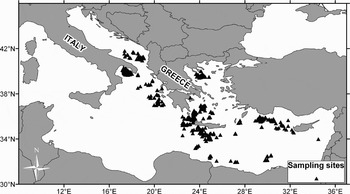
Fig. 1. Map showing the areas used for sampling blue shark, Prionace glauca, during 1998–2003 in the Mediterranean Sea (Adriatic Sea, Ionian Sea, Aegean Sea and Levantine basin). Sampling sites are indicated as black triangles.
Size, sex-ratio and sexual maturity
Size measurements were recorded for each specimen including total length (LT), total weight (WT) and dressed weight (WD). Total length was measured in a straight line from the snout tip to the end of the upper caudal lobe (the straight measurement). Whenever length measurements were not possible—because fish were gutted, finned and decapitated—the following conversion formula was used to derive total length from dressed weight:
For 501 blue sharks, the reproductive system (claspers and testis in males and ovaries and uteri in females) was inspected visually to determine the sex and stage of sexual maturity. The sex-ratio (females:males) was analysed by area and size. The χ2 goodness-of-fit test (a = 0.05; Zar, Reference Zar1996) was used to examine significant differences in the observed sex-ratios from the expected 1:1 sex-ratio. Sexual maturity in males was evaluated on the basis of the morphology and rigidity of claspers and comparing clasper length to pelvic fin length. Macroscopic observations of ovaries and uteri were taken into consideration for assessing the maturity of females (Holden & Raitt, Reference Holden and Raitt1974; Pratt, Reference Pratt1979). Ovary weight in grams and oocyte diameters in mm using Quantimet 500/W (Leica, UK) image analyser were measured for 13 females.
Tissue samples from testes (N = 50, 90–235 cm LT) and ovaries (N = 50, 70–299 cm LT) were fixed in Bouin's solution for histological analysis. Then, they were dehydrated in ethanol and embedded in paraffin wax. Sections, 5 µm thick, were stained with haematoxylin and eosin, Mallory's trichrome, and periodic acid-Schiff reagent (Pas).
Length at 50% maturity was determined through the fitting of maturity gives. The percentages of mature individuals per length-class were estimated for males and females separately. The specimens in Stages I and II were considered as ‘immature’ while the specimens in other stages were considered as ‘mature’. A logistic curve was fitted to the data and the total length at which 50% of individuals were calculated (King, Reference King1995) using the equations:
where Pi is the proportion of mature individuals in length-class i and a and b are fitted parameters which can change during the life cycle. The mean length at sexual maturity was calculated as L50 = a/b (Spare & Venema, Reference Spare and Venema1992).
Age and growth estimation
A section of the vertebral column, usually 5–6 vertebrae from the precaudal notch area, was removed and frozen in a plastic bag labelled with data for that individual. The individual vertebrae were subsequently separated, and the centra were cleaned and left to dry at room temperature.
Two methods were applied for age reading: (a) X-ray radiography (Cailliet et al., Reference Cailliet, Martin, Kusher, Wolf, Welden, Prince and Pulos1983; Cailliet, Reference Cailliet, Pratt, Gruber and Taniuchi1990). All centra were X-rayed with the corpus calcareum facing the tube at a set distance of 100 cm. At 50 mA, exposure times ranged from 0.3 to 0.8 seconds and voltage from 28 to 34 kV. The X-radiographs were then scanned with an HP scanner and Adobe photoshop software. Image Analysis Pro Plus 3.1 (Media Cybernetics) was used to enhance the images; and (b) digital enhanced images of whole vertebrae. Whole vertebral face images were obtained under reflected light using a SONY Exwave HAD digital camera at a resolution of 768*576 pixels and Image Analysis Pro Plus 3.1 video capturing software. The images were subsequently digitally enhanced. Growth band reading was easier examining whole vertebrae face images than the X-radiograph images and therefore this method was selected for age estimation (Figure 2).

Fig. 2. (A) Image of a blue shark vertebra using X-ray radiography. Growth bands are annotated; (B) digital enhanced image of a blue shark whole vertebral face obtained under reflected light. The birth band is shown.
Growth rings on vertebrae were defined as band pairs of one opaque (calcified) and one translucent (less-calcified) band. Birthmark, the first opaque band distal to the focus, coincides with a slight angle change on the centrum face. Band pairs were counted to assign an age to each specimen by two readers, since tagging experiments with oxytetracycline in the North Atlantic confirmed their annual periodicity in formation (Skomal & Natanson, Reference Skomal and Natanson2003). For samples in which readings did not coincide, the procedure was repeated. If no agreement was achieved the sample was omitted from analysis. From a total of 54 samples examined, four (7.4%) were excluded from the analysis.
To convert length measurements to age estimates growth parameters were calculated based on the Von Bertalanffy growth model: Lt = L∞ [1–exp(–k(t–to))] where Lt is length at age t, L∞ is the asymptotic length, k is the growth parameter and t0 is a theoretical age at which the length is equal to zero. The equation was fitted using the non-linear regression procedure of Statgraphics (Statistical Graphics Corp.). The life span was defined as the time required to attain 95% of the L∞ using the Taylor's equation: A95 = to + [loge(1–0.95)]k−1 (in Skomal & Natanson, Reference Skomal and Natanson2003).
RESULTS
Length distributions and sex-ratio
A total of 870 blue sharks were measured during the period 1998–2003, ranging in total length from 70–349 cm (Figure 3). Out of 501 specimens sexed, 323 (64.3%) were males and 178 (35.7%) females. Overall males outnumbered females by a sex-ratio (females:males) of 1:1.8, which was significantly different from a 1:1 sex ratio (χ 2 test, P < 0.05). Sex-ratio favoured males in all areas examined ranging from 1:1.3–1:3.0 (Figure 4A). Males ranged in total length from 74–330 cm (mean = 162.8 cm) and females from 70–349 cm (mean = 156.3 cm). There was a statistically significant difference between the two length–frequency distributions (males–females) (Kolmogorov–Smirnov test, P < 0.05) and sex-ratio showed an increase in the proportion of males in larger size-groups (Figure 4B).

Fig. 3. (A) Length–frequency distribution for the blue shark, Prionace glauca, sampled in the Mediterranean Sea during 1998–2003; (B) length–frequency distribution for the blue shark sampled in the Mediterranean Sea during 1998–2003 by area.

Fig. 4. Sex-ratio for the blue shark, Prionace glauca, sampled in the Mediterranean during the period 1998–2003; (A) per area (1, Aegean Sea; 2, Levantine basin; 3, Ionian Sea; 4, Adriatic Sea.); (B) by length-class. *Denotes statistically significant difference from 1:1.
Reproductive organs and histological observations
(a) Testis: the histological sections showed the presence of a complete spermatogenesis with spermatozoa and deiscent cysts in all the 50 samples examined. Each spermatocyst was constituted by spermatoblasts at the same stage of differentiation bounded by a basement membrane. A single Sertoli cell and its cohort of germ cells constituted the spermatoblast.
The primary spermatogonia, located in the germinal zone, did not occur within spermatocysts while secondary spermatogonia, located within spermatocysts, were surrounded by Sertoli cell processes. Sertoli cells nuclei lined the spermatocyst lumen (Figure 5 A, B). More developed spermatogonial cysts showed the migration of the Sertoli cell nuclei from their position lining the lumen to lie just internal to the basement membrane. The spermatocysts in the following development stage contained primary spermatocytes showing nuclei with granular, dark staining chromatin. Secondary spermatocytes, contained in secondary spermatocyte cysts, could be identified for their smaller and compact nuclei (Figure 5C). The cysts in the following stage, containing spermatids, were characterized by nuclei further condensed (Figure 5D). The last differentiation stage of the spermatocysts showed Pas positive sperm heads, progressively packed, arranged around the periphery of the cyst (Figure 5E). Finally, spermatocysts containing mature spermatozoa disintegrate (Figure 5F), discharging their contents into the interstitial spaces of the testis.

Fig. 5. Histological sections of blue shark, Prionace glauca, testis showing: (A) the germinal zone (gz) and spermatocysts in different stages of differentiation (arrows). Haematoxylin and eosin. Scale bar = 200 µm; (B) secondary spermatogonia spermatocysts. Sertoli cells nuclei (arrows) line the cyst lumen. Arrowhead, basement membrane. Haematoxylin and eosin. Scale bar = 30 µm; (C) two spermatocysts containing primary spermatocytes (left) and secondary spermatocytes (right). Arrows, basement membranes. Haematoxylin and eosin. Scale bar = 30 µm; (D) spermatocysts containing spermatids. Arrow, basement membrane; arrowhead, Sertoli cell nucleus. Haematoxylin and eosin. Scale bar =30 µm; (E) spermatocysts containing spermatozoa. Arrows, Pas positive sperm heads. Scale bar = 30 µm; (F) deiscent spermatocyst (ds) and spermatocysts containing spermatozoa progressively packed. Haematoxylin and eosin. Scale bar = 30 µm.
(b) Ovary: the histological observations of 50 samples demonstrated the presence of oocyte follicles in different stages of development in a connective tissue stroma (Figure 6A). The follicles wall consisted of the zona radiata and a granulosa composed by a pseudo-stratified columnar epithelium (Figure 6B). The theca, separated by a basement membrane from the granulosa, was constituted by an inner layer of blood vessels and connective tissue cells, a thin investment of collagenous fibres and fibroblast, a layer of flat tubules lined by contiguous cells resembling a simple cuboidal epithelium (which is continuous with the outer tubule lining of squamous cells) and outermost blood vessels in loose membranes of connective tissue. Developing oocytes gradually accumulated yolk vesicles in the ooplasm (Figure 6C). The presence of ‘corpora lutea’ has been shown in all the ovaries observed. An empty, collapsed and contracted follicle with a thick theca and a folded granulosa characterized ‘corpora lutea’.

Fig. 6. Section of a blue shark, Prionace glauca, ovary showing: (A) unyolked oocytes in different stages of development (arrows) in a connective tissue stroma. Haematoxylin and eosin. Scale bar = 30 µm; (B) a yolked oocyte. Arrow, granulosa cells; n, nucleus. Mallory's trichrome. Scale bar = 30 µm; (C) a particular of the yolked follicle. Arrows, yolk vesicles; arrowhead, zona radiata; gc, granulosa cells. Haematoxylin and eosin. Scale bar = 30 µm.
Sexual maturity
Observations on the reproductive organs in relation to body length revealed that all females smaller than 120 cm LT had immature ovaries with no maturing oocytes, while mature ovaries with visible yolky oocytes were present in specimens larger than 203 cm LT (Figure 7). Ovary weight varied from a minimum of 4 g for immature to a maximum of 137 g for mature specimens. In the ovary of mature females a cohort of about 100 large oocytes of almost equal diameter dominated the hundreds of smaller oocytes. Maximum oocyte diameter measured was 21.1 mm in mature females.

Fig. 7. Blue shark Prionace glauca, ovaries in different maturity stages. 1: Stage I, immature ovary with no visible oocytes; 2: Stage II, ovary with small, visible oocytes; 3: Stage III, mature ovary with large yellow oocytes. Scale bar = 50 mm.
All males smaller than 125 cm LT were immature presenting non-calcified claspers that did not reach the posterior end of the pelvic fins. Males larger than 187 cm were mature, having heavily calcified claspers, which extended beyond the posterior end of the pelvic fins. Sexual maturity data showed that males mature at a slightly smaller length than females. Length-at-first maturity was 187 cm LT for males and 203 cm LT for females, while length at 50% maturity (L50) was estimated to be 202.9 cm LT for males and 214.7 cm LT for females (Figure 8).

Fig. 8. The logistic relathionships between the percentage of sexually mature specimens and total length for male and female blue shark, Prionace glauca, sampled in the Mediterranean Sea during 1998–2003.
Age and growth
Vertebrae from a total of 54 blue sharks, ranging from 81.7 to 315 cm in LT, were examined. Determining the birthmark was straightforward for all cases and a thick opaque band was deposited just after the edge of the mark. Age estimates ranged from 1 to 12 years. The oldest fish in our sample was a male of 315 cm LT, while the youngest one was a female of 81.7 cm LT. Mean observed lengths at age are shown in Figure 9 while growth parameters with 95% confidence intervals and correlation matrix are shown in Table 1. Using Taylor's method the longevity was estimated at 22.4 years. Converting length–frequency to age–frequency distribution it was found that most of the blue shark sampled was juveniles from one to three years old (Figure 10). Age at 50% maturity was estimated at 4.9 years for males and 5.5 years for females.

Fig. 9. Length at age estimates for 50 blue sharks, Prionace glauca, sampled in the Mediterranean Sea during 1998–2003. Mean total lengths at estimated ages are shown.

Fig. 10. Age–frequency distribution for the blue shark, Prionace glauca, sampled in the Mediterranean Sea during 1998–2003.
Table 1. Von Bertalanffy growth parameters for the blue shark, Prionace glauca, sampled in the Mediterranean Sea.

DISCUSSION
This study provides the first data on the biological and demographic characteristics of blue shark in the Mediterranean Sea. Our findings revealed that the blue shark enters the swordfish and albacore longline fishery in its first year of life, and the catches consist of both juveniles and adults of both sexes. Especially, more than 80% of the blue shark catches in the eastern Mediterranean Sea were composed of immature and maturing specimens from 1–4 years old, while less than 20% were composed of mature specimens. Several studies in the Atlantic Ocean showed different catch compositions in relation to geographical area. Catch records from the eastern North Atlantic largely comprise neonates and juveniles of both sexes and adult females (Aasen, Reference Aasen1966; Stevens, Reference Stevens1975, Reference Stevens1976; Connett, Reference Connett, Casey, Pratt, Kohler and Stillwell1987) while other studies in the western North Atlantic have shown that the catches were dominated by juveniles, adult males and sub-adult females (Pratt, Reference Pratt1979; Casey, Reference Casey, Grosslein and Azarovitz1982).
The sex-ratio observed in the Mediterranean, constantly in favour of males, was similar to that reported by Buencuerpo et al. (Reference Buencuerpo, Rios and Moron1998) in the eastern North Atlantic Ocean and the Strait of Gibraltar but inverse to those reported in the western North Atlantic (Pratt, Reference Pratt1979) and eastern North Atlantic in British and Ireland waters (Stevens, Reference Stevens1976; Henderson et al., Reference Henderson, Flanery and Dunne2001). Particularly, the sex-ratio reported in Irish coastal waters was heavily biased in favour of females (Whelan, Reference Whelan1991), agreeing with Pratt's (Reference Pratt1979) speculation that male blue sharks move inshore only upon attaining sexual maturity. Our data showed that the sex composition of blue shark catches varied spatially. Males dominated in catches, especially in the Levantine basin where the average total length (216.4 cm) was also higher than in the other areas. We assumed that variations in sex composition of catches might partly reflect different natural distributions of the sexes and sizes, possibly resulting from sexual differences in reproductive behaviour.
Gravid females were not recorded in our sample, however, the substantial presence of mature male and females as well as personal observations of gravid females with embryos during previous investigations in the Ionian Sea support the opinion that the Mediterranean is an area of reproduction for blue shark. Earlier studies in the Mediterranean Sea have shown that the Adriatic is a nursery area and gravid females have been observed both in the Adriatic and Ionian Seas (Bianchi personal communication; Bianchi et al., Reference Bianchi, Clò and Costantini1997; Pomi, Reference Pomi1997). In the Mediterranean, Megalofonou et al. (Reference Megalofonou, Yannopoulos, Damalas, De Metrio, Deflorio, de la Serna and Macias2005b) reported a minimum size of 55 cm for free swimming blue sharks. Pratt (Reference Pratt1979), in discussing the reproduction of blue shark in the Atlantic Ocean, suggested that gravid females occupied a niche that is different from that of the rest of the blue shark specimens. Unfortunately, very little is known of this important part of the blue shark life cycle.
The histological observation of the gonads revealed that gametogenesis in the blue shark occurs according to the model already described in other elasmobranchs (TeWinkel, Reference TeWinkel1972; Wourms, Reference Wourms1977; Parsons & Grier, Reference Parsons and Grier1992). The microscopic anatomy of ovaries showed the presence of ‘corpora lutea’ in all the samples observed, including those macroscopically classified as immature. This observation, in agreement with preceding findings on other elasmobranch species (Hisaw & Hisaw, Reference Hisaw and Hisaw1959; Lance & Callard, Reference Lance and Callard1969), makes obvious that ‘corpora lutea’ can be originated both by pre- and post-ovulatory follicles. Even so, the presence of mature oocytes in the ovary was a reliable indicator of sexual maturity in female blue shark. The microscopic anatomy of the testis showed that the various stages of spermatocyst development, as described by Maruska et al. (Reference Maruska, Cowie and Tricas1996) were present in our samples. Especially, a complete spermatogenesis was revealed in all the testes examined, including those belonging to specimens macroscopically immature (small cylindrical testes, reproductive tract not completely differentiated and undeveloped claspers). Therefore, it was deduced that the microscopic appearance of the immature testis is quite similar to that of the mature testis. The absence of variation in the testicular appearance makes it necessary to have a different approach to determine the length at sexual maturity, such as state of the claspers and/or spermatophore development, as noted by Pratt (Reference Pratt1979).
Cortés (Reference Cortés2000), who examined 164 shark species, concluded that, on average, shark maturity begins at about 75% of their maximum size. In this study, blue shark did not follow this general value, with both males and females maturing at a smaller size, especially in the case of males, where mean length at first maturity (202.9 cm LT) was estimated to be 55.1% of the maximum observed size (368 cm LT) in the Mediterranean (Megalofonou et al., Reference Megalofonou, Yannopoulos, Damalas, De Metrio, Deflorio, de la Serna and Macias2005b). Female blue shark seemed to mature at a relatively larger size and therefore mean length at first maturity (214.7 cm LT) was estimated to be 58.3% of the maximum observed size in the case of females. The fact that size at first maturity was higher for females than males seems to be common for sharks in general, and had previously been observed for blue shark in the Atlantic Ocean (Pratt, Reference Pratt1979). However, prior reported estimates of body size at sexual maturity for blue shark in the Pacific and Atlantic Oceans were slightly larger compared with ours. Regional differences in size-at-maturity have been reported for a number of other elasmobranch species, including widely distributed species such as the scalloped hammerhead shark, Sphyrna lewini (Chen et al., Reference Chen, Leu, Joung and Lo1990), dusky shark, Carcharhinus obscurus (Natanson & Kohler, Reference Natanson and Kohler1996) and shortfin mako, Isurus oxyrinchus (Mollet et al., Reference Mollet, Cliff, Pratt and Stevens2000).
According to the estimations of Stevens (Reference Stevens1976), Pratt (Reference Pratt1979) and Castro & Mejuto (Reference Castro and Mejuto1995), North Atlantic female blue shark do not mature until after 220 cm LT. Pratt (Reference Pratt1979) proposed that sexual maturity in male blue sharks occurs at a size of 215 cm LT (183 cm LF) when 50% possessed spermatophores, and this would coincide with an age of 4–5 years. Females pass through a subadult phase (145–185 cm LF) when the organs for copulation and sperm storage are developed but the ova are undeveloped, becoming fully mature by 5 years, at a minimum size of 185 cm LF (217 cm LT)[J1]. Taking into consideration our results on age-at first maturity, it can be noted that sexual maturity in the Mediterranean blue shark occurs almost at the same age as in the Atlantic blue shark. In conclusion our results on size and age at first maturity, suggest that blue shark in the Mediterranean Sea possibly reach sexual maturity in a smaller size than blue shark in the Atlantic Ocean but at a similar age.
Our dataset for age and growth estimations suffered from the absence of very small and very large blue sharks. The estimation of the growth function parameters is quite poor, owing to small sample size, and perhaps difficulty in interpretation. Nevertheless, a comparison among 5 different growth curves from four earlier studies in the Atlantic and Pacific Oceans indicated that our curve is similar, being closer to Nakano's curve for Pacific blue sharks (Figure 11; Tables 2 & 3). The maximum size reported for blue shark in the Atlantic Ocean (400 cm LT; in Froese & Pauly, Reference Froese and Pauly2005) is very close to our estimation. Stevens (Reference Stevens1975) used both centrum bands counts and Aasen's (Reference Aasen1966) size–frequency data to generate growth curves and to estimate asymptotic lengths for sexes combined. He found L∞ values of 395 and 423 cm LT respectively. Both values are quite similar to the asymptotic length we found (401.5 cm LT). Likewise, the coefficient k estimated in this study is found inside the range of the values (0.11 to 0.17) estimated by other researchers. Hence, Mediterranean blue sharks seem to have quite similar growth rates to that reported in the Atlantic and Pacific Oceans.

Fig. 11. Comparison of Von Bertalanffy growth curves estimated through vertebral analysis for the blue shark, Prionace glauca, from literature and present study.
Table 2. Von Bertalanffy growth parameters and age-range in the blue shark, Prionace glauca, derived from vertebral band studies by location and sex.

* , total length was derived from fork length conversion TL = 1.203*FL –1.67 (Skomal et al., Reference Skomal and Natanson2003);
** , total length was derived from body length conversion BL = 0.746*TL (Compagno, Reference Compagno1984).
Table 3. Estimates of the total length at age in the blue shark, Prionace glauca, from vertebral band studies by location and sex.

* , body length converted to total length using BL = 0.746*TL (Compagno, Reference Compagno1984); **, fork length converted to total length using TL = 1.203*FL –1.67 (Skomal et al., Reference Skomal and Natanson2003).
Various authors have studied age and growth of the blue shark, but very few have estimated the lifespan. The maximum age and lifespan of the blue shark in the Mediterranean Sea seems to be higher than those in the Atlantic Ocean. Skomal & Natanson (Reference Skomal and Natanson2003) using the Taylor method found that the age at which 95% of the L∞ is reached was 16.5 years.
Calculated size at birth was 30.6 cm LT, corresponding to a t 0 of –0.62. For large carcharinids like blue shark, the Von Bertalanffy t 0 parameter could be considered as an indication of the gestation period, being 0.62 years or almost 8 months. Compagno (Reference Compagno1984), after consulting several researchers, suggested that the gestation period is about 9 months and size at birth is more likely to be in the range of 35–44 cm LT.
Of course, blue sharks living under different oceanic conditions could exhibit different growth and life history characteristics. The examination of a larger sample in the future with greater samples per age-group could provide better results in age estimations and growth parameters. Moreover, population genetic studies and further tagging programmes could give more definitive answers to the questions about the stock structure of blue shark in the Mediterranean Sea. The differences found in growth and maturity between this and previous studies for the blue shark are most likely because the Mediterranean population is discrete from that elsewhere. Already, for fisheries management purposes, the population in the Mediterranean is considered independent of the North and South Atlantic population by the International Commission for the Conservation of Atlantic Tunas (ICCAT) and the European Scientific, Technical and Economic Committee for Fisheries (STECF), however, the extent of exchange between these populations (if any) is poorly understood (Soldo et al., Reference Soldo, Megalofonou, Bianchi, Macias, Cavanagh and Gibson2007). Unfortunately, the Mediterranean Sea seems to be a relatively overlooked area of research for blue shark and sharks in general. Therefore, our results, which indicate that the main bulk of the blue shark caught in the Mediterranean has not reached maturity, are of great concern and reinforce the need for global assessment and management measurements in the area.
ACKNOWLEDGEMENTS
This study was performed with the financial aid of the Commission of the European Communities and does not necessarily reflect the views of the European Commission. We wish to thank all the fishermen and observers who assisted in data collection, both on-board fishing boats and at landing locations.


